


Abstract
Background. Available statistical data from 2015 show that 28% of pregnancies in developed countries end in cesarean section (CC). Discomfort associated with the scar after surgery is a common complication.
Objectives. This study aimed to evaluate the changes in the structure of the cesarean scar after the application of a scheme of manual therapy.
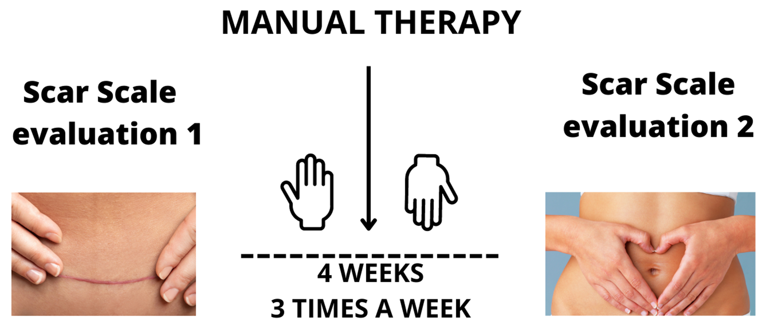
Materials and methods. The study included 15 women in the treatment group (TG) and 15 in the control group (CG). The scars were evaluated twice at 5-week intervals with the use of quantitative scales: the Vancouver Scar Scale (VSS), the Manchester Scar Scale (MSS) and the Patient and Observer Scar Assessment Scale (POSAS). During each examination, the scar was compared, using the specified criteria, to the physiological skin, i.e., the tissues directly bordering the incision. During therapy, 8 manual techniques were used during a 4-week program consisting of 30-minute sessions 3 times per week.
Results. Patients in the TG showed a statistically significant improvement in all of the analyzed characteristics of the scar. A statistically significant difference was also observed between the results obtained during the 2nd examination (after the therapy) in the TG and the CG.
Conclusions. As a result of the therapy, the condition of the scar in the TG significantly improved. Onerous scar-related symptoms were alleviated. The vascularity, hyperpigmentation and distortion of the scar were reduced. The elasticity and pliability of the scar increased, and the height of the scar decreased. The texture, finish and contour of the scar improved. Obtained results suggest that manual therapy of the scar after CC should be a part of the treatment in women during the postpartum period.
Key words: cesarean section, women’s health, scar therapy
Background
According to the recommendations of the World Health Organization (WHO), the percentage of pregnancies delivered by cesarean section (CC) should not exceed 10–30%. Statistical data show that in Organization for Economic Co-operation and Development (OECD) countries (36 highly developed countries of the world), the number of deliveries ending in CC increased from 20% in 2000 to 27.9% in 2015.1 Globally, the CC rate has also been rising – it grew from 12.1% in 2005 to 21.1% in 2015. In Western Europe, 19.6% of pregnancies were delivered by CC in 2000, and by 2015, this rate increased to 26.9%.2
The decision to perform a CC is made more frequently due to the early detection of hazards, an increasing number of multiple gestations and preterm births, as well as late births (in terms of the mother’s age). In addition to obstetric indications, the procedure is performed for indications related to ophthalmic, orthopedic, cardiac, neurological, and psychiatric issues.1, 3 Pregnant women are often convinced that a CC is the easier, safer and painless method of delivery, better than natural childbirth. The most frequently employed technique is the transperitoneal CC, involving a transverse lower abdominal incision through the skin, the subcutaneous tissue, the peritoneum over the uterus, and the uterus. Cutting through muscle fibers is currently avoided.4 The findings of Cromi et al.3 demonstrated that the type and technique of surgical closure used following the procedure has no impact on the appearance of the scar evaluated using the Vancouver Scar Scale (VSS) and the Patient and Observer Scar Assessment Scale (POSAS).
The wound healing and scar formation processes begin with hemostasis, followed by inflammation, proliferation and remodeling. They take place over a period of up to 2 years, and their duration depends on the etiology of the wound. The process consists of the conversion of type III collagen to type I collagen. The finer and less ordered structure of type I collagen both strengthens the tissue and decreases its elasticity. This process can be distorted by several factors.5
Remodeling abnormalities can cause the formation of hypertrophic, keloid or atrophic scars. Women who underwent a CC are at an increased risk of developing pathological lesions due to hormonal activity, taking care of an infant, a change in the body’s center of gravity, and genetic makeup.
Despite a growing interest in pregnancy and childbirth, women often overlook the needs of their own bodies, focusing instead on the everyday chores related to taking care of a baby. Such situation can cause the changes in the locomotor system that solidify during pregnancy. Painful scar and a reflex to protect the injured area cause the woman to assume antalgic positions, enhancing and solidifying incorrect posture patterns.6 Additionally, pain predisposes the woman to assume a forward flexion posture and avoid tensing the abdominal wall muscles, which causes further weakening of the muscles that become stretched during pregnancy. Women report various scar-related complaints, such as itching, pulling, pain, numbness (hypoesthesia) or hyperesthesia in the scar area, pricking, burning, tingling, and stinging.
There are no uniform standards for procedures in cesarean scar therapy. Literature mentions the beneficial impact of massage on the rate of scar remodeling and pain alleviation.7, 8 Wong et al. reported the effectiveness of delicate scar mobilization in combination with exercises for chronic pain.9
Scar-related complaints can cause physical problems and psychological issues, consisting of malaise and a lack of acceptance of the scar and oneself. In critical situations, the patients see the scar as a non-existent, unaesthetic element of their body that restricts their functioning.10 Physical complaints and a lack of acceptance of oneself can also cause women to limit or discontinue physical exercises.11 Postoperative scars are often overlooked in the postoperative period. Currently, numerous operations are carried out in the abdominal area. Not all possible ailments caused by the presence of scars are unequivocally investigated. Cesarean section is one of the most numerous operations. Each interruption in tissue communication is traumatic for the body and results in psychosocial and purely physical ailments. One of the research directions related to scar treatment is manual therapy, which, due to its nature, is one of the simplest, cheapest and most accessible methods. According to the available literature, there are indications of the effects of manual therapy on scars, but the research results are inconsistent. The evidence for the use of scar massage is weak, the regimens vary, and the measured results are neither normalized nor objective. Although scar massage is anecdotally effective, there is limited scientific data in the literature to support it.12 The review by Wasserman et al. shows preliminary strong evidence of the benefit of soft tissue mobilization for symptoms associated with acute postoperative adhesions, preliminary moderate evidence of the benefit of soft tissue mobilization for symptoms of chronic inoperable adhesions, and moderate evidence of the benefit of using soft tissue mobilization for symptoms associated with chronic postoperative adhesions.13 Although most postoperative adhesions are clinically silent, the consequences of adhesion formation can be a lifelong problem, including chronic abdominal pain, recurrent bowel obstructions requiring multiple hospitalizations, and infertility. Moreover, adhesion disease can become a chronic condition with significant morbidity, and lacks effective treatment. Despite recent advances in surgical techniques, there is no reliable strategy to treat postoperative adhesions.14 It is vital to look at the human body comprehensively. With the increasing number of CCs, the impact of scarring on the overall health of patients is increasingly observed. Abdominal stretch marks and cesarean scars are considered important predictors of intraperitoneal adhesions. Women with significant stretch marks had thick intraperitoneal adhesions. Women with intraperitoneal adhesions had more vascularized, discolored, less flexible, and raised scars. Therefore, verifying the effect of scars and the possibility of their modification, for example, to reduce adhesions, is of utmost importance.15 Tapes, used in the prevention of surgical scars, effectively reduce scars and display features of growth, color and itching.16, 17 An overview of the methods used in physical therapy on scars is discussed in the review by Deflorin et al.18 Overall, 1 meta-analysis showed that the effect on the scar by different physiotherapeutic means has a significantly positive influence on pain, pigmentation, suppleness, itching, surface area, and scar thickness.18 There are reports stating that therapy has a clear positive effect on the state of scars and the overall wellbeing of patients.19 The current state of research does not allow for a direct transfer of the above research results to clinical treatment of patients with big scars. However, the continued clinical implementation of the results obtained in studies with respect to the mechanical sensitivity of isolated fibroblasts and continuous adaptation of manual techniques created an evidence basis for manual scar therapy. Manual doses are adapted to the physiology of the tissue and the respective phases of wound healing. Clinical observations show improved mobility of affected areas and fewer relapses of the inflammatory phase due to mechanical overload.20
Objectives
This study aimed to evaluate the changes in the structure of the cesarean scar as a result of an applied scheme of manual therapy.
Materials and methods
Study design
This study was designed to investigate the influence of our scheme of manual therapy on scar recurrence after a CC. In this study, vascularity, pigmentation, pliability, height pain, itching, color, stiffness, thickness, texture, and overall condition of the scar were evaluated in patients using subjective scales.
Setting
The trial has been registered and allocated in the Australian New Zealand Clinical Trials Registry (No. ACTRN12620000115932, registration date: February 7, 2020). Data were collected from January 7, 2019 to May 12, 2019. The first participant’s enrollment date was January 7, 2019, and the enrollment date of the last participant was April 12, 2019.
Participants
The criterion for inclusion in the treatment group (TG) was the lack of any prior work on the CC scar. All participants (n = 30) were randomly assigned to 2 same-sized groups – the TG that participated in the therapy of the scar and the control group (CG) that did not undergo therapy. Demographic characteristics of both groups are presented in Table 1. Patients were recruited according to the diagram depicted in Figure 1, but the sample size was established a priori using G*Power 3.1 software (v. 3.1.9.2; G*Power, Kiel, Germany).21 The expected effect size (ES) was set at 0.85 (Cohen’s f), the α level was set at 0.05 and the power was set at 0.8.22 The minimum group size was 10 participants, but the final recruitment consisted of 15 women in each group.21
Variables
The experiment consisted of 2 examination meetings and 12 therapy sessions. During the examination meetings, the scar of each participant was assessed visually and by touch using quantitative scar evaluation scales: VSS and Manchester Scar Scale (MSS). Additionally, the scar was assessed by each patient using the POSAS Patient Scale. The VSS was used to evaluate vascularity, pigmentation, pliability, and height. The MSS evaluated color, finish, contour, distortion, and texture. The subjective POSAS Patient Scale was utilized to evaluate pain, itching, color, stiffness, thickness, texture, and the overall condition of the scar.23
Data measurement
The patients from the TG underwent scar therapy 3 times per week for 4 weeks. The therapy consisted of twelve 30-minute sessions carried out 3 times per week. During each session, the therapist applied 8 manual techniques in the following order: stroking, superficial rubbing using spiral and transverse motions, deep rubbing using spiral and transverse motions, moving, rolling, breaking up, and pinching. The order and intensity of the techniques were adjusted to the condition and the response of the treated tissues. The visualization of the scars before and after therapy was conducted using a thermal imaging camera FLIR T335 (Merlin Lazer, Crowborough, UK).
Bias
The only criterion for inclusion in the study was the absence of any previous work on the cesarean scar to avoid misinterpretation of the obtained results.
Study size
The study size was determined by the preliminary character of this project. An investigation conducted on a larger group of patients is currently under consideration as a part of a dedicated grant.
The therapy and all examinations were carried out at the Scientific and Research Laboratory of the Faculty of Physiotherapy at Wroclaw University of Health and Sport Sciences (Poland). The trial was approved by the Senate Committee on Ethics of Scientific Research at the Wroclaw University of Health and Sport Sciences (approval No. ACTRN12620000115932).
Quantitative variables
The MSS and VSS were used to evaluate the scar before and after the therapy. On both scales, each characteristic of the scar was given a specific score. The higher the final score, the worse the condition of the scar. The maximum score on the MSS was 18 points, and on the VSS it was 13 points. The subjective scar-related experiences of the participants were evaluated twice with the use of the POSAS Patient Scale.23 Each described characteristic was given a score from 1 to 10 (the higher the score, the worse the result). The maximum score on that scale was 70. The median was calculated for the results obtained before and after the therapy in both evaluated groups.
Statistical analyses
The statistical calculations were made using Statistica v. 13.1 (StatSoft, Inc., Tulsa, USA). The Shapiro–Wilk test was used to check the distribution of all quantitative variables. Due to the ordered nature of the survey results and the lack of normality in the distribution of the quantitative variables, the median was used as a measure of central tendency, and the interquartile range (IQR) as a measure of dispersion.
The results were analyzed with the use of statistical tests for ordinal variables: the Mann–Whitney U test and the Wilcoxon matched-pairs test. The statistical significance of the differences in the appearance and structure of the scar in both groups before and after the therapy, as well as between the TG and CG before and after the therapy, were assessed. Due to the occurrence of multiple comparisons, the Bonferroni correction was applied to reduce the risk of a type I error. The assumed significance level (0.05) was divided by the number of comparisons (4). The adjusted significance level was set at 0.0125.
Results
The median was calculated for each analyzed parameter in both evaluated groups (Figure 2, Figure 3). Using the MSS, we observed that the color, finish, contour, distortion, and texture were statistically significantly improved. According to the VSS, vascularity, pigmentation, pliability, and height were statistically significantly improved. According to the POSAS Patient Scale, color, stiffness, thickness, texture, and overall opinion were statistically significantly improved. Pain and itching did not change. The median was calculated for the results obtained before and after the therapy in both evaluated groups (Figure 4).
The MSS, VSS and POSAS Patient Scale scores obtained during the first examination were not statistically significantly different between the TG and CG. Statistically significant differences were observed between the groups during 2nd examination. The scores obtained during both examinations for each of the scales were different in a statistically significant manner only in the TG (Table 2, Table 3). Representative photos of the results of manual therapy are presented in Figure 5. Generally, as a result of therapy, the condition of the scar in the TG significantly improved. Onerous scar-related symptoms were alleviated. The vascularity, hyperpigmentation and distortion of the scar were reduced, the height of the scar decreased, and the elasticity and pliability of the scar increased. The texture, finish and contour of the scar improved.
Following the results from the thermal imaging camera, these were pilot studies. So far, the thermal imaging camera has been used, for example, to verify the effect of thermal factors on the scar and the course of the wound healing process,24, 25 but not to verify the effect of manual therapy. The effects of therapy on temperature changes cannot be unequivocally determined due to the different types of scars.
Atrophic, hypertrophic and keloid scars differ in their initial temperature distribution. In addition, the use of manual methods varies in proportion over time. To draw specific conclusions, the study group should be broadened and differentiated according to the types of scars.
No statistically significant changes were observed, but a certain trend in temperature change was noted, which requires further investigation. In all cases, the temperature of the scar area after treatment was close to that of the surrounding tissues.
We are planning to conduct a research in a larger group of patients, taking into account various diagnostic and therapeutic methods to compare their effectiveness.
Discussion
Globally, 21.1% of pregnancies were delivered by CC in 2015.2 Women are not educated on scar care and the possible side effects of failing to undergo or discontinuing therapy. The scar- and adhesion-related symptoms are not associated with the presence of those lesions in any way by the physician or patient.26 Onerous symptoms included a pulling sensation and pain during the performance of everyday chores, as well as pain originating in the lumbar vertebral column, the gastrointestinal tract and the scar itself. Furthermore, an important complaint related to the cesarean scar is the aesthetic aspect, i.e., the lack of acceptance of a part of the patient’s own body that has a negative impact on everyday functioning.10
According to the research performed by Chochowska et al.,26 the aforementioned complaints are alleviated or reduced after the scar therapy. Twenty-four months after the procedure, scar treatment may be less effective. Chochowska et al.26 noticed that postoperative adhesions cannot be eliminated during therapy, and each subsequent surgery increases the risk of their formation. They cause onerous symptoms resulting from the decreased glide between tissues, which may give rise to pain in the head, vertebral column, pelvis, and other regions. Pilot studies on the use of Myofascial Induction Therapy (MIT) showed that the aforementioned activities effectively change the structure of the scar despite the completion of the remodeling process.27
The most recent research suggests that it is possible to change the structure of adhesions.28 Ault et al. focused on hypertrophic burn scars.29 They claimed that massage is an effective treatment that impacts the height, vascularity, elasticity, pain, itching, and depression of scars. Anthonissen et al. compared the effectiveness of various types of therapy: pressure, silicone, massage, moisturizing creams, physical activity, and mobilization.30 They questioned the accuracy of the analyzed projects due to the small size of the evaluated groups and the lack of a detailed description of the therapy. Many authors suggest that both objective and subjective scales should be used.3, 29, 30 In our research, the changes that occurred during therapy were assessed using the VSS, MSS and POSAS Patient Scale.
The improvement in the parameters such as pigmentation, color or elasticity may result from the massage stimulating the reconstruction of connective tissue by increasing the number of collagen fibers while reducing their diameter and cross-section.31 In our research, according to the VSS, MSS and POSAS Patient Scale, scar therapy improved all evaluated parameters. It is possible that pain and itching require a different type of treatment than the ones used. O’Reilly et al. observed a reduction in itching within the studied scars.16 In our studies, no statistically significant changes were observed in these parameters. According to the results of other experiments, pain decreased after scar therapy.12, 13, 18 However, these results are not conclusive. The evidence for the use of scar massage is still not sufficient, the regimes vary, and the measured outcomes are neither standardized nor objective. However, the effectiveness of the therapy appears to be greater in postoperative scars than in traumatic or post-burn scars. Although scar massage is known to be effective, there are little scientific data in the literature to support it.12 Recently, the clinical impact of scar management has been studied more intensively.19, 20 It is important to analyze and continue research in order to determine the best procedures affecting scars. Moreover, it is worth clarifying and differentiating which therapy protocol is most effective as well as verifying what affects the effectiveness of the performed treatments, in order to improve the abovementioned parameters and the comfort of a patient’s life.
Andrzejewski noticed increased blood flow in the skin and muscles, as well as the normalization of the autonomic nervous system after scar massage.32 The results achieved during therapy indicated that massage in the form of rubbing increases the level of fibroblast growth factor (FGF) in the tissue, which causes structural changes in collagen fibers.
Another method used in scar therapy is Kinesiology Taping.16, 17, 33 The research on various types of pathological scars was conducted in 54 patients. Following a 7-day application, a change in the tissue color, an increase in its mobility and improved satisfaction were demonstrated in the majority of subjects.33
It is becoming increasingly common to use various types of topical formulations that the patients can self-administer on the scar. However, there are no solid scientific studies that would corroborate the positive influence of such formulations on the tissues.
A few studies concerning cesarean scar therapy and an increasing rate of surgical births encourage further attempts to develop standards for cesarean scar management. Abdominal striae and a cesarean scar on intraperitoneal adhesions were significant predictors of scar adhesion type. It has been observed that women with severe stretch marks had thicker intraperitoneal adhesions, whereas women with intraperitoneal adhesions had more vascularized, discolored, less pliable, and raised scars.15 Results obtained by our team could have implications for physiotherapy practice. Currently, the use of various topical preparations of varying efficacy is becoming more common in scar therapy. In many cases, they are not sufficient due to persistent scar-related ailments. The growing percentage of surgical deliveries and a small number of scientific studies on the treatment of scars after CC prompts the development of therapeutic management standards. We have obtained interesting results using a thermal imaging camera to assess scar conditions after CC. Siah and Childs noted that heat is a sign of surgical wound infection in the wound assessment criteria, but no diagnostic tool is used in clinical practice to assess the skin temperature of surgical wounds.24 Observations made by other authors showed that thermovision is a valuable tool in scar management25 and, in our opinion, it has clinically useful potential in the rehabilitation of cesarean scars. It is worth noting that postoperative adhesions, although clinically silent, can cause chronic pain and infertility14; thus, new methods of scar management are needed.
The results of our research indicate significantly positive effects of using manual techniques in scar therapy. They can be used in perinatal care, as well as in broadly understood surgery, plastic surgery, orthopedics, and traumatology, wherever scars are common.
Limitations
Our work presents the results of a preliminary study, and for this reason, the sample size was not numerous. It is worth expanding the groups of patients in the future studies and checking whether the time elapsed since the CC affects the influence of therapy over long-time follow-up. In conducted research, subjective assessment methods were used, which should be complemented by objective (quantitative) methods, allowing for a better comparison with results obtained by other researchers.
Conclusions
As a result of the manual therapy, the condition of the scar in the TG significantly improved. The presented scheme of manual therapy has the potential to be a simple and effective treatment for women after a CC.


