


Abstract
Background. Neuropathy affects 25% of people with diabetes mellitus. The evaluation of disease severity is still a challenge for modern medicine. Many screening instruments are based primarily on clinical criteria. There is a lack of a simple, reliable and precise scoring system that could improve the classification of neuropathy and monitor disease progression using not only clinical criteria but also electroneurography. There is a need to find sensitive neurography parameters that reflect peripheral nerve impairments in this group of patients.
Objectives. This study aimed to create a scoring system for diabetic neuropathy, based on electroneurography criteria, that reflects the natural course of the disease. A new scoring system will improve the treatment of patients with diabetes mellitus.
Materials and methods. A total of 113 patients with distal symmetrical polyneuropathy (DSPN) were involved in the study. Median, ulnar, sural, tibial, and peroneal nerves were examined. Parameters such as amplitude, conduction velocity, distal latency, and F wave latency were analyzed. The results of nerve conduction studies in the investigated group were compared to those of the control group, which consisted of 61 healthy volunteers.
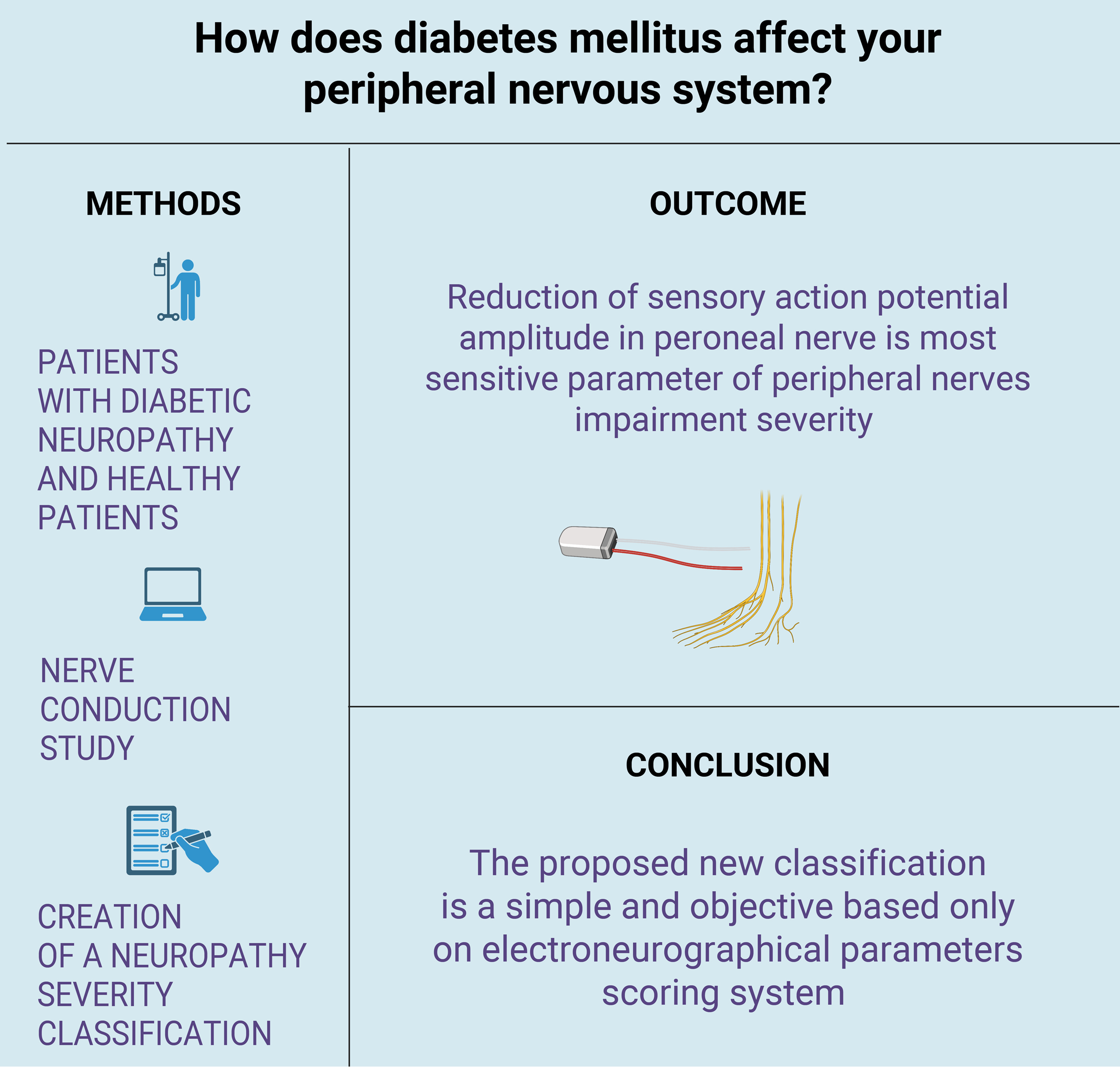
Results. The most sensitive parameter of peripheral nerve impairment severity was a reduction of the sensory action potential amplitude in the peroneal nerve by 72.8% (p < 0.05). The observation of changes in sensory action potential amplitudes in the peroneal nerve is the most important element of our scoring system.
Conclusions. A new electroneurography scoring system of DSPN severity should be based on sensory and motor action potential amplitudes that reflect axonal loss in the examined nerves and the nature of the disease.
Key words: diabetes mellitus, classification, peroneal nerve, nerve conduction studies, diabetic neuropathies
Background
Neuropathy affects 25% of people with diabetes mellitus. This disease is the leading known cause of neuropathy in developed countries and is implicated in 50–75% of nontraumatic amputations.1, 2, 3, 4 Distal symmetrical polyneuropathy (DSPN) is the most common presentation of the disease. A glove–stocking distribution of numbness, sensory loss, dysesthesia, loss of reflexes, and nighttime pain are the most common symptoms.5, 6 Ulcers, infections, as well as fractures of the knees, ankles or feet are the most common complications of the disease. Distal symmetrical polyneuropathy is associated with significant morbidity and increased mortality. The most important risk factor for developing DSPN in type 1 diabetes mellitus is poor glycemic control.7, 8, 9 Pathophysiology of the disease remains elusive, but nerve biopsies from patients with diabetic neuropathy revealed degeneration of both myelinated and unmyelinated fibers.10, 11, 12 Peripheral nerve impairment in patients with diabetes mellitus may be connected to mitochondrial malfunction. This organelle is responsible for the production of energy. Its dysfunction is connected to the activation of the tryptophan–kynurenine metabolic system that has an impact on tryptophan metabolism.13, 14, 15 Tryptophan is an endogenous amino acid necessary for protein synthesis and a precursor to the synthesis of bioactive molecules, including nicotinamide adenine dinucleotide.16 It undergoes transformations, leading to the formation of kynurenines, the role of which is to regulate the excitability of neurons as well as the response of immune cells.17 Kynurenine has a role in processing neurogenic inflammation by affecting glutamate receptors.18, 19, 20
In another study, the increased plasma levels of proinflammatory factors such as tumor necrosis factor alpha (TNF-α) and intercellular adhesion molecule 1 (ICAM-1) were connected to polyneuropathy incidence in diabetes patients.21 Metabolic changes induced by hyperglycemia lead to dysregulation of cytokine control. Another study revealed that in patients with DSPN, interleukin (IL)-1, IL-6, leukemia inhibitory factor (LIF), ciliary neurotrophic factor (CNTF), and transforming growth factor beta (TGF-β) production is more intense than in healthy patients.22, 23 A study by Behse and Buchthal revealed that in the blood of patients with diabetic polyneuropathy, the levels of IL-10 were lower than in healthy individuals.24 A clinical investigation of nerve biopsies in neuropathic patients revealed upregulated TNF-α expression in human Schwann cells.25
The diagnosis of DSPN is primarily clinical, and its most important part is the physical examination, including neurologic testing. Nerve conduction studies (NCSs) play an important role in the diagnostic process, especially in patients with minimal or no objective symptoms, and are the most useful in patients with large fiber neuropathy.26, 27, 28, 29 Other forms of neuropathy that occur in diabetes mellitus include mononeuropathies, cranial neuropathies, plexopathies, radiculopathy, amyotrophy, autonomic neuropathy, and small fiber neuropathy. Moreover, chronic inflammatory demyelinating neuropathy is more common in diabetic patients.30, 31, 32
Many screening instruments based mainly on clinical criteria have been developed to evaluate the severity of diabetic neuropathy. Most of them are time-consuming and require a precise neurological examination. There is a lack of a simple, objective scoring system using electroneurographic parameters that could help monitor disease progression with the use of objective criteria, especially in patients in the initial stage of DSPN.33, 34
Objectives
This study aimed to confirm the hypothesis that DSPN affects primarily sensory nerve fibers in the legs and leads mainly to axonal loss.35, 36 Our study is an attempt to create a simple scoring system with the use of objective parameters of nerve conduction that will reflect the natural course of the disease.37 We propose a simple, clear system of the classification of the severity of neuropathy that can be used by neurologists and other clinical practitioners to evaluate the severity of neuropathy and monitor disease progression in patients with DSPN.38
Materials and methods
This retrospective study investigated a group of 113 patients (53 females and 60 males) with DSPN due to diabetes mellitus. All patients in the investigated group were diagnosed at the Electromyography and Nerve Conduction Laboratory in Warsaw, Poland. The mean age in the group was 59.27 years (standard deviation (SD) ±13.66). Additionally, a control group of 61 healthy volunteers with a mean age of 41.70 (±18.37) years were included. The size of the study and control groups was estimated based on statistical methodology. Patients with alcohol addiction, post-oncologic treatment, kidney and thyroid diseases, and other potential causes of peripheral nerve impairments were excluded from the study. In all patients, a NCS was performed after obtaining the approval of the Bioethical Committee of Military Institute of Medicine (approval No. 14/WIM/2021). The median, ulnar, sural, tibial, and peroneal nerves were examined. Parameters such as amplitude, conduction velocity, distal latency, and F wave latency were analyzed.
Sensory studies were performed antidromically. In the case of the ulnar nerve sensory action potential, the recording electrode was placed on the 5th finger, while for the median nerve, it was placed on the 2nd finger. The sural nerve was stimulated on the posterolateral aspect of the lower third of the leg, and the distance between the recording electrode and the stimulating one was approx. 14 cm. For ulnar nerve motor fibers, the recording electrode was placed over the abductor digiti minimi muscle. In the case of median nerve motor fibers, the recording electrode was placed over the abductor pollicis brevis muscle. For the peroneal nerve, the recording electrode was placed over the extensor digitorum brevis, whereas the abductor hallucis muscle was used for the placement of the recording electrode for the tibial nerve. The distance between the recording electrode and the stimulating electrode was 8 cm in the testing of all motor nerves. Locations of stimulating and recording electrodes in the case of all examined nerves were typical, as presented in previous studies.39, 40 Motor and sensory NCSs were performed in an environment with a temperature of 21–25°C, as are the optimal parameters for electroneurography.41
The results of the NCSs in the investigated group were compared to those of the controls. Nerves were ranked depending on the degree of impairment to create the scoring system. Normal distribution of variables was obtained in the case of the conduction velocity in sensory fibers, as well as F wave latency of the median nerve, F wave latency of the ulnar nerve, conduction velocity in sensory fibers of the peroneal nerve, and conduction velocity in sensory fibers of the sural nerve. In the remaining cases, the distribution of variables was non-normal. Student’s t-test or Mann–Whitney U tests were used to statistically analyze the data.
Results
The analysis with the use of the Mann–Whitney U test revealed that both amplitude and conduction velocity of sensory action potentials of the sural, peroneal, median, and ulnar nerves were reduced in the investigated group when compared to controls. Moreover, the analysis with the use of the same test revealed a reduction in the amplitude and conduction velocity, as well as elongation of distal latency and F wave latency of motor action potentials in the tibial, peroneal, median, and ulnar nerves in comparison to controls. Based on that data, we have created diagnostic criteria for the impairment of all investigated nerves (Table 1, Table 2, Table 3, Table 4, Table 5). In the case of the conduction velocity of sensory action potentials, the differences between the 2 analyzed groups were compared using the Student’s t-test (regarding the peroneal and sural nerves). The Mann–Whitney U test was used in the case of all other nerves (Table 3).
The mean amplitude of sensory action potentials in the investigated group was significantly lower in comparison to controls. The most significant reduction of amplitude in the investigated group compared to controls was observed in the peroneal nerve (72.8%, p < 0.05 using the Mann–Whitney U test).
Moreover, the values of sensory action potential amplitudes for the sural, peroneal, median, and ulnar nerves where impairment was present are depicted in Table 1 and Figure 1.
The mean amplitude of motor action potentials in the investigated group was statistically significantly lower in comparison to controls. The most significant reduction of amplitude in the investigated group compared to controls was observed in the peroneal nerve (45%, p < 0.05 using the Mann–Whitney U test). Moreover, the values of motor action potential amplitudes for tibial, peroneal, median, and ulnar nerve impairments are presented in Table 2.
Mean conduction velocity of sensory action potentials in the investigated group was statistically significantly lower in comparison to controls. The most significant reduction of conduction velocity in the investigated group compared to controls was observed in the median nerve (19.6%, p < 0.05, Mann–Whitney U test). Moreover, the values of sensory action potential conduction velocities for sural, peroneal, median, and ulnar nerve impairments are presented in Table 3.
The mean conduction velocity of motor action potentials was statistically significantly lower in the investigated group when compared to controls. The most significant reduction of conduction velocity in the investigated group compared to controls was observed in the median (12.38%) and peroneal (12.17%) nerves (p < 0.05, Mann–Whitney U test). Values for tibial, peroneal, median, and ulnar nerve impairments are presented in Table 4.
The mean distal latency of motor action potentials in the investigated group was statistically significantly lower in comparison to controls. The most significant reduction of distal latency in the investigated group compared to controls was observed in the median nerve (24.1%, p < 0.05, Mann–Whitney U test; data not shown).
The mean F wave latency of motor action potentials in the investigated group was statistically significantly longer in comparison to controls. The most significant reduction of F wave latency in the investigated group compared to controls was observed in the median nerve (13%, p < 0.05, Mann–Whitney U test; data not shown).
In the present study, the biggest difference in conduction parameters between the examined nerves in the investigated group was observed in the amplitude of both motor and sensory action potentials. The most sensitive parameter of the severity of peripheral nerve impairments was the reduction of sensory action potential amplitude in the peroneal nerve. Similar observations were made for the amplitude of sensory and motor action potentials in other examined nerves. In the case of the conduction velocity as well as distal and F wave latencies, that connection was not so evident.
Taking those data into consideration, we have created a simple scoring system presented in Table 5. We propose the working name “SAP (sensory amplitude potential) – reduction scale” for this scoring scale with stratification of severity.
Discussion
The evaluation of severity of neuropathy remains a challenge for modern medicine.42 Many scales have been proposed to resolve this problem, but each of them has its limitations. In many cases, a relationship between a patient’s clinical status and NCS results does not exist. Severely disabled patients with neuropathies can have mild NCS results, and the converse relationship is also often observed. Moreover, the ideal scoring system should reflect the natural disease course and include only objective parameters. Distal symmetrical polyneuropathy is the most common polyneuropathy among patients with diabetes mellitus. Electromyography plays an important role in the diagnosis and disease progression monitoring, especially in patients in the initial phases of neuropathy when symptoms can be subtle.43, 44, 45 Moreover, clinical evaluation of disease progression can also be subjective and new objective and more precise methods should be evaluated. Our study is an attempt to create a simple scoring system based on electroneurography criteria that could improve the standards of medical care and positively impact the cooperation between electromyography administrators and neurologists, as well as other specialists such as cardiologists, nephrologists and rehabilitation therapists.
Many screening instruments with numerous composite scores are used to evaluate the severity of neuropathy. One of the most frequently used systems of classification is Neuropathy Disability Score (NDS) that examines temperature, vibration perception and presence of reflexes.46, 47 Another scale is the Neuropathy Impairment Score (NIS) which includes the examination of sensory functions and reflexes but also the evaluation of motor function impairments.48, 49 Disease symptoms, reflexes and sensory symptoms are found in the Toronto Clinical Neuropathy Scoring System.50, 51 Using the systems of classification presented above allows physicians to assign patients to different disease levels; however, it is important to notice that the clinical features of one patient may not be directly comparable to those of another one, even if the same degree of neuropathy has been established in both cases. The explanation of this observation derives from the fact that symptoms of neuropathy may vary from patient to patient. Foot deformities are often seen in neuropathies, especially hereditary ones. Additionally, asymmetric motor and sensory symptoms can make the evaluation more difficult.52, 53 Many scores were developed for screening for a particular type of neuropathy or to evaluate disease severity. For example, the Michigan Neuropathy Screening Instrument questionnaire (MNSIq) contains symptoms, sensory disturbances and reflex disturbances, and is used in screening for diabetic neuropathy, similar to the Clinical Neuropathy Examination (CNE).54, 55, 56 The indubitable advantage of those scales is that they do not require specific knowledge or appropriate professional training, and can be performed by clinicians other than neurologists. Unfortunately, they also include some criteria that can make the objective assessment of disease progression difficult.
In the present study, among all the parameters evaluated in the NCS, the most significant reduction was observed in the case of sensory and motor action potential amplitudes. It confirms the hypothesis that in cases of DSPN, axonal loss plays an important role in the pathogenesis of the disease. Features of the demyelinating process were also observed in these patients, but they were far less increased. Due to that, conduction velocity and distal and F wave latencies, which are the parameters of demyelination, were not reduced as much as the amplitude of motor and sensory action potentials. The presented results also confirm the hypothesis that sensory nerves are more affected by the disease than motor nerves. In our study, the most sensitive parameter of peripheral nerve impairment severity was the reduction of sensory action potential amplitude in the peroneal nerve. It seems that the reduction of sensory action potential amplitude in this nerve is the first sign of disease. Patients with diabetes and isolated impairment of peroneal nerve sensory fibers should be monitored for DSPN. Symptoms of DSPN usually start in the lower limbs and sensory fibers. In our study, the most significant reduction was observed in both the amplitude of sensory action potentials and conduction velocities. The presented scale seems to reflect the nature of the disease with the proposed sensory nerve evaluation. Our scoring system may be very useful not only for neurologists but also for diabetologists. Electromyography studies are safe for the patient and can be performed in every hospital.
Limitations
The limitation of our study is that the correlation between our scoring system and classifications was based on clinical criteria that were not evaluated. The next step of our investigations will be the assessment of this classification system in different types of neuropathies.
Conclusions
Proper polyneuropathy systems of classification seem to be inevitable in routine neurological practice. Not only could they improve the organization of medical research, but also they could help monitor disease progression and treatment effects. The majority of scores involve sensory perception, motor functions, reflexes, and disease symptoms. If the scale is too subtle or does not reflect the severity of the disease, the symptomatology alone is insufficient. Electroneurographic studies have a high sensitivity in the diagnosis of polyneuropathy, as well as play a big role in the evaluation of disease progression. Although numerous, the attempts of creating new scores have failed to be widely used in clinical practice. The presented classification is a simple and objective scoring system based only on electroneurographic parameters and can be used in patients with DSPN who have experienced disease progression. In our study, the most significant reductions were in the amplitude of sensory action potentials. This finding is connected to axonal degeneration and Schwann cell damage that were observed during nerve biopsies in patients with diabetes mellitus polyneuropathy in previous studies.57, 58, 59, 60 There is a strong correlation between electroneurographic results and histopathological findings; thus, a scoring system based on electroneurographic criteria reflects the natural disease course. The presented paper is a pilot study, and further studies evaluating the correlation between our classification and those based on clinical criteria are in progress.
