Advances in Clinical and Experimental Medicine

Title abbreviation: Adv Clin Exp Med
JCR Impact Factor (IF) – 2.1 (5-Year IF – 2.0)
Journal Citation Indicator (JCI) (2023) – 0.4
Scopus CiteScore – 3.7 (CiteScore Tracker – 4.2)
Index Copernicus  – 171.00; MNiSW – 70 pts

ISSN 1899–5276 (print)
ISSN 2451-2680 (online)
Periodicity – monthly

Download original text (EN)

Advances in Clinical and Experimental Medicine

2024, vol. 33, nr 2, February, p. 183–192

doi: 10.17219/acem/166535

Publication type: review

Language: English

License: Creative Commons Attribution 3.0 Unported (CC BY 3.0)

Download citation:

  • BIBTEX (JabRef, Mendeley)
  • RIS (Papers, Reference Manager, RefWorks, Zotero)

Cite as:


Janecka A, Stefanowicz J. Use of salusin β for predicting atherosclerosis and components of the metabolic syndrome. Adv Clin Exp Med. 2024;33(2):183–192. doi:10.17219/acem/166535

Use of salusin β for predicting atherosclerosis and components of the metabolic syndrome

Aleksandra Janecka1,2,A,B,C,D, Joanna Stefanowicz1,2,3,A,B,C,E,F

1 Department of Paediatrics, Haematology and Oncology, Faculty of Medicine, Medical University of Gdansk, Poland

2 Department of Paediatrics, Haematology and Oncology, University Clinical Centre, Gdańsk, Poland

3 Faculty of Health Sciences with Institute of Maritime and Tropical Medicine, Medical University of Gdansk, Poland

Graphical abstract


Graphical abstracts

Abstract

Salusin β is a bioactive peptide, detectable in many tissues and body fluids, first identified nearly 20 years ago. Since then, many studies have been performed to define the role of salusin β, concentrating on its role in atherosclerosis and conditions leading to vascular injury such as hypertension, diabetes and hyperlipidemia, in which salusin β seems to play a proatherogenic role. Previous literature has evaluated salusin as a predictor of atherosclerosis. Herein, we performed online research using 5 databases, namely PubMed, Ovid, Web of Science, Scopus, and Cochrane Library. Inclusion criteria were articles published in the years 2017–2022, concerning the association between salusin β and obesity, atherosclerosis, hypertension, and hyperglycemia. The aim of the review was to provide comprehensive data regarding the latest studies in this area. The latest research confirms that salusin β plays an important role in the development of vascular remodeling, inflammation, hypertension, and atherosclerosis. Additionally, the peptide is associated with hyperglycemia and lipid disorders, and its widespread activity makes it a potential therapeutic target. However, there is a need for additional studies to confirm the potential role of salusin β as a novel target for treatment. Many of the reports were performed in animal models, while research conducted in humans was generally based on small groups of patients and not always compared with healthy controls; studies enrolling children are rare.

Key words: salusin β, atherosclerosis, hypertension, obesity, type 2 diabetes mellitus

Introduction

Salusins are endogenous bioactive peptides first identified by Shichiri et al. in 2003. Salusin α consists of 28 amino acids, and salusin β consists of 20 amino acids. Both peptides are translated from alternative splicing of mRNA from the target of rapamycin 2A (TOR2A) gene. Preprosalusin mRNA is ubiquitously expressed and found in many tissues and organs, such as the nervous system, endothelium, muscles, liver, lungs, kidneys, bone marrow, lymph nodes, spleen, thymus, adrenal glands, small intestine, stomach, salivary glands, and testes, and in fluids, such as plasma and urine.1

It was discovered that when salusins were intravenously administered to rats, they affected cardiac function, causing both rapid bradycardia and hypotension.2 However, endogenous salusins play important roles in the development of atherosclerosis and foam cell formation by influencing cholesteryl ester accumulation and acetyl-CoA acetyltransferase 1 (ACAT-1) protein expression.3

Salusin α shows anti-atherogenic effects, as it reduces atherosclerotic plaques, and its expression is decreased in patients with hypertension and lipid disorders.4, 5 Conversely, salusin β plays a proatherogenic role. The available literature has evaluated salusin α and β as predictors for atherosclerosis, and salusin β seems to be a better indicator of atherosclerosis development than salusin α.4

Salusin β has been shown to increase nicotinamide adenine dinucleotide phosphate (NAD(P)H) oxidase activity and reactive oxygen species (ROS) production in cells. It activates the release of inflammatory cytokines, such as interleukin (IL)-1β, IL-6 and tumor necrosis factor alpha (TNF-α).5, 6 Inflammation stimulates vascular smooth muscle cell (VSMC) proliferation and atherosclerotic lesion formation. Furthermore, high glucose levels seem to induce the production of salusin β,7 and increased salusin β expression is observed in conditions that are components of the metabolic syndrome, such as obesity, hypertension, diabetes mellitus (DM)/hyperglycemia, and lipid disorders.

Objectives

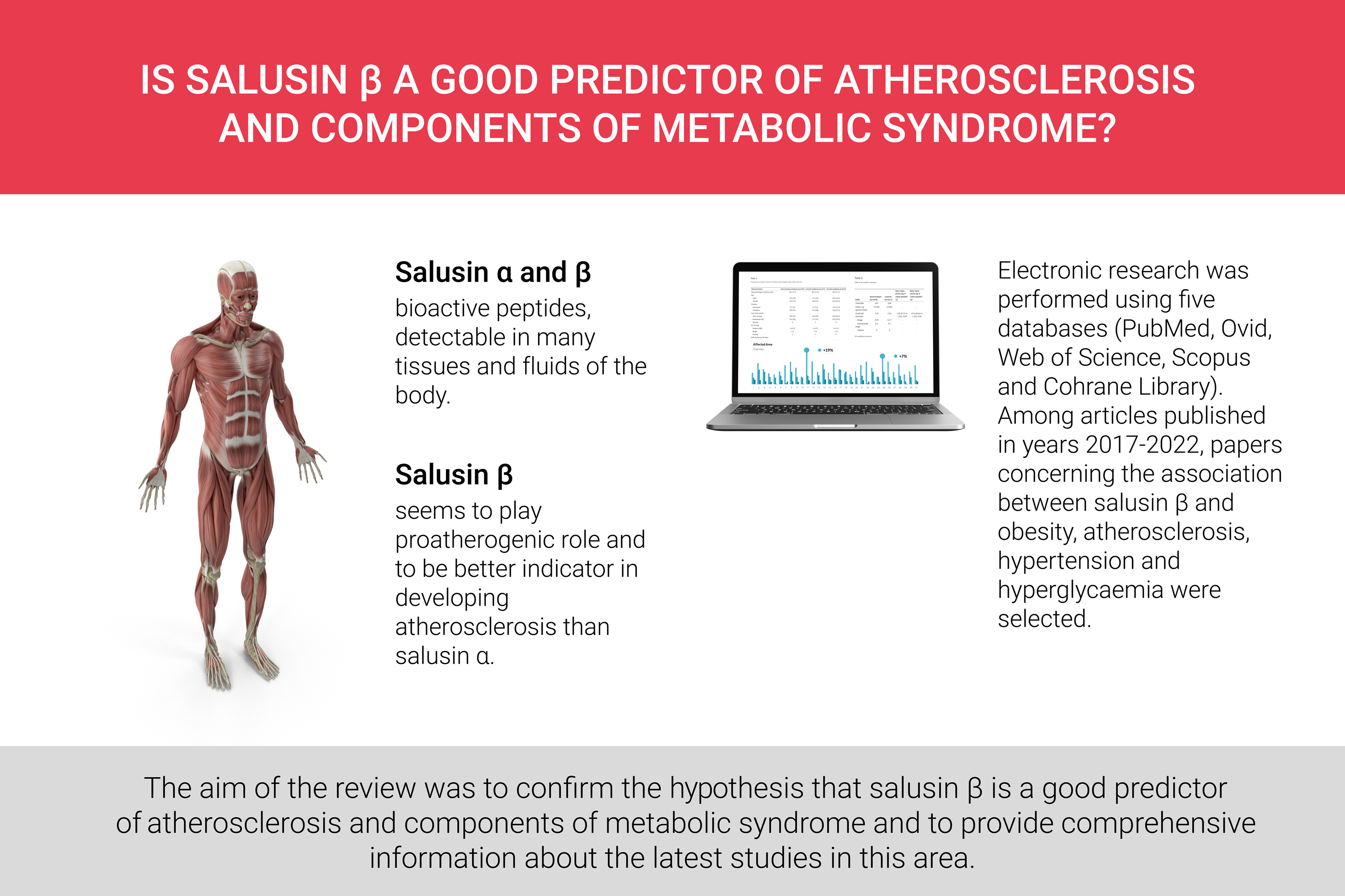
The aim of this review was to confirm the hypothesis that salusin β is a good predictor of atherosclerosis and components of the metabolic syndrome, and to provide comprehensive information about the latest studies in this area.

Materials and methods

The research was performed using 5 online databases, namely PubMed, Ovid, Web of Science, Scopus, and Cochrane Library. Only articles published in the years 2017–2022 and those concerning the association between salusin β and atherosclerosis, hypertension, obesity, hyperlipidemia, and hyperglycemia/DM were selected. Only original papers were included, with 33 articles meeting the inclusion criteria. Data were double-checked independently by 2 authors. The process of selection comprised the removal of duplicates, and the elimination by title, abstract and full-text review, following Preferred Reporting Items for Systematic Reviews and Meta-Analyses (PRISMA) guidelines. The selection process is presented in Figure 1.

Results

Contrary to salusin α, the expression of salusin β is increased in patients with atherosclerosis, hypertension and metabolic syndrome. Increased levels of salusin β are observed in patients with cardiovascular disease (CVD) and cerebrovascular disease. Endogenous expression of salusin β is the lowest in the morning. Furthermore, the stimulation of the parasympathetic nervous system and insulin secretion decreases the level of free salusin β.8 In the central nervous system, salusin β stimulates or depresses sympathetic and vagal activity depending on the location within the brain. It reduces blood pressure and heart rate in the intermediate dorsal motor nucleus of the vagus.6

Salusin β has been shown to stimulate the proliferation of VSMCs and fibroblasts through the activation of immediate response genes such as c-Myc and Fos in rats and humans. It also activates the release of inflammatory cytokines, including IL-1β, IL-6 and TNF-α. Consequently, oxidative stress markers increase and monocyte-endothelial adhesion is promoted.9 This occurs within lesions of the blood vessel endothelium and in atherosclerosis.

Atherosclerosis

Salusin β is a pro-inflammatory agent. Human umbilical vein endothelial cells (HUVECs) incubated in salusin β increased the production of mRNA and protein levels of IL-6, IL-8, IL-18, and reduced the level of IL-1Ra.10 Furthermore, salusin β has an influence on the formation of macrophage foam cells. The peptide promotes the growth of atherosclerotic plaques and stimulates the adhesion of monocytes to the endothelium. Salusin β boosts the production of ACAT-1, the enzyme that breaks down fatty acids into acetyl coenzyme A in human monocytes/macrophages. In addition, salusin β encourages the storage of lipid droplets, increases the intracellular cholesterol content and stimulates monocyte adhesion. The proatherogenic role of the peptide was shown by silencing salusin β. The knockdown of the peptide improved cardiac function and cardiovascular remodeling in myocardial infarction-induced heart failure in rats and alleviated cardiac inflammation in diabetic rats.2, 9

The relationship between salusin β and atherosclerosis was also confirmed among patients suffering from coronary artery disease (CAD). A study by Awad et al. compared patients undergoing transcatheter therapy, and found that salusin β levels were significantly higher in patients with CVD before therapy compared to healthy controls. Moreover, after therapy, salusin β expression was significantly lower than before the intervention, or when compared to the control group. In addition, patients and controls varied in fasting blood glucose levels, insulin levels, body mass index (BMI), systolic blood pressure (SBP), and lipid profile.11 Salusin β levels were significantly lower in patients with stenosis or dilatation in coronary angiography than in healthy volunteers.12, 13 A similar finding was observed in patients with slow coronary flow that seemed to be caused by microvascular atherosclerosis.14

Risk factors of atherosclerosis are similar to abdominal aortic aneurysm (AAA). However, an evaluation of 48 patients with AAA revealed lower salusin β levels compared to 47 healthy controls. The levels of salusin β were also negatively correlated with abdominal aortic diameter.15 The role of salusin β in the pathogenesis of atherosclerosis is presented in Figure 2.

Diabetes mellitus

Many studies have confirmed that the serum level of salusin β is increased in DM. High glucose not only increases the level of salusin β mRNA but also upregulates its production by stimulating prosalusin protein expression. In cell culture experiments, the exposure of HUVECs to high glucose reduced proliferation and migration, and the knockdown of salusin β reversed these abilities. Adenosine monophosphate-activated protein kinase (AMPK) participates in the signaling pathway of salusin β.7 Parallel overexpression of salusin β in proximal tubular (HK-2) cells from both human and mouse models as well as human retinal capillary endothelial cells incubated in high glucose induced inflammatory cytokines and oxidative stress. In human research, inflammation and apoptosis are activated by ROS-dependent signaling pathways.16, 17, 18, 19

In the study performed by Argun et al., the group of patients suffering from type 2 diabetes mellitus (T2DM) had significantly higher levels of salusin β, but only when their hemoglobin A1C (HbA1C) level was higher than 9%.20 Deyekh et al. also confirmed higher serum salusin β levels in patients with T2DM compared to those with prediabetes or healthy controls, while there was no significant difference in salusin β levels between the prediabetes and control groups. Although each group consisted of 30 persons, a detailed description was not provided.21 Additionally, Aldulimya and Alaaraji revealed higher levels of salusin β in women with T2DM than in healthy volunteers, and the levels of salusin β correlated with fasting serum glucose. However, a limitation of this study was the significant difference in age between the examined groups.22 In a study by Wang et al., the level of salusin β was higher in patients with diabetic retinopathy than in healthy controls.16 Moreover, Yassien et al. revealed a correlation between the mean carotid intima-media thickness and left ventricular hypertrophy.23 These conclusions suggest that salusin β participates in the development of complications associated with chronic hyperglycemia.

Sipahi et al. reported that the serum levels of salusin β were elevated in patients undergoing hemodialysis compared to healthy controls.24 In patients also suffering from DM, the salusin β/salusin α ratio was higher, and the levels of salusin α were significantly lower. However, there was no evidence of a correlation between salusin β and diabetes in this group of patients.

An opposite outcome was observed in a study performed among diabetic patients with and without diabetic foot.25 Interestingly, among healthy volunteers, salusin β levels were significantly higher than in diabetic patients. Moreover, salusin α was significantly higher in healthy controls, which raises doubts regarding the methodology of the study.

Hypertension

Salusin β is elevated in patients with hypertension, inducing VSMC proliferation and fibrosis within the vascular wall. Moreover, it activates intimal hyperplasia after vascular injury and can trigger vascular constriction. This occurs via the activation of endothelial nitric oxide synthase (eNOS), the release of nitric oxide (NO) and an increase in NAD(P)H oxidase activity. Acute intravenous administration of salusin β increased the mean arterial pressure in spontaneously hypertensive rats (SHRs), while injections of anti-salusin β IgG reduced the mean blood pressure and heart rate. The important role of salusin β in the development of hypertension and the subsequent vascular remodeling was verified by salusin β knockdown, after which the vascular function was augmented in hypertensive rats.26, 27, 28

Contrary to patients that normally present with physiological night-time blood pressure reduction, those with newly diagnosed non-dipper hypertension demonstrate elevated levels of salusin β. In addition, salusin β positively correlates with the left ventricle mass index and negatively correlates with diastolic parameters of the left ventricle. These results were not compared with healthy controls, which is a limitation of the study.29

The interaction between salusin β and hypertension was confirmed in a study performed in patients treated with anti-hypertensive medicines.30 The therapy consisting of felodipine and enalapril was more effective and brought a more relevant reduction of salusin β level than felodipine alone. This is the first study that demonstrated practically this piece of theoretical knowledge.

The correlation between the level of salusin β and hypertension has been confirmed in both adults and children. The study by Kołakowska et al. performed in adolescents revealed that the serum level was significantly higher in patients with essential hypertension when compared to those with white coat hypertension.31

Finally, salusin β has an influence on the consequences of hypertension. Ageing SHRs have higher concentrations of the peptide in the hypothalamic paraventricular nucleus, myocardium and mesenteric artery. The knockdown of salusin β improved cardiac function and reduced the levels of p38 MAPK, ERK1/2, JNK, and NAD(P)H, which are likely to be involved in the signaling pathway.32 Furthermore, the level of this peptide is significantly increased in the calcified VSMCs of rats. The calcification has been shown to stimulate the expression of salusin β, and the overexpression of salusin β has been shown to promote spontaneous calcification. This process is probably mediated by ROS and NAD(P)H oxidase. Furthermore, the overexpression of salusin β decreases the levels of Klotho, which is an anti-inflammatory and anti-oxidative stress protein.33

Obesity/lipid disorders

Previous studies revealed that obesity is another condition that leads to increased serum salusin β levels. In patients with T2DM, salusin β correlated with both BMI and waist circumference, and was significantly higher than in healthy controls.23 The correlation between salusin β and BMI was also revealed in patients suffering from CVD.11

However, research performed on a group of 75 obese children aged 6–18 years did not confirm this theory.34 No correlation was found between salusin β and BMI, blood pressure, carotid intima-media thickness, and epicardial adipose tissue thickness. There was a negative correlation between salusin α and diastolic blood pressure (DBP). Additionally, in a group of 48 children with Down syndrome, the connection between salusin β and excessive body weight (obesity and overweight) was not proved.35 Nevertheless, physical training decreases the level of serum salusin β in patients with high body weight.

In this regard, moderate- and high-intensity interval training (HIIT) were compared between obese and overweight women. It was revealed that both types of training improved lipid parameters and decreased serum salusin β levels.36

Similar studies were performed among obese or overweight males with a mean age of 11 years. The levels of salusin β were reduced after 12 weeks of training, with HIIT demonstrating more significant results than aerobic training. Apart from salusin β and α, other parameters also improved, such as the lipid profile and markers of inflammation.37, 38 A summary of the studies on salusin β used in this review is included in Table 1.

Discussion

Our research encompassed 12 articles based on studies performed on animals or cell culture systems and 21 articles concerning humans; 15 of these included healthy controls, and only 5 articles involved children. Four studies enrolled more than 100 patients, and 11 enrolled more than 50 patients. Therefore, most studies were based on relatively small study groups, which hampers a definitive evaluation. Nonetheless, most of the studies confirmed the thesis that salusin β has a relevant function in atherosclerotic development, and is correlated with components of the metabolic syndrome. Furthermore, Genç Elden et al. demonstrated an association between sudden hearing loss, atherosclerosis and salusin β, and all groups presenting with similar atherosclerotic parameters. The collected material confirmed that salusin β is a prognostic factor in hearing loss. However, the results of the above study did not indicate sufficient evidence for the development of atherosclerosis in the study group, and hence it was not included in the review.39

The analyzed data indicate that there is much that remains to be discovered about salusin β. Preprosalusin mRNA is ubiquitously expressed, being found in many tissues and organs, such as the nervous system, endothelium, muscles, liver, lungs, kidneys, bone marrow, lymph nodes, spleen, thymus, adrenal glands, small intestine, stomach, salivary glands, and testes, as well as in fluids, such as plasma and urine. A significant number of recent studies on salusin β have concentrated on its role in biochemical pathways in hypertension, atherosclerosis, hyperglycemia, and obesity. Currently available data indicate that salusin β could be used in diagnostics of developing atherosclerosis and hypertension, and appears to be a promising novel target for treatment.

Studies performed on animal models provide important knowledge but in a narrow range. Nevertheless, they indicate that salusin β could be a direct target in the treatment of hypertension and heart failure, or an indicator of treatment efficacy. Initial attempts have already been made to use these findings in clinical practice. One promising study on the evaluation of the treatment of hypertension with 110 patients provides interesting and valuable results.30

Recent studies do not directly confirm the connection between elevated levels of salusin β and obesity. However, physical activity in overweight or obese patients brings about a reduction of salusin β and improvement of both lipid profile and inflammatory factors. These data suggest that salusin β could be valuable in the treatment of obesity.

Cohort studies are necessary to confirm the scope and bring us closer to the practical application of salusin β.

Limitations

This review has some limitations. The available literature on salusin β is quite limited and based on a variety of methods, which hinders a systematic review. The division of included papers into sections was based on the main topics of the studies and seemed to be artificial, as each study contained multiple components and had a high degree of overlap. The aim of this classification was to organize the data. Moreover, research conducted in humans is generally based on small groups of patients.

Conclusions

Recent studies confirm that salusin β plays an important role in the development of vascular remodeling, inflammation, hypertension, and atherosclerosis. Additionally, the peptide is related to hyperglycemia and lipid disorders. The widespread activity of salusin β makes it a potential therapeutic target. However, there is a need for additional studies to confirm the potential role of salusin β as a novel target for treatment.

While it could be useful in the prophylaxis or the treatment of the abovementioned disorders, many reports have been performed in animal models, and those conducted in humans are based on small groups of patients and are not always compared with healthy controls. Studies enrolling children are rare. There is a lack of studies conducted in humans confirming the thesis that salusin β is a good predictor of atherosclerosis and metabolic syndrome, hence further investigations are necessary.

Tables


Table 1. Summary of the studies on salusin β included in the review

No.

Author, year of publication

Title

Subject

Results

Conclusions

Atherosclerosis

1.

Esfahani et al. (2018)10

The effect of salusin-β on expression of pro- and anti-inflammatory cytokines in human umbilical vein endothelial cells

HUVECs

Incubation in salusin β stimulates the production of mRNA and proteins of IL-6, IL-8 and IL-18, and suppresses the production of IL-1Ra.

Salusin β is a proinflammatory agent.

2.

Sipahi et al. (2019)24

Relationship of salusin-alpha and salusin-beta levels with atherosclerosis in patients undergoing haemodialysis

180 adult patients, 90 healthy controls

The levels of salusin β are higher in patients undergoing hemodialysis. There is a positive correlation between the duration of hemodialysis and salusin  β levels. There is no correlation between salusin β and pulse wave analysis. Among patients without DM, the salusin α/salusin β ratio was significantly lower than in the patients also suffering from DM.

Patients undergoing hemodialysis had increased levels of salusin β and an increased salusin α/salusin β ratio.

3.

Wang et al. (2020)4

Salusin-β is superior to salusin-α as a marker for evaluating coronary atherosclerosis

256 patients, 37 healthy controls

Both salusin α and β have substantial impact on SYNTAX scores, but the effect of salusin β is more significant.

There is a connection between salusins and coronary artery injury. Salusin β is superior to salusin α.

4.

Awad et al. (2020)11

Assessment of serum levels of salusin α and salusin β in cardiovascular disease patients undergoing transcatheter therapy

30 patients with CVD and 30 healthy controls

Before therapy, salusin β levels in patients with CVD are significantly higher than in healthy controls. After therapy, salusin β levels in patients are significantly lower than in the control group and before therapy. The group of CVD patients has higher fasting blood glucose, insulin levels, BMI, SBP, and lipid levels than the control group.

Serum salusin β levels are higher in patients with CVD and decrease after transcatheter therapy.

5.

Xu et al. (2021)2

Knockdown of salusin-β improves cardiovascular function in myocardial infarction-induced chronic heart failure rats

rats

Knockdown of salusin β increases hemodynamic parameters, lumen diameter of arteries, microvascular density in infarcted area, and decreases the media thickness of arteries, expression of eNOS, ROS and NAD(P)H oxidase, as well as plasma levels of leptin and visfatin.

Salusin β influences endothelial dysfunction, cardiovascular remodeling and cardiac dysfunction in chronic heart failure.

6.

Arkan et al. (2021)13

The importance of circulating levels of salusin-α, salusin-β, and heregulin-β1 in atherosclerotic coronary arterial disease

113 patients, 55 healthy controls

Serum salusin β levels are lower in patients with CAD than in controls; heregulin-β1 and hsCRP were significantly different among examined groups.

Salusin β seems to be one of the biomarkers of atherosclerosis.

7.

Akyüz et al. (2019)14

Relationship of serum salusin beta levels with coronary slow flow

39 patients, 42 healthy controls

Salusin β is a predictor of CSF.

Salusin β seems to play an important role in the development of CSF.

8.

Yildirim and Kucukosmanoglu (2021)12

Relationship between serum salusin beta levels and coronary artery ectasia

71 patients with CAE and 72 healthy subjects

Mean SBP and DBP values are significantly higher in the CAE group than in the control group, and the mean LVEF is significantly lower. The serum salusin β levels are significantly higher in the CAE group compared to the control group.

Serum salusin β levels are increased in patients with CAE.

9.

Karagöz et al. (2022)15

A new insight into pathophysiological mechanism of abdominal aortic aneurysm with novel parameters salusin-β and arterial stiffness

48 patients with AAA and 47 healthy controls

Salusin β levels are significantly lower in patients with AAA. There is a significant negative correlation between salusin β levels and abdominal aorta diameter.

The pathophysiologic mechanism of AAA is unclear.

Diabetes mellitus

10.

Sun et al. (2017)19

Salusin-β is involved in diabetes mellitus-induced endothelial dysfunction via degradation of peroxisome proliferator-activated receptor gamma

mice

High glucose upregulates the expression of salusin β. Salusin β blockade prevents the overproduction of ROS and inflammatory molecules that are regulated by PPARΥ.

Salusin β takes part in endothelial dysfunction in DM.

11.

Zhao et al. (2017)9

Salusin-β contributes to oxidative stress and inflammation in diabetic cardiomyopathy

rats

Salusin β increases the expression of inflammatory cytokines; high glucose increases the levels of salusin β and prosalusin; knockdown of salusin β reduces the expression of inflammatory cytokines and oxidative stress in cardiomyocytes and left ventricle function, and fails to reduce glucose levels and insulin resistance.

Salusin β induces cardiac inflammation in DM. The knockdown of salusin β improves cardiac function in DM.

12.

Zhu et al. (2017)7

Salusin-β mediates high glucose-induced endothelial injury via disruption of AMPK signaling pathway

HUVECs

HUVECs exposed to high glucose present with the inhibition of proliferation, migration, angiogenesis, as well as retarded cell cycle. The knockdown of salusin β reverses the changes via the AMPK signaling pathway.

Salusin β contributes to endothelial dysfunction, which is related to high glucose by inactivating the AMPK signaling pathway.

13.

Sun et al. (2019)33

Salusin-β promotes vascular calcification via nicotinamide adenine dinucleotide phosphate/reactive oxygen species-mediated klotho downregulation

VSMCs of rats

The levels of salusin β are increased by calcification; the overexpression of salusin β induces calcification and the spontaneous conversion of VSMCs, decreases Klotho protein levels, and increases the expression of oxidative stress molecules.

Salusin β plays an important role in vascular calcification.

14.

Yassien et al. (2020)23

Serum salusin-β in relation to atherosclerosis and ventricular dysfunction in patients with T2DM

60 patients, 25 healthy controls

Serum levels of salusin β are higher in patients with DM and positively correlate with obesity, the insulin resistance index, dyslipidemia, carotid intima-media thickness, and left ventricular hypertrophy, and negatively correlate with left ventricular function.

Salusin β levels correlate with left ventricular dysfunction and atherosclerosis in DM.

15.

Deyekh et al. (2020)21

Evaluation of salusin β in patients with prediabetes and T2DM

30 patients with prediabetes, 30 patients with T2DM, 30 healthy volunteers

Serum salusin β levels in patients with T2DM are significantly higher than in the prediabetes group and healthy subjects. There is no significant difference in salusin β levels between prediabetes group and healthy control subjects.

Serum salusin β is increased in T2DM.

16.

Argun et al. (2021)20

Evaluation of salusin-α and salusin-β levels in patients with T2DM and determination of the impact of severity of hyperglycemia on salusin levels

55 patients, 35 healthy controls

Salusin β positively correlates with fasting glucose and HbA1c levels. The group with HbA1c > 9% has significantly higher levels of salusin β than the group with HbA1c < 9%.

Salusin β levels are higher in patients with DM than in healthy controls and the changes are more significant in those with worse glycemic control.

17.

Wang et al. (2021)18

Salusin-β participates in high glucose-induced HK-2 cell ferroptosis in a Nrf-2-dependent manner

human proximal tubular (HK-2) cells

The overexpression of salusin β exacerbates the iron overload triggered by high glucose and the production of ROS and lipid peroxidation. The knockdown of salusin β reverses these effects.

The positive correlation between salusin β and ferroptosis promotes injury of renal tubular cells in DM.

18.

Wang et al. (2021)16

Salusin-β mediates high glucose-induced inflammation and apoptosis in retinal capillary endothelial cells via a ros-dependent pathway in diabetic retinopathy

60 patients, 20 healthy controls

Salusin β levels are higher in patients with DM and significantly higher in patients with diabetic retinopathy, both proliferative and nonproliferative. High glucose increases the levels of salusin β in HRECs. Salusin β increases inflammatory molecules, apoptosis and ROS production; the knockdown of salusin β has the opposite effects.

Salusin β induces inflammation and apoptosis via an ROS-dependent signaling pathway.

19.

Chen and Jin (2021)17

Downregulation of salusin-β protects renal tubular epithelial cells against high glucose-induced inflammation, oxidative stress, apoptosis and lipid accumulation via suppressing miR-155-5p

mice

Salusin β silencing increases inflammatory factors and oxidative stress, suppresses apoptosis and lipid accumulation in high glucose-induced cells, and reduces the expression of miR-155-5p.

The downregulation of salusin β protects cells against inflammation, oxidative stress, apoptosis, and excessive lipid accumulation that is induced by high glucose.

20.

Sağmak Tartar et al. (2021)25

Association between dermcidin, salusin-α, salusin-β molecules and diabetic foot infections

40 diabetic patients without DFI, 50 patients with DFI, 40 healthy controls

Salusin β levels were significantly higher in controls than in patients with diabetes. Salusin β levels were higher in DFI than in diabetes without DFI; the difference was not statistically significant. There was a significant difference in dermcidin levels between both groups of patients and between DFI and controls.

Dermcidin seems to be associated with DFI.

21.

Aldulimya and Alaaraji (2021)22

Study the association of β-salusin with some anthropometric measurements in Iraqi type II diabetics women

60 women with T2DM and 24 healthy women

β-salusin levels are higher in the patients with T2DM than in healthy controls.

Salusin β level increases in T2DM.

Hypertension

22.

Ren et al. (2017)28

Silencing salusin-β attenuates cardiovascular remodeling and hypertension in spontaneously hypertensive rats

rats

Silencing salusin β reduced left ventricular weight, cardiomyocyte hypertrophy, plasma norepinephrine and tyrosine hydroxylase levels, plasma angiotensin II levels, the expression of type 1 receptors of angiotensin in myocardium and mesenteric artery, and prevented media thickness in the arteries of hypertensive rats.

Increased salusin β contributes to the pathogenesis of hypertension and cardiovascular remodeling.

23.

Kołakowska et al. (2018)31

Correlation of salusin beta with hs-CRP and ADMA in hypertensive children and adolescents

58 children with essential hypertension, 30 children with white coat hypertension

Salusin β levels are significantly higher in patients with hypertension and positively correlate with the levels of hs-CRP and ADMA.

Salusin β correlates with hs-CRP and ADMA in hypertensive adolescents.

24.

Li et al. (2019)32

Silencing salusin β ameliorates heart failure in aged spontaneously hypertensive rats by ROS-relative MAPK/NF-κB pathways in the paraventricular nucleus

rats

Aging hypertensive rats with heart failure have massively increased salusin β expression. The knockdown of salusin β improves cardiac and vascular functions; there is no result in rats without hypertension. Silencing of salusin β reduces paraventricular nucleus proinflammatory cytokines and ROS levels in aging hypertensive rats with heart failure.

Central salusin β knockdown deteriorates cardiac and vascular functions in ageing hypertensive rats with heart failure via a ROS-related pathway in the hypothalamic PVN.

25.

Alpsoy et al. (2021)29

Assessment of salusin alpha and salusin beta levels in patients with newly diagnosed dipper and non-dipper hypertension

88 patients

Salusin β levels are higher in patients with non-dipper hypertension.

Increased salusin β may be a predictor of non-dipper hypertension and a poor cardiovascular prognosis.

26.

Pan et al. (2021)26

Improvement of vascular function by knockdown of salusin-β in hypertensive rats via nitric oxide and reactive oxygen species signaling pathway

rats

Silencing of salusin β enhances vascular relaxation and remodeling, decreases blood pressure and vasoconstriction in hypertension by the activation of eNOS, the release of NO, and the inhibition of NAD(P)H oxidase and ROS.

The knockdown of salusin β improves vascular functions and prevents vasculopathy in hypertension.

27.

Sun et al. (2021)27

A TOR2A gene product: Salusin-β contributes to attenuated vasodilatation of spontaneously hypertensive rats

rats

The intravenous administration of salusin β and anti-salusin β IgG in hypertensive rats has an impact on blood pressure, heart rate and basal vascular tone. The administration of salusin β decreases eNOS activity and NO levels and increases NAD(P)H oxidase activity in hypertensive rats, while anti-salusin β has a reverse impact.

The overexpression of salusin β plays a role in impaired vasodilatation in hypertension by activating NAD(P)H oxidase and inhibiting NO release.

28.

Zhang et al. (2022)30

Efficacy of felodipine and enalapril in the treatment of essential hypertension with coronary artery disease and the effect on levels of salusin-β, apelin, and PON1 gene expression in patients

110 patients with essential hypertension and CAD

Better effectiveness and lower levels of salusin β were observed in patients who were administered felodipine and enalapril than in patients who were administered felodipine alone.

Combination of felodipine and enalapril is more effective in treatment of hypertension with CAD. It decreases the level of salusin β.

Obesity/lipid disorders

29.

Dervişoğlu et al. (2019)34

Salusin-α levels are negatively correlated with diastolic blood pressure in children with obesity

75 obese children, 101 healthy children

No significant correlation between salusin β and heart rate, SBP, DBP, BMI, carotid intima-media thickness, epicardial adipose tissue thickness, and left ventricular mass index was found; there was a negative correlation between salusin α and DBP.

Salusin α seems to be an earlier predictor of cardiovascular disorders than salusin β in obesity.

30.

Nazari et al. (2020)36

Effects of two types of moderate- and high-intensity interval training on serum salusin-α and salusin-β levels and lipid profile in women with overweight/obesity

80 women divided in 2 groups according to the type of training, 40 women – control group

Both types of training improved the lipid profile and increased salusin β levels, but in moderate interval training, the changes were more significant.

Moderate intensity interval training is more effective in improving the lipid profile than HIIT.

31.

Stefanowicz-Bielska et al. (2020)35

Obesity and overweight and accompanying metabolic disorders occur in children with Down syndrome

48 patients with DS (26 girls and 22 boys), aged from 7 to 18 years, divided into 2 groups – 39 with normal weight or underweight and 9 obese or overweight

Higher values of HDL cholesterol were found in patients with normal body mass and underweight than in patients with obesity and overweight (p = 0.009). Higher values of uric acid were found in the group of patients with obesity and overweight than in the normal mass and underweight group (p = 0.012). The children who are physically active have normal body weight (p = 0.039). There was no significant difference in salusin β levels between both groups.

The role of salusin β as an early indicator of metabolic disorders in children with DS was not demonstrated.

32.

Paahoo et al. (2020)38

Effect of two chronic exercise protocols on pre-atherosclerotic and anti-atherosclerotic biomarkers levels in obese and overweight children

30 obese and overweight boys divided into 2 groups according to the type of training, 15 boys – control group

A significant increase of serum levels of salusin α and NO, and a decrease of serum levels of salusin β, body weight, BMI, and TG/HDL ratio in both training groups, but more significant after HIIT.

Both HIIT and continuous aerobic training have positive impact on cardiometabolic parameters, but HIIT seems to be more effective.

33.

Paahoo et al. (2021)37

Effectiveness of continuous aerobic versus high-intensity interval training on atherosclerotic and inflammatory markers in boys with overweight/obesity

30 boys divided into 2 groups according to the type of training, 15 boys – control group

Both types of training improved the lipid profile, increased the levels of inflammatory factors and salusin β and increased the salusin ratio; in HIIT, the changes were more significant.

HIIT is more effective in improving the lipid profile and inflammatory factors then aerobic training in obese boys.

T2DM – type 2 diabetes mellitus; eNOS – endothelial nitric oxide synthase; ROS – reactive oxygen species; NAD(P)H – nicotinamide adenine dinucleotide phosphate; CAD – coronary artery disease; CSF – coronary slow flow; PPARΥ – peroxisome proliferator-activated receptor γ; AMPK – adenosine monophosphate-activated protein kinase; HUVECs – human umbilical vein endothelial cells; VSMCs – vascular smooth muscle cells; HRECs – human retinal capillary endothelial cells; DFI – diabetic foot infections; ADMA – asymmetric dimethylarginine; PVN – paraventricular nucleus; AAA – abdominal aortic aneurysm; DS – Down syndrome; IL – interleukin; BMI – body mass index; hsCRP – high-sensitivity C-reactive protein; LVEF – left ventricular ejection fraction; CAE – coronary artery ectasia; ROS – reactive oxygen species; HbA1c – hemoglobin A1C; IgG – immunoglobulin G; HDL – high-density lipoprotein; TG – triglycerides; HIIT – high-intensity interval training; SBP – systolic blood pressure; DBP – diastolic blood pressure; CVD – cardiovascular disease.

Figures


Fig. 1. Study selection process
Fig. 2. The role of salusin β in the pathogenesis of atherosclerosis
TNF-α – tumor necrosis factor alpha; LPS – lipopolysaccharides; VSMCs – vascular smooth muscle cells; eNOS – endothelial nitric oxide synthase; NAD(P)H – nicotinamide adenine dinucleotide phosphate; IL – interleukin; ACAT-1 – acetyl-coenzyme A acetyltransferase 1; VCAM-1 – vascular cell adhesion molecule-1; NF-kB – nuclear factor kappa B.

References (39)

  1. Shichiri M, Ishimaru S, Ota T, Nishikawa T, Isogai T, Hirata Y. Salusins: Newly identified bioactive peptides with hemodynamic and mitogenic activities. Nat Med. 2003;9(9):1166–1172. doi:10.1038/nm913
  2. Xu Y, Pan Y, Wang X, et al. Knockdown of salusin-β improves cardiovascular function in myocardial infarction-induced chronic heart failure rats. Oxid Med Cell Longev. 2021;2021:8896226. doi:10.1155/2021/8896226
  3. Sun HJ, Zhao MX, Liu TY, et al. Salusin-β induces foam cell formation and monocyte adhesion in human vascular smooth muscle cells via miR155/NOX2/NFκB pathway. Sci Rep. 2016;6(1):23596. doi:10.1038/srep23596
  4. Wang Y, Wang S, Zhang J, et al. Salusin-β is superior to salusin-α as a marker for evaluating coronary atherosclerosis. J Int Med Res. 2020;48(2):30006052090386. doi:10.1177/0300060520903868
  5. Watanabe T, Sato K, Itoh F, et al. The roles of salusins in atherosclerosis and related cardiovascular diseases. J Am Soc Hypertens. 2011;5(5):359–365. doi:10.1016/j.jash.2011.06.003
  6. Wu LL, Bo JH, Zheng F, et al. Salusin-β in intermediate dorsal motor nucleus of the vagus regulates sympathetic-parasympathetic balance and blood pressure. Biomedicines. 2021;9(9):1118. doi:10.3390/biomedicines9091118
  7. Zhu X, Zhou Y, Cai W, Sun H, Qiu L. Salusin-β mediates high glucose-induced endothelial injury via disruption of AMPK signaling pathway. Biochem Biophys Res Commun. 2017;491(2):515–521. doi:10.1016/j.bbrc.2017.06.126
  8. Fujimoto K, Hayashi A, Kamata Y, et al. Circulating levels of human salusin-β, a potent hemodynamic and atherogenesis regulator. PLoS One. 2013;8(10):e76714. doi:10.1371/journal.pone.0076714
  9. Zhao MX, Zhou B, Ling L, et al. Salusin-β contributes to oxidative stress and inflammation in diabetic cardiomyopathy. Cell Death Dis. 2017;8(3):e2690. doi:10.1038/cddis.2017.106
  10. Esfahani M, Saidijam M, Najafi R, Goodarzi MT, Movahedian A. The effect of salusin-β on expression of pro- and anti-inflammatory cytokines in human umbilical vein endothelial cells (HUVECs). ARYA Atheroscler. 2018;14(1):1–10. doi:10.22122/arya.v14i1.1602
  11. Awad A, Ali H, Al-Rufaie M. Assessment of serum levels of salusin α and salusin β in cardiovascular disease patients undergoing transcatheter therapy. Indian J Med Forensic Med Toxicol. 2020;14(2):303–308. doi:10.37506/ijfmt.v14i2.2807
  12. Yildirim A, Kucukosmanoglu M. Relationship between serum salusin beta levels and coronary artery ectasia. Acta Cardiol Sin. 2021;37(2):130–137. doi:10.6515/ACS.202103_37(2).20200910A
  13. Arkan A, Atukeren P, Ikitimur B, et al. The importance of circulating levels of salusin-α, salusin-β, and heregulin-β1 in atherosclerotic coronary arterial disease. Clin Biochem. 2021;87:19–25. doi:10.1016/j.clinbiochem.2020.10.003
  14. Akyüz A, Aydın F, Alpsoy Ş, Gur DO, Guzel S. Relationship of serum salusin beta levels with coronary slow flow. Anatol J Cardiol. 2019;22(4):177–184. doi:10.14744/AnatolJCardiol.2019.43247
  15. Karagöz A, Kurt D, Günaydın ZY, et al. A new insight into pathophysiological mechanism of abdominal aortic aneurysm with novel parameters salusin-β and arterial stiffness. Tex Heart Inst J. 2022;49(6):e217561. doi:10.14503/THIJ-21-7561
  16. Wang H, Zhang M, Zhou H, et al. Salusin-β mediates high glucose-induced inflammation and apoptosis in retinal capillary endothelial cells via a ros-dependent pathway in diabetic retinopathy. Diabetes Metab Syndr Obes. 2021;14:2291–2308. doi:10.2147/DMSO.S301157
  17. Chen H, Jin G. Downregulation of salusin-β protects renal tubular epithelial cells against high glucose-induced inflammation, oxidative stress, apoptosis and lipid accumulation via suppressing miR-155-5p. Bioengineered. 2021;12(1):6155–6165. doi:10.1080/21655979.2021.1972900
  18. Wang WJ, Jiang X, Gao CC, Chen ZW. Salusin β participates in high glucose induced HK 2 cell ferroptosis in a Nrf 2 dependent manner. Mol Med Rep. 2021;24(3):674. doi:10.3892/mmr.2021.12313
  19. Sun HJ, Chen D, Wang PY, et al. Salusin-β is involved in diabetes mellitus-induced endothelial dysfunction via degradation of peroxisome proliferator-activated receptor gamma. Oxid Med Cell Longev. 2017;2017:6905217. doi:10.1155/2017/6905217
  20. Argun D, Argun F, Borku Uysal B. Evaluation of salusin-α and salusin-β levels in patients with type 2 diabetes mellitus and determination of the impact of severity of hyperglycemia on salusin levels. Ir J Med Sci. 2021;190(4):1403–1411. doi:10.1007/s11845-021-02674-4
  21. Deyekh S, Hamzah M, Ateaai J. Evaluation of salusin β in patients with prediabetes and type 2 diabetes mellitus Medico Legal Update. 2020;20(1):651–654. doi:10.37506/mlu.v20i1.438
  22. Aldulimya BHA, Alaaraji SFT. Study the association of β-salusin with some anthropometric measurements in Iraqi type II diabetics women. Egypt J Chem. 2021;64(11):6515–6522. doi:10.21608/ejchem.2021.78221.3830
  23. Yassien M, Fawzy O, Mahmoud E, Khidr EG. Serum salusin-β in relation to atherosclerosis and ventricular dysfunction in patients with type 2 diabetes mellitus. Diabetes Metab Syndr. 2020;14(6):2057–2062. doi:10.1016/j.dsx.2020.10.025
  24. Sipahi S, Genc A, Acikgoz S, et al. Relationship of salusin-alpha and salusin-beta levels with atherosclerosis in patients undergoing haemodialysis. Singapore Med J. 2019;60(4):210–215. doi:10.11622/smedj.2018123
  25. Sağmak Tartar A, Uğur K, Tuncer Kara K, Akbulut A, Demirdağ K, Aydin S. Association between dermcidin, salusin-α, salusin-β molecules and diabetic foot infections. Int J Low Extrem Wounds. 2021:153473462110655. doi:10.1177/15347346211065527
  26. Pan Y, Sun S, Wang X, et al. Improvement of vascular function by knockdown of salusin-β in hypertensive rats via nitric oxide and reactive oxygen species signaling pathway. Front Physiol. 2021;12:622954. doi:10.3389/fphys.2021.622954
  27. Sun S, Zhang F, Pan Y, et al. A TOR2A gene product: Salusin-β contributes to attenuated vasodilatation of spontaneously hypertensive rats. Cardiovasc Drugs Ther. 2021;35(1):125–139. doi:10.1007/s10557-020-06983-1
  28. Ren XS, Ling L, Zhou B, et al. Silencing salusin-β attenuates cardiovascular remodeling and hypertension in spontaneously hypertensive rats. Sci Rep. 2017;7(1):43259. doi:10.1038/srep43259
  29. Alpsoy S, Dogan B, Ozkaramanli Gur D, et al. Assessment of salusin alpha and salusin beta levels in patients with newly diagnosed dipper and non-dipper hypertension. Clin Exp Hypertens. 2021;43(1):42–48. doi:10.1080/10641963.2020.1797086
  30. Zhang W, Zhang J, Jin F, Zhou H. Efficacy of felodipine and enalapril in the treatment of essential hypertension with coronary artery disease and the effect on levels of salusin-β, apelin, and PON1 gene expression in patients. Cell Mol Biol (Noisy-le-grand). 2022;67(6):174–180. doi:10.14715/cmb/2021.67.6.24
  31. Kołakowska U, Kuroczycka-Saniutycz E, Olański W, Wasilewska A. Correlation of salusin beta with hs-CRP and ADMA in hypertensive children and adolescents. Curr Pharm Des. 2018;24(30):3551–3557. doi:10.2174/1381612824666180607124531
  32. Li HB, Yu XJ, Bai J, et al. Silencing salusin β ameliorates heart failure in aged spontaneously hypertensive rats by ROS-relative MAPK/NF-κB pathways in the paraventricular nucleus. Int J Cardiol. 2019;280:142–151. doi:10.1016/j.ijcard.2018.12.020
  33. Sun H, Zhang F, Xu Y, et al. Salusin-β promotes vascular calcification via nicotinamide adenine dinucleotide phosphate/reactive oxygen species-mediated Klotho downregulation. Antioxid Redox Signal. 2019;31(18):1352–1370. doi:10.1089/ars.2019.7723
  34. Dervişoğlu P, Elmas B, Kösecik M, İşgüven ŞP, Büyükavcı M, Köroğlu M. Salusin-α levels are negatively correlated with diastolic blood pressure in children with obesity. Cardiol Young. 2019;29(10):1225–1229. doi:10.1017/S1047951119001173
  35. Stefanowicz-Bielska A, Wierzba J, Stefanowicz J, Owczarzak A, Chamienia A. Obesity and overweight and accompanying metabolic disorders occur in children with Down syndrome. Acta Med. 2020;36(4):2473–2479. doi:10.19193/0393-6384_2020_4_384
  36. Nazari M, Minasian V, Hovsepian S. Effects of two types of moderate- and high-intensity interval training on serum salusin-α and salusin-β levels and lipid profile in women with overweight/obesity. Diabetes Metab Syndr Obes. 2020;13:1385–1390. doi:10.2147/DMSO.S248476
  37. Paahoo A, Tadibi V, Behpoor N. Effectiveness of continuous aerobic versus high-intensity interval training on atherosclerotic and inflammatory markers in boys with overweight/obesity. Pediatr Exerc Sci. 2021;33(3):132–138. doi:10.1123/pes.2020-0138
  38. Paahoo A, Tadibi V, Behpoor N. Effect of two chronic exercise protocols on pre-atherosclerotic and anti-atherosclerotic biomarkers levels in obese and overweight children. Iran J Pediatr. 2020;30(2):e99760. doi:10.5812/ijp.99760
  39. Genç Elden S, Yılmaz MS, Altındiş M, Köroğlu M, Elden H. The role of serum salusin alpha and beta levels and atherosclerotic risk factors in idiopathic sudden hearing loss pathogenesis. Eur Arch Otorhinolaryngol. 2022;279(3):1311–1316. doi:10.1007/s00405-021-06804-7