Abstract
Background. Cyclophosphamide is a drug used in various types of cancer. It can cause oxidative and inflammatory ovarian damage and infertility. Thiamine pyrophosphate (TPP) to be investigated for its effect on cyclophosphamide-induced ovarian damage and reproductive dysfunction in the present study is the active metabolite of thiamine. It has been shown that TPP protects organs and tissues from oxidative stress and proinflammatory cytokine damage.
Objectives. To investigate the effect of TPP against the ovarian damage and reproductive dysfunction caused by cyclophosphamide in rats.
Materials and methods. Albino Wistar type female rats were divided into healthy control (HG), cyclophosphamide (CYC) and TPP + cyclophosphamide (TPPC) groups (for each group, n = 12). Thiamine pyrophosphate at a dose of 25 mg/kg was injected intraperitoneally (ip.) in the TPPC group, and 0.9% NaCI solution was injected ip. in the CYC and HG groups. One hour after the injection, 75 mg/kg of cyclophosphamide was administered ip. in the TPPC and CYC groups. This procedure was repeated once a day for 30 days. At the end of this period, 6 rats from each group were euthanized with a high dose of anesthetic (50 mg/kg of sodium thiopental). Biochemical and histopathological examinations were performed on the extracted ovarian tissue. The remaining animals were kept in the laboratory with mature male rats for 2 months for reproduction.
Results. Thiamine pyrophosphate significantly decreased the cyclophosphamide-induced increase in the levels of the oxidant parameter malondialdehyde (MDA), proinflammatory nuclear factor kappa B (NF-κB), tumor necrosis factor alpha (TNF-α), and interleukin 1 beta (IL-1β). In addition, TPP decreased the severe histopathological damage associated with cyclophosphamide in the ovarian tissue and prevented infertility.
Conclusions. Our experimental results have suggested that TPP could be beneficial in the treatment of cyclophosphamide-induced ovarian injury and infertility.
Key words: infertility, rat, cyclophosphamide, ovarian
Background
The Food and Drug Administration (FDA) has approved the use of cyclophosphamide in the treatment of lymphomas, multiple myeloma, breast cancer, diffuse neuroblastomas, retinoblastoma, pediatric minimal change nephrotic syndrome, and ovarian adenocarcinoma.1 Cyclophosphamide is also used in the treatment of autoimmune diseases, such as multiple sclerosis, and in the prevention of transplant rejection.2, 3 In the clinic, cyclophosphamide is initiated intravenously (iv.) at doses of 40–50 mg/kg, depending on tolerance.4 However, serious adverse reactions of cyclophosphamide during the treatment are considered. The common adverse reactions reported in clinical studies include hemorrhagic cystitis, amenorrhea, myelosuppression, alopecia, nausea, and vomiting.5, 6 Renal tubular necrosis, pulmonary fibrosis, cardiotoxicity, and infertility have been shown among other toxic effects associated with cyclophosphamide.7 It is stated that cyclophosphamide-induced infertility is caused by ovarian failure.8 Infertility due to ovarian failure is the most common and serious side effect of cyclophosphamide and occurs in 30–70% of women receiving the treatment.9, 10 It has been reported that cyclophosphamide causes ovarian failure through oxidative and inflammatory damage.11 Nair et al. showed that cyclophosphamide-induced damage in the ovarian tissue was caused by a decrease in the endogenous antioxidant glutathione (GSH) and an increase in proinflammatory tumor necrosis factor alpha (TNF-α).12 In a study by Khedr, it was emphasized that cyclophosphamide-induced infertility was associated with an increase in malondialdehyde (MDA) – a lipid peroxidation (LPO) product in the ovarian tissue.13 The information obtained from the literature suggests that antioxidant and anti-inflammatory drugs can be beneficial in the prevention or treatment of cyclophosphamide-induced infertility.
Thiamine pyrophosphate (TPP) to be investigated in this study for its effect on cyclophosphamide-induced ovarian damage and reproductive dysfunction is the active metabolite of thiamine. It is also the cofactor of enzymes playing a role in maintaining the cell redox state by synthesizing nicotinamide adenine dinucleotide phosphate (NADPH) and GSH.14 Thiamine pyrophosphate has been reported to prevent the LPO and oxidative DNA damage caused by ischemia/reperfusion damage.15 Moreover, there are findings in the literature showing that TPP prevents the oxidative damage and infertility caused by cisplatin in rats.16 It has also been noted in another study that TPP suppresses the overproduction of TNF-α, interleukin 1 beta (IL-1β) and other proinflammatory cytokines.17 The data indicate that TPP can be beneficial in the treatment of cyclophosphamide-induced ovarian injury and infertility.
Objectives
To investigate the effect of TPP against the possible ovarian damage and reproductive dysfunction induced by cyclophosphamide in female rats, and to examine the ovarian tissue biochemically and histopathologically.
Materials and methods
Experimental animals
In our study, a total of 36 albino Wistar type female and 6 male rats weighing 235–250 g and aged 3.5–4 months were obtained from the Atatürk University Medical Experimental Application and Research Center (Erzurum, Turkey). The animals were kept at normal room temperature (22°C) in the laboratory of the same center and fed with standard animal feed. The animal experiments were performed in accordance with the ethical standards laid down in the 1964 Declaration of Helsinki and its later amendments. This study was specifically approved by the local Animal Experimentation Ethics Committee (meeting No. 77040475-641.04-E.2000115834, as of May 4, 2020).
Chemicals
Cyclophosphamide used in the experiments was supplied by Eczacıbaşı Pharmaceuticals Marketing Co. (EIP) (Istanbul, Turkey) (a vial containing 1 g of solution powder to be used for infusion), sodium thiopental was supplied by E. Ulagay İlaç Sanayii Türk A.Ş (Istanbul, Turkey) and TPP (50 mg of solution powder) was obtained from BioPharma (Moscow, Russia).
Experimental groups
The rats to be used in the experiment were divided into cyclophosphamide (CYC), TPP + cyclophosphamide (TPPC) and healthy control (HG) groups (for each group, n = 12).
Drug preparation
Preparation of cyclophosphamide solution
A total of 1 g of cyclophosphamide solution powder was dissolved in the vial with 10 mL of normal saline (0.9% NaCl). The cyclophosphamide solution was taken into injectors at a dose of 75 mg/kg. This dose resulted in 18.2 mg given to an animal with an average weight of 242.5 g. A total of 0.18 mL of cyclophosphamide solution was taken into each injector for this dose to be administered to each animal.
Preparation of thiamine pyrophosphate solution
A total of 50 mg of TPP solution powder was diluted with 2 mL of 0.9% NaCl. This solution was taken into injectors at a dose of 25 mg/kg. The dose was calculated to be 6 mg for an animal with an average weight of 242.5 g. In order to apply this dose to each animal, 0.24 mL of TPP solution was taken into each injector.
Experimental procedure
For the experiment, TPP was injected intraperitoneally (ip.) into the anterior part of the abdomen in the TPPC group at a dose of 25 mg/kg. This dose of TPP has been reported to protect the tissue from oxidative damage in previous studies.18 Normal saline (0.9% NaCl) was used as a solvent for the CYC and HG groups. One hour after the injection, 75 mg/kg of cyclophosphamide was administered to the anterior part of the abdomen in the TPPC and CYC groups once a week. Previous studies have shown that this dose of cyclophosphamide causes ovarian damage.12 This procedure was performed once a day for 30 days. At the end of this period, 6 rats from each group were euthanized with a high dose of anesthetic (50 mg/kg of sodium thiopental), and biochemical and histopathological examinations were performed on the extracted ovarian tissue. The remaining animals (6 female rats from each group) were kept in the laboratory with mature male rats for 2 months for reproduction. The rats which became pregnant during this period were taken to separate cages and kept alone in a suitable environment. The rats which did not get pregnant and give birth within 2 months were considered infertile. In addition, maternity duration was calculated by subtracting the standard duration of gestation (21 days) from the period between the day the female rats met the male rats until the time of birth (A) (maternity period = A − 21). All biochemical, histopathological and reproductive test results obtained in the TPPC and HG groups were compared with those obtained in the CYC group.
Biochemical analyses
Prior to dissection, all tissues were rinsed with phosphate-buffered saline (PBS) solution. The ovarian tissues were homogenized in ice-cold phosphate buffers (50 mM, pH 7.4) that were appropriate for the variable to be measured.19 The tissue homogenates were centrifuged at 5000 rpm for 20 min at 4°C, and the supernatants were extracted to analyze MDA, total glutathione (tGSH), total antioxidant system (TAS), total oxidant system (TOS), and protein concentration levels. The protein concentration of the supernatant was measured using the method described by Bradford.20 The drug concentrations in all the tissues were expressed per 1 g of protein.20 All spectrophotometric measurements were recorded using a microplate reader (BioTek, Winooski, USA).
MDA analysis
The MDA measurement was based on the spectrophotometric measurement (at 532 nm) of the absorbance of the pink-colored complex formed by thiobarbituric acid and MDA at high temperature (95°C).21
tGSH analysis
The 5,5’-Dithiobis (2-nitrobenzoic acid) in the measurement medium is a disulfide chromogen, and is readily reduced by compounds with sulfhydryl groups. The resulting yellow color was measured spectrophotometrically at 412 nm.22
NF-κB, TNF-α and IL-1β analysis
The rat-specific sandwich enzyme-linked immunosorbent assay (ELISA) was used to measure the concentrations of NF-κB, TNF-α and IL-1β in the tissue homogenates: rat NF-κB ELISA kits (Cat. No. 201-11-0288; Shanghai Sunred Biological Technology Co., Ltd., Shanghai, China); rat TNF-α and rat IL-1β ELISA kits (Cat No. YHB1098Ra; Shanghai LZ Biotech Co., Ltd., Shanghai, China).
Histopathological examination
After a routine tissue follow-up, five-micrometer sections were obtained for histopathological evaluation. These sections were stained with hematoxylin and eosin (H&E), and the ovarian tissues were evaluated with a light microscope (Olympus BX 51; Olympus Corp., Tokyo, Japan) by a pathologist who was blinded to the treatment protocol; the photographs were taken with a digital camera (Olympus DP 71; Olympus Corp.). The histopathological damage severity in each ovarian tissue section was scored as grades 0–3 (0 – normal; 1 – mild damage; 2 – moderate damage; and 3 – severe damage).
Statistical analyses
The results obtained in the experiments were expressed as mean and standard deviation (M ±SD) or median (minimum–maximum) (Me (min–max)). The normality of variables was checked with the Shapiro–Wilk normality test. The homogeneity of variances was evaluated with Levene’s test. The significance of the difference between groups was determined using the one-way analysis of variance (ANOVA), followed by Tukey’s post hoc tests. While comparing groups, the Kruskal–Wallis test and the Dunn’s post hoc test were used, and adjusted p-values were presented. All statistical procedures were performed using the IBM SPSS Statistics for Windows software, v. 20.0 (IBM Corp., Armonk, USA), and a p-value <0.05 was considered significant.
Results
Biochemical results
MDA and tGSH analysis results
As can be seen in Figure 1, the amount of MDA in the ovarian tissue from the CYC group was found to be significantly higher as compared to the HG and TPPC groups (p < 0.0001). The amount of MDA in the TPPC group was higher than in the HG group (p < 0.0001). However, the difference in the MDA level between the TPPC group and the HG group was statistically nonsignificant. Cyclophosphamide caused a decrease in the amount of tGSH in the ovarian tissue. Thiamine pyrophosphate significantly prevented the reduction of tGSH by cyclophosphamide (p < 0.0001). The difference in the tGSH level between the TPPC group and the HG group was statistically non-significant (Table 1, Table 2).
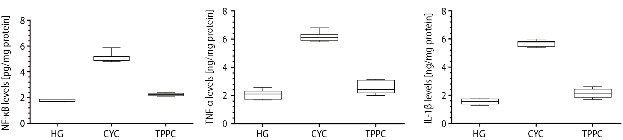
NF-κB, TNF-α and IL-1β analysis results
Cyclophosphamide caused an increase in the NF-κB, TNF-α and IL-1β levels in the ovarian tissues of animals. The NF-κB, TNF-α and IL-1β levels in the ovarian tissues of the animals treated with cyclophosphamide (the CYC group) were significantly higher, as compared to the HG and TPPC groups (p < 0.0001) (Table 1). Thiamine pyrophosphate prevented the increase in the levels of NF-κB, TNF-α and IL-1β induced by cyclophosphamide (p < 0.0001). The differences in the levels of these proinflammatory cytokines between the TPPC and HG groups were found to be statistically nonsignificant (Figure 2, Table 2).
Histopathological results
As seen in Figure 3, there were no histopathological findings in the ovarian tissue of the HG group, which was evaluated as grade 0, except mild fluid accumulation with cystic changes in the lumen of follicule structures.
In the ovarian tissue of the CYC group, grade 3 vacuolized and degenerated follicle cells, a cystic change with fluid accumulation in the lumen of the follicle structure, congestion, and hemorrhages were observed (Figure 4).
Vacuolized and degenerated follicle cells in the developing follicle structures were not detected in the ovarian tissue of the TPPC group (grade 0). However, other histopathological findings, such as fluid accumulation with cystic changes in the lumen of follicle structures, congestion and hemorrhages in the corpus luteum and other areas, were observed in the TPPC group and evaluated as grade 1 (Figure 5) (Table 3, Table 4).
Reproductive test results
As can be seen in Table 5, all 6 animals in the HG group gave birth at around 25–28 days. In the TPPC group, 4 out of 6 animals gave birth on day 35–37, but 2 animals in this group did not give birth within the waiting period (2 months). In the CYC group, none of the 6 animals taken for breeding gave birth within 2 months.
Discussion
In this study, the protective effect of TPP against possible cyclophosphamide-induced ovarian damage and reproductive dysfunction in female rats was investigated. In addition, it was evaluated whether reproductive dysfunction was associated with the severity of ovarian damage. The ovarian damage induced by cyclophosphamide was determined by measuring the MDA, tGSH, NF-κB, TNF-α, and IL-1β levels in the ovarian tissues, and the histopathological examination of 6 out of 12 animals treated with cyclophosphamide. Our experimental results showed that cyclophosphamide administered at a dose of 75 mg/kg once a week (4 doses in total) increased the oxidant and proinflammatory cytokine levels, and decreased the antioxidant level in the ovarian tissue as compared to the healthy and TPP-treated groups. In addition, reproductive dysfunction was observed in the CYC group with high oxidant and cytokine levels, together with a low antioxidant level. In a previous study, cyclophosphamide was used at a higher dose (200 mg/kg) to create toxic effects on the ovarian tissue in rats.23 There is evidence in the literature supporting our biochemical findings related to cyclophosphamide.11, 12, 13 As it is known, MDA is a toxic product formed as a result of the oxidation of cell membrane lipids by reactive oxygen species (ROS).24 Hamzeh et al. histopathologically demonstrated that the increased ROS and MDA production induced by cyclophosphamide was associated with ovarian damage.25 It has also been reported that this oxidative stress is due to phosphoramide mustard, one of the metabolites of cyclophosphamide.23
In previous studies, cyclophosphamide was shown to cause an increase in MDA and a decrease in GSH levels in the ovarian tissue of animals.26 The literature data being consistent with our experimental results show that cyclophosphamide disturbs the oxidant/antioxidant balance in the ovarian tissue in favor of oxidants. As it is known, the oxidant/antioxidant balance is provided by the dominance of antioxidants in physiological conditions. The disruption of this balance in favor of oxidants causes tissue damage. This condition is defined as oxidative stress.24 Our biochemical findings revealed that the amount of tGSH in the ovarian tissue of the CYC group significantly decreased as compared to that of the HG and TPPC groups. In the literature, a decrease in GSH is explained by the inadequacy of antioxidants in neutralizing oxidants.27 Having an important role in the antioxidant defense system in living organisms, GSH directly reacts with ROS to oxidize thiol groups and preserve cell integrity.28, 29
Furthermore, cyclophosphamide was found to increase the levels of proinflammatory cytokines, such as NF-κB, TNF-α and IL-1β, in the ovarian tissue in our study. In a recent study, it was emphasized that these proinflammatory cytokines played a role in the pathogenesis of cyclophosphamide toxicity.30 Elkady et al. reported that cyclophosphamide caused an increase in NF-κB in the ovarian tissue.31 In a study by Nair et al., it was stated that proinflammatory TNF-α as well as oxidative stress were responsible for cyclophosphamide-induced ovarian damage.12 In another study, the role of IL-1β in ovarian injury was presented, and it was also argued that the suppression of the increase in the IL-1β production caused a decrease in ovarian tissue damage.23 In our study, the levels of NF-κB, TNF-α and IL-1β increased in the CYC group, where MDA, a ROS product, has also increased. There is evidence in the literature reporting that ROS increase the NF-κB production.32 In addition, it has been shown that NF-κB induces the secretion of inflammatory mediators, such as IL-1β and TNF-α.33, 34
In our study, the biochemical and histopathological findings regarding the ovarian tissue of the entire animal group were found to be consistent with the reproductive test results. The CYC group presented with severe histopathological damage in the ovarian follicles and tissue, and none of the animals in this group gave birth. In previous studies, animals that were kept in a breeding environment and did not give birth within 2 months were considered infertile.35 Supporting our experimental results, Saleh et al. reported that cyclophosphamide caused infertility in animals.23 It was argued that this infertility due to cyclophosphamide was caused by oxidative stress and inflammation in the ovaries; in addition, it was reported that ovarian damage increased with antioxidant and anti-inflammatory therapy.33 Moreover, in a study on cyclophosphamide supporting our histopathological results, it was reported that degenerative damage developed in the ovarian follicles at decreased antioxidant levels and increased cytokine (TNF-α) levels.12 In another study, cyclophosphamide-associated stromal and follicle degeneration, edema, vacuolization, and the severity of vascular congestion decreased with the elimination of inflammation and oxidative stress.25 As can be seen from our experimental results, the attenuation of the cyclophosphamide-induced oxidant and the proinflammatory cytokine increase with TPP resulted in a decrease in histopathological damage and the number of infertile animals. It is known from previous studies that TPP prevents infertility caused by ovarian ischemia/reperfusion damage.36 It has been stated in the literature that the protective effect of TPP on the ovary is due to an increased LPO reaction and the inhibition of DNA oxidation.15 However, no information has been found in the literature stating that TPP inhibits the proinflammatory cytokine production in the ovarian tissue. However, in a recent experimental study, it was reported that TPP suppressed the ethanol-induced increase in TNF-α and IL-1β in the optic nerve tissue.17
Limitations
This study has some limitations. Firstly, more studies are needed to clarify the mechanism of action of TPP in the ovarian damage caused by cyclophosphonamide. Secondly, the estrogen and progesterone levels can also be measured in relation to infertility. Finally, the pathology can be measured at the molecular level.
Conclusions
Cyclophosphamide caused an increase in the oxidant and proinflammatory parameters, and a decrease in antioxidants in the ovarian tissue of animals. It was observed that the ovarian tissue damage caused by cyclophosphamide resulted in infertility. Decreasing the severity of the cyclophosphamide-associated oxidative and inflammatory ovarian injury with TPP resulted in a decrease in the number of infertile animals. Our experimental results suggest that TPP can be beneficial in the treatment of cyclophosphamide-induced ovarian injury and infertility.