Abstract
Background. In cases of alveolar bone atrophy prior to implant treatment, sinus floor elevation remains the treatment of choice. During a sinus lift procedure, a stabilizing grafting material is required. Among the many available options, xenografts and allografts are widely used. Owing to their different properties, they can be applied in various clinical settings. Low-level laser therapy (LLLT) is an increasingly popular adjunctive tool in regenerative dentistry. Its beneficial effects on the healing process are well established. However, its ability to influence bone healing following sinus floor elevation remains insufficiently investigated.
Objectives. The primary objective was to compare 2 bone grafts of different origins (allograft and xenograft) used in sinus lift procedures. The secondary objective was to evaluate the influence of LLLT on postoperative recovery in terms of pain, trismus, and soft tissue response.
Materials and methods. Twenty patients were randomly allocated to 2 groups. A xenograft was used in half of the patients, whereas an allograft was used in the remaining patients. In each group, 5 patients were randomly selected to receive adjunctive LLLT using a diode laser with a wavelength of 635 nm and an energy density of 6 J/cm2. All patients underwent clinical and radiological examinations before surgery. Subsequently, a standard lateral sinus lift procedure was performed. Follow-up visits were conducted on postoperative days 3 and 7, during which pain, trismus, and swelling were assessed. After 135 days, cone-beam computed tomography (CBCT) and clinical examinations were performed to evaluate bone gain, density, and homogeneity.
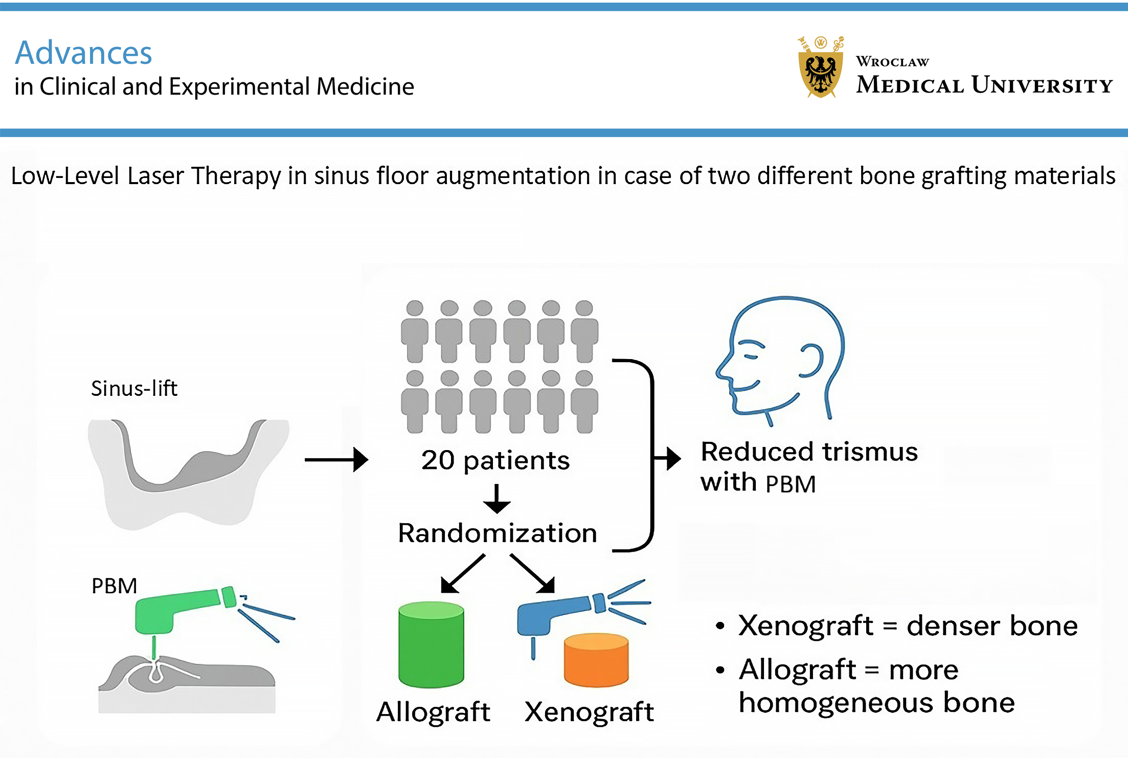
Results. Low-level laser therapy significantly reduced trismus. Trismus was also less pronounced in male patients. Low-level laser therapy was associated with increased facial swelling. Pain levels were low, with no significant differences between the study groups. After 135 days, allografts demonstrated greater homogeneity, whereas xenografts exhibited higher radiographic density.
Conclusions. Low-level laser therapy may improve recovery following sinus lift surgery by reducing postoperative trismus. No significant differences were observed between the grafting materials in terms of bone gain. However, differences in radiodensity suggest distinct resorption patterns, with xenografts exhibiting higher density values after 135 days.
Key words: laser, xenograft, allograft, sinus-lift, biomodulation
Background
Despite the rapid pace at which dentistry has developed over recent years, tooth loss remains a significant challenge that requires restoration of the missing dentition. Among the available treatment options, dental implant therapy is considered the most desirable due to the high level of comfort it provides to patients. It also demonstrates a very high success rate and does not require the involvement of adjacent teeth in the treatment, which is a major drawback of conventional prosthetic crowns and bridges. Despite its obvious benefits, implant-prosthetic rehabilitation of missing dentition also has limitations, mainly associated with gradual bone loss following tooth extraction, resulting from remodeling of the alveolar process of the maxilla and pneumatization of the maxillary sinuses.1, 2, 3
Bone volume, if insufficient for implant treatment, can be restored through augmentation procedures, which commonly involve the use of bone grafting materials of autogenous, allogenous, or xenogenous origin. Autogenous grafts are considered the gold standard due to their osteogenic, osteoinductive, and osteoconductive properties. Nonetheless, their availability is often limited, and their use requires an additional surgical procedure at the donor site, which may increase morbidity.4 Therefore, bone substitute biomaterials have been introduced into regenerative medicine. They are characterized by chemical and biological biocompatibility, osteoconductivity, and, in some cases, even osteoinductivity.5 Xenogenous bone grafts are commercially available; however, they possess only osteoconductive properties. These biomaterials are derived from other species, most commonly bovine, porcine, or equine, and exhibit favorable biomechanical properties.6 As allografts, in addition to being osteoconductive, may also possess osteoinductive potential, they are often preferred, and their use is becoming increasingly common.7, 8, 9 The osteoconductive potential of xeno- and allografts may be enhanced through the additional application of growth factors. One of the possible options in this regard, particularly preferred in oral surgery, is platelet concentrates. Human platelets are a source of numerous growth factors, including platelet-derived growth factor (PDGF), vascular endothelial growth factor (VEGF), and transforming growth factor beta (TGF-β), which play a role in the initial stages of bone defect healing. They also act as attractants for immune cells, which may serve as a source of additional growth factors during later stages of healing.10 When it comes to surgical techniques, in cases of severe atrophy of the lateral aspects of the maxillary alveolar ridge, sinus lift surgery is considered the treatment of choice. Initially introduced by Tatum in 1975, it involves surgically creating an opening in the lateral wall of the maxillary sinus and carefully elevating the Schneiderian membrane to create a space for the bone grafting material. A cavity prepared in this manner exhibits highly osteoconductive properties because it is surrounded predominantly by osseous and well-vascularized tissues. Although highly predictable in terms of long-term outcomes, this method may lead to several early complications, including pain, trismus, and swelling caused by hematoma formation, which can significantly reduce patient satisfaction.11
Among the available methods for reducing these symptoms, the use of lasers has recently gained considerable attention. So-called low-level laser therapy (LLLT) is known to modulate the inflammatory proceess, reduce pain intensity and swelling, and promote tissue healing without causing adverse effects.12, 13 It has been evaluated in many oral surgery procedures, particularly surgical tooth extractions. Most authors report its positive influence on soft tissue healing.14, 15 Although its effectiveness has been well documented in numerous dental procedures, there is still a lack of evidence regarding the effects of LLLT in sinus lift surgery.
Objectives
The main objective of this study was to evaluate the influence of adjunctive LLLT following sinus lift surgery on patient recovery in cases involving 2 grafting materials of different origins, as assessed by trismus, pain scores, and midfacial swelling. An additional objective was to compare 2 different grafting materials used in sinus floor elevation procedures with regard to new bone height, density, and homogeneity based on cone-beam computed tomography (CBCT) examinations.
Materials and methods
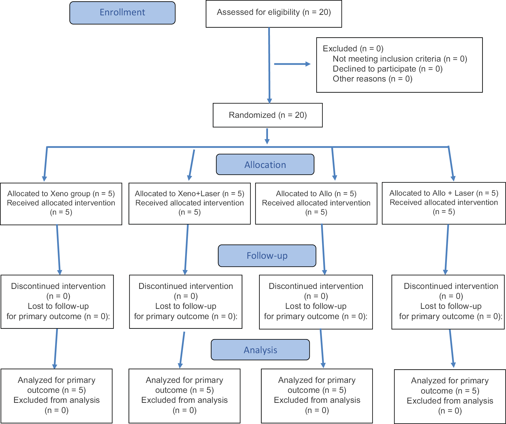
This prospective study was conducted at the Department of Oral Surgery, Wroclaw Medical University (Poland), between 2023 and 2024. It included 20 patients requiring a lateral sinus lift procedure prior to implant-prosthetic treatment. The assignment of the medical intervention was not at the discretion of the investigators but was determined by the need for implant-based restoration in cases of reduced maxillary alveolar bone height. The study was approved by the Local Bioethics Committee of Wroclaw Medical University (approval No. KB 10/2023N). All participants provided 2 forms of written informed consent: the 1st for the lateral sinus lift procedure and the 2nd for participation in the study. The study was conducted in full compliance with the Declaration of Helsinki and adhered to personal data protection regulations under the General Data Protection Regulation (GDPR). The study followed the CONsolidated Standards Of Reporting Trials (CONSORT) guidelines, and the flow diagram presents patient enrollment (Figure 1).16
Inclusion and exclusion criteria
Patient recruitment was performed by an investigator who was not involved in the surgical procedures (A.B.). The inclusion criteria were as follows: 1) adults capable of providing informed consent for the surgical procedure; 2) single or multiple missing teeth in the posterior region of the maxillary dental arch; 3) alveolar ridge height of less than 5 mm and width greater than 7 mm; and 4) at least 2 mm of keratinized tissue height (HKT) in the region of interest.
The exclusion criteria were as follows: 1) poor oral hygiene (Plaque Index (PI) >20%); 2) pregnancy; 3) breastfeeding; and 4) general or local contraindications to surgical procedures, including active sinus infection, large sinus cysts, previous radiation therapy to the head and neck region, and severe systemic conditions such as uncontrolled diabetes, immunocompromised states, and severe cardiovascular disease (CVD). The study group consisted of 20 adult Caucasian patients (Figure 1). Subsequently, each participant was randomly assigned to 1 of the study groups and underwent the procedure according to the protocol of the allocated group.
Study group design
Patients were randomly divided into 2 equal groups, each consisting of 10 patients. The division was based on the type of bone grafting material used during the sinus lift procedure. In half of the patients, an allogenic bone graft in the form of cortico-cancellous bone granules “S” (granulometry: 0.5 mm; Biobank, Lieusaint, France) was used. In the remaining patients, a xenogenic grafting material in the form of cancellous bone granules (granulometry: 0.25–1.0 mm; Geistlich, Wolhusen, Switzerland) was used. From each group, 5 patients were randomly selected to receive LLLT during the postoperative period. As a result, 4 equal groups, each consisting of 5 patients, were created. The groups were as follows: 1) xenograft; 2) xenograft + LLLT; 3) allograft; and 4) allograft + LLLT.
Protocol of the experiment
Presurgical examination (T1)
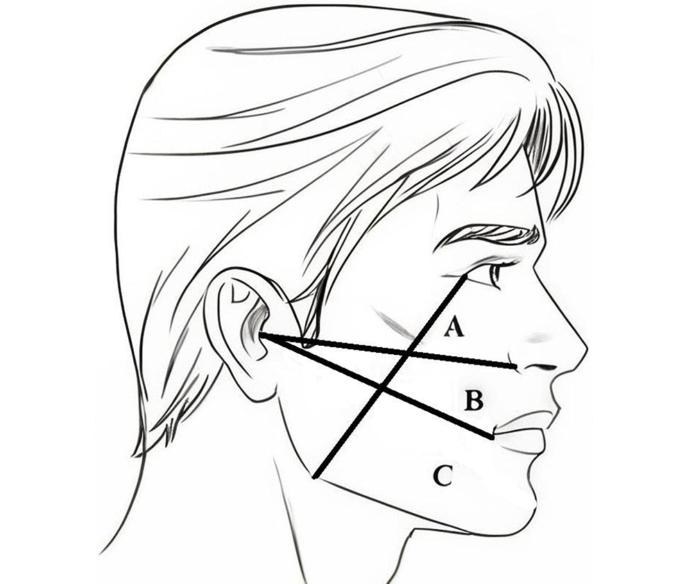
Every patient included in the study underwent a clinical examination. Measurements were performed by one of the researchers who was not involved in the subsequent clinical procedures (A.B.). Particular emphasis was placed on the evaluation of the future surgical site; therefore, the following parameters were recorded: height (HKT) and width (WKT) of the keratinized gingiva, alveolar ridge dimensions, and the PI according to Silness and Löe. Additionally, maximum jaw opening was measured as the distance between the upper and lower central incisors. Facial swelling was also assessed by measuring 3 reference lines. This method was a modification of the technique originally introduced by Laskin. As the surgical field in the present study involved the midface rather than the lower face, as assessed by Laskin, the measurement lines were modified accordingly.17 The lines used in this study were as follows:
– Line A – a straight line extending from the lateral canthus of the eye to the angle of the mandible;
– Line B – a straight line extending from the midpoint of the tragus of the ear to the base of the nasal ala (Camper’s plane);
– Line C – a straight line extending from the midpoint of the tragus of the ear to the corner of the mouth (Figure 2).
Following the clinical examination, every patient was referred for radiological assessment. Cone-beam computed tomography scans were acquired using the Planmeca Viso® G7 system (Planmeca®, Helsinki, Finland). A field of view (FOV) centered on the maxilla, measuring 300 mm in width and 200 mm in height, was used. The scans were analyzed using Romexis® software (Planmeca®), and the foll day owing parameters were measured and assessed: residual bone height (RBH); sinus condition, including Schneiderian membrane thickening; the presence of sinus polyps, cysts, or septa; and periapical inflammatory lesions associated with adjacent teeth. All measurements were performed by a researcher who was not involved in the surgical procedures (C.O.).
Surgical procedure for the lateral sinus-lift
Regardless of study group allocation, all patients were treated according to the same surgical protocol. Under local anesthesia with 4% articaine and epinephrine 1:200,000 (Septanest® 1:200,000; Septodont, Saint-Maur-des-Fossés, France), an envelope-type full-thickness mucoperiosteal flap with a distal releasing incision was elevated to expose the lateral wall of the maxillary sinus. Subsequently, a standardized osteotomy and elevation of the Schneiderian membrane were performed using the DASK® kit (Dentium® Co., Ltd, Suwon, South Korea). First, a bony window was created using identical 8-mm diamond burs under constant saline irrigation and minimal pressure. Next, DASK® elevators were used to prepare an appropriate recipient site for the bone grafting material. The augmented area was covered with a collagen membrane (Bio-Gide®) to prevent epithelial ingrowth into the regenerated region. The flap was repositioned and sutured without tension in the primary position using non-resorbable 5-0 nylon monofilament sutures (Atramat®, Mexico City, Mexico).
Follow-up procedures
Low-level laser therapy
In the appropriate groups (+LLLT), diode laser therapy was applied using a laser device (Lasotronix®, Piaseczno, Poland) with a wavelength of 635 nm, a beam diameter of 0.5 cm2, and an energy density of 6 J/cm2 for 1 min in continuous-wave mode. Low-level laser therapy was performed intraorally, with the laser tip positioned directly over the osteotomy site and the overlying soft tissues immediately after the sinus lift procedure, as well as 3 and 7 days postoperatively.
Postoperative treatment
Every patient, regardless of group allocation, was prescribed Eludril Classic® mouthwash for 2 weeks (twice daily) and an antibiotic (2.0 g/day; Duomox®; Astellas Pharma, Tokyo, Japan). The sutures were removed after 7 days. The 1st follow-up visit was scheduled for postoperative day 3 (T2), and the 2nd for postoperative day 7 (T3). On both occasions, a clinical examination was performed in the same manner as before surgery. On postoperative day 135, a follow-up examination was performed (T4). Clinical and radiological evaluations were repeated in the same manner, with the inclusion of the following additional parameters:
– Bone gain (BG) measured within the region of interest (ROI) as the difference between the postoperative radiographic height of the alveolar process and the RBH.
– Homogeneity of the augmented area, assessed according to the following features:
• initial union observed at the junctional area;
• appearance of mottled radiographic shadows throughout the grafted area;
• differentiation of newly formed bone into a trabecular pattern;
• absence of highly radiopaque particles of non-resorbed graft material.
The presence of at least 3 of the abovementioned features was considered indicative of high graft homogeneity, whereas the presence of fewer than 3 features was considered indicative of low graft homogeneity.
– Bone density, expressed in Hounsfield units (HU), was assessed as follows: First, the imaging plane was positioned at the graft site and aligned parallel to the panoramic curve to visualize the area in which the greatest bone gain had been recorded. Next, an oval region of measurement was carefully selected to include as much of the grafted area as possible. The mean HU value generated by the software was then recorded for subsequent analysis.
Statistical analyses
Due to the relatively small sample size, nonparametric tests were conducted using the nparLD package (https://CRAN.R-project.org/package=nparLD) in R (R Foundation for Statistical Computing, Vienna, Austria). Fisher’s exact test is a nonparametric test of statistical significance used, among other purposes, to examine hypotheses concerning the association between 2 nominal-scale variables, often dichotomous in nature. Its advantages lie in its applicability to small sample sizes. An extension of this test is the Fisher–Freeman–Halton test, which is designed for larger contingency tables and, unlike Fisher’s exact test, allows for analyses involving more extensive data sets. Friedman’s rank test is a nonparametric statistical test based on rank ordering, used to assess differences among at least 3 dependent (i.e., correlated) groups. The rank corresponds to the sequential number assigned to a statistical observation within a sample after ordering the observations according to the value of a given variable. On the other hand, the Kruskal–Wallis test is a rank-based statistical test employed to compare the distributions of a variable across populations. Particularly suitable for small sample sizes, this test does not assume normality of distributions and is regarded as a nonparametric alternative to one-way analysis of variance (ANOVA) for comparisons across groups.
Table 1 presents the results of comparisons for parameters measured once, whereas Table 2 contains the results for parameters measured at 4 time points. In addition, to assess the potential impact of interactions between independent variables on dependent variables, nonparametric statistical analyses for longitudinal data in mixed designs were performed. The results are presented in Table 3.
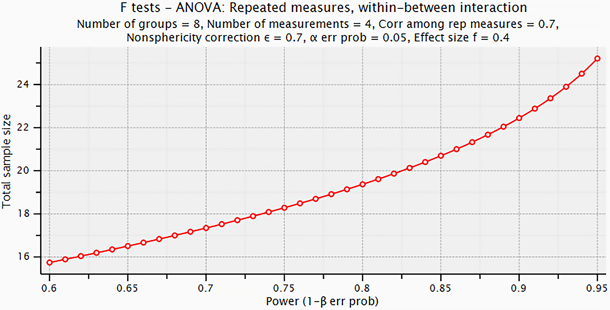
To evaluate the effects of the between-subject factors (graft and laser) and the within-subject factor (time) on quantitative dependent variables (Jaw Opening, Line A, Line B, Line C, visual analogue scale (VAS), and PI), nonparametric statistical analyses for longitudinal data in mixed designs were carried out. The nparLD package in R was used for this purpose. The results are summarized in Table 3. To evaluate the statistical significance and effect size of 3 independent factors on the dependent variables, repeated-measures ANOVA was performed. The sample size (n = 20) was determined using the G*Power software (https://www.psychologie.hhu.de/arbeitsgruppen/allgemeine-psychologie-und-arbeitspsychologie/gpower), assuming a significance level of α = 0.05, statistical power of at least 1 − β = 0.80, 8 groups (2 × 2 × 2 design), 4 repeated measurements, an inter-measurement correlation of r = 0.7, and a nonsphericity correction factor of ε = 0.7 (Figure 3).
Results
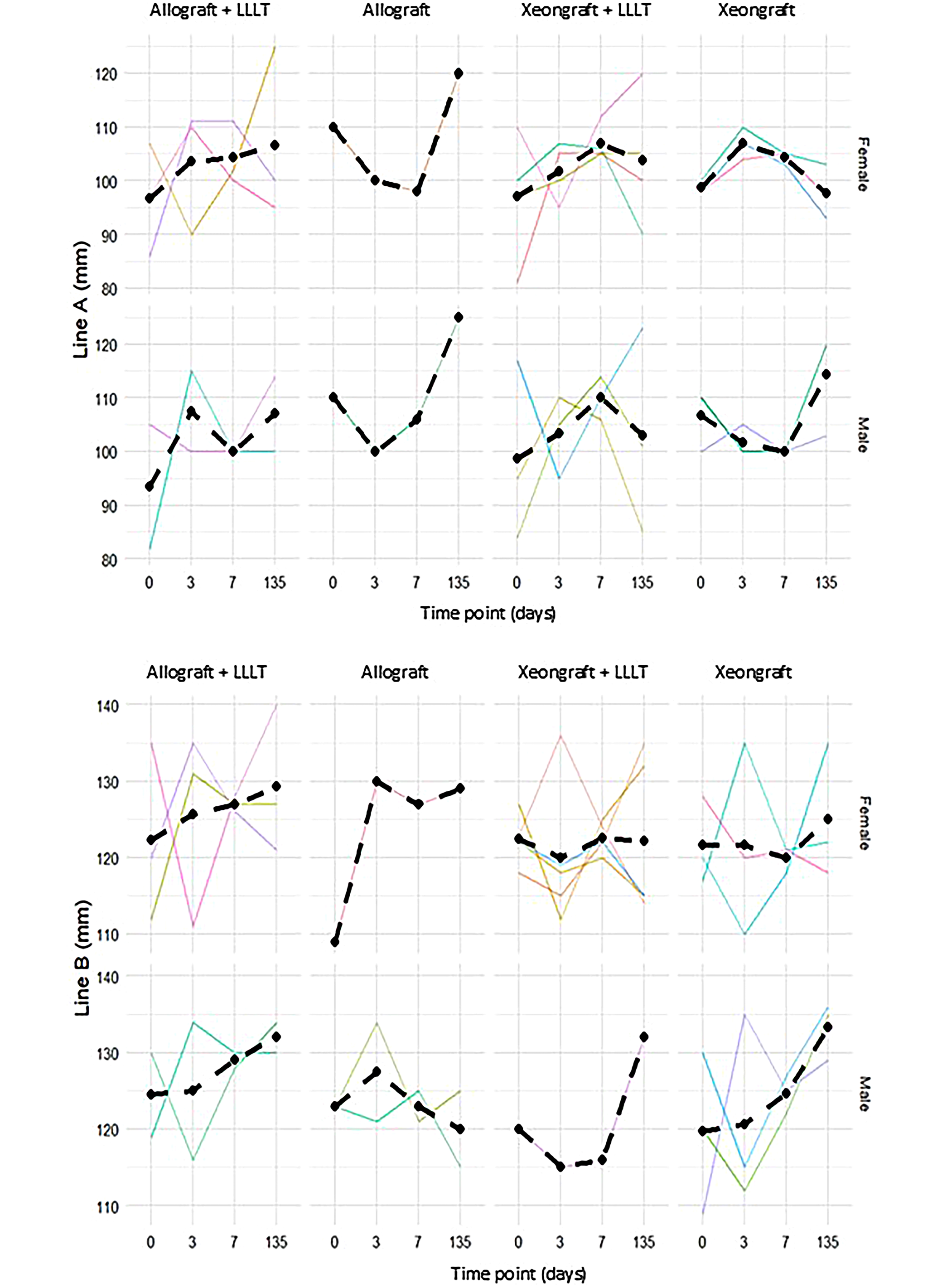
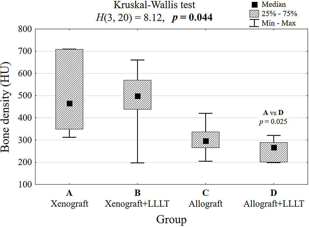
The mean age of the participants was 50.7 years. The study groups were homogeneous with respect to sex distribution, as 9 of the 20 included patients (45%) were women. Detailed data on patient age and sex distribution among the subgroups are presented in Table 1. After completion of the follow-up period for all included patients, a statistical analysis of the collected data was performed. On the day of surgery, patients in all 4 groups were homogeneous with respect to the analyzed clinical parameters (Table 1). The analysis of jaw opening indicated that the measured values depended on whether laser photobiomodulation had been applied after surgery. Jaw opening values were higher in both patient groups that received photobiomodulation compared with those that did not. Although LLLT appeared to increase jaw opening after surgery, the observed association did not reach statistical significance (Table 2, Table 3). The analysis of the lengths of Lines A, B, and C showed higher values in male patients than in female patients and in patients who received allografts (Table 2, Table 3; Figure 4). Pain scores were not associated with either graft type or sex. Photobiomodulation increased VAS scores on postoperative day 3 but did not influence pain levels at later stages of healing. The main finding for this variable was that pain intensity was time-dependent in patients from both groups. Patients reported significantly higher pain scores on day 3 than on days 7 and 135 after surgery (p < 0.05; Table 3). The analysis of radiological parameters based on CBCT examinations revealed no statistically significant differences between the groups in bone gain after 135 days. However, the analysis of HU in the regenerated bone clearly demonstrated higher values in the xenograft group than in the allograft group, and the standard deviation (SD) was also significantly higher in the xenograft group (Figure 5). We also observed that the allograft was more homogeneous after 135 days compared with the xenograft. This finding is likely related to the longer resorption time characteristic of xenografts compared with allografts.
Discussion
The positive effect of photobiomodulation on postoperative healing has been reported previously in oral surgery procedures. Landucci et al., in a study involving third-molar surgery, demonstrated that laser therapy is effective in reducing postoperative pain, trismus, and swelling.18 In contrast, Momeni et al. and Alan et al. found that LLLT had no significant effect on edema or trismus; however, it was associated with reduced postoperative pain levels.19, 20
In the present study, a positive effect of LLLT on trismus was observed, whereas no statistically significant effects on pain levels or swelling following sinus lift surgery were found. This may be explained by differences in the surgical site (i.e., the distal region of the mandibular alveolar process vs the lateral wall of the maxilla). Previous studies evaluated surgical sites within the oral cavity characterized by strong muscle attachments adjacent to the elevated flap,18, 19, 20 making trismus more likely to occur. Another important consideration is that the VAS scores recorded immediately after surgery in our study were already remarkably low. Therefore, the additional application of LLLT may not have provided a clinically meaningful benefit in terms of pain reduction for our patients.
Despite numerous reports on the influence of LLLT in oral surgery procedures, as mentioned above, its role in recovery following sinus lift surgery remains poorly described in the literature. In a study investigating this procedure, Ozturan et al. reported a positive effect of LLLT on swelling and quality of life. However, it is worth noting that, similarly to our findings, no effect of LLLT on postoperative pain intensity was observed.21 Trismus is a common symptom during postsurgical recovery. Although it is recognized as part of the physiological healing process, its severity may indicate the influence of additional factors on recovery. More severe trismus may be associated with inflammation, extensive surgical trauma, or prolonged operative time; however, anatomical factors may also contribute. In our study, we observed that male patients exhibited greater jaw opening ranges regardless of the use of LLLT or the grafting material applied. Most studies related to this topic have been conducted in the context of third molar extraction; nevertheless, certain similarities can be identified. In a study by Vranckx et al., the influence of sex on postsurgical healing was evaluated among other factors. Consistent with our findings, the authors reported that women exhibited more pronounced trismus than men.22 However, reduced jaw opening following surgical procedures is not always associated with patient sex. In contrast, Kiencało et al. found no correlation between sex and trismus after evaluating 339 wisdom tooth extractions.23
The aim of every bone grafting procedure is to achieve new bone formation. This is particularly important in procedures followed by implant placement, as direct contact between the implant surface and bone tissue fulfills the definition of osseointegration. New bone formation can be confirmed with certainty only through histological examination; however, this requires a trephine biopsy and may significantly increase the risk to the patient. Therefore, most studies have employed less invasive radiographic methods, particularly CBCT, to assess bone graft remodeling and turnover.24, 25, 26
It is also a routine method for assessing the outcomes of sinus lift procedures in everyday dental practice. It is generally accepted that the presence of a trabecular pattern and the absence of voids within the grafted area on CBCT images are indicative of bone maturation, whereas increases in radiodensity following bone grafting should be considered a normal stage of healing, as more mature bone typically exhibits a denser appearance.24, 25, 26
In studies with a follow-up period similar to ours, the origin of the bone graft usually does not influence the degree of bone gain, which is consistent with our findings. A similar conclusion was reached by Al-Moraissi et al., whose large meta-analysis of 52 studies found no differences in new bone height among different grafting materials.27 The density and homogeneity of the augmented bone, however, are closely related to graft origin. Autologous and allogenic bone grafts are more likely to mimic the properties of the recipient site’s residual bone at an earlier stage. Xenografts and alloplastic materials, on the other hand, exhibit prolonged remodeling times, which may explain the differences in homogeneity and density of the newly formed bone. In our study, after 135 days, allogenic bone grafts tended to be more homogeneous, whereas xenografts achieved higher values on the Hounsfield scale. Tabrizi et al. reported similar findings, as xenografts in their study demonstrated significantly higher CBCT density values compared with allogenic material 9 months after maxillary sinus augmentation.28
Limitations of the study
The main limitation of this study is the small sample size, which resulted from funding constraints associated with the Doctoral Subsidy program, limiting enrollment to a maximum of 20 patients and the observation period to no more than 12 months. Additional limitations include the fact that new bone formation can be confirmed with absolute certainty only through histological examination, which was not performed in the present study. Furthermore, 2 grafting materials of different origins were used, and these materials exhibit distinct characteristics in radiological evaluation, which may have influenced the interpretation of the radiographic findings.
Conclusions
Low-level laser therapy may improve patient recovery following sinus lift surgery by reducing postoperative limitations in jaw opening. The observation period used in this study corresponds to the average waiting time before implant placement after maxillary sinus floor elevation. The data obtained from this study suggest that different types of bone substitutes do not influence the amount of newly formed bone. However, differences in radiodensity between allografts and xenografts suggest distinct resorption patterns of the 2 materials.
Data Availability Statement
The datasets supporting the findings of the current study are openly available in Zenodo at https://doi.org/10.5281/zenodo.15447878.
Consent for publication of personal information
Not applicable.
Use of AI and AI-assisted technologies
Not applicable.