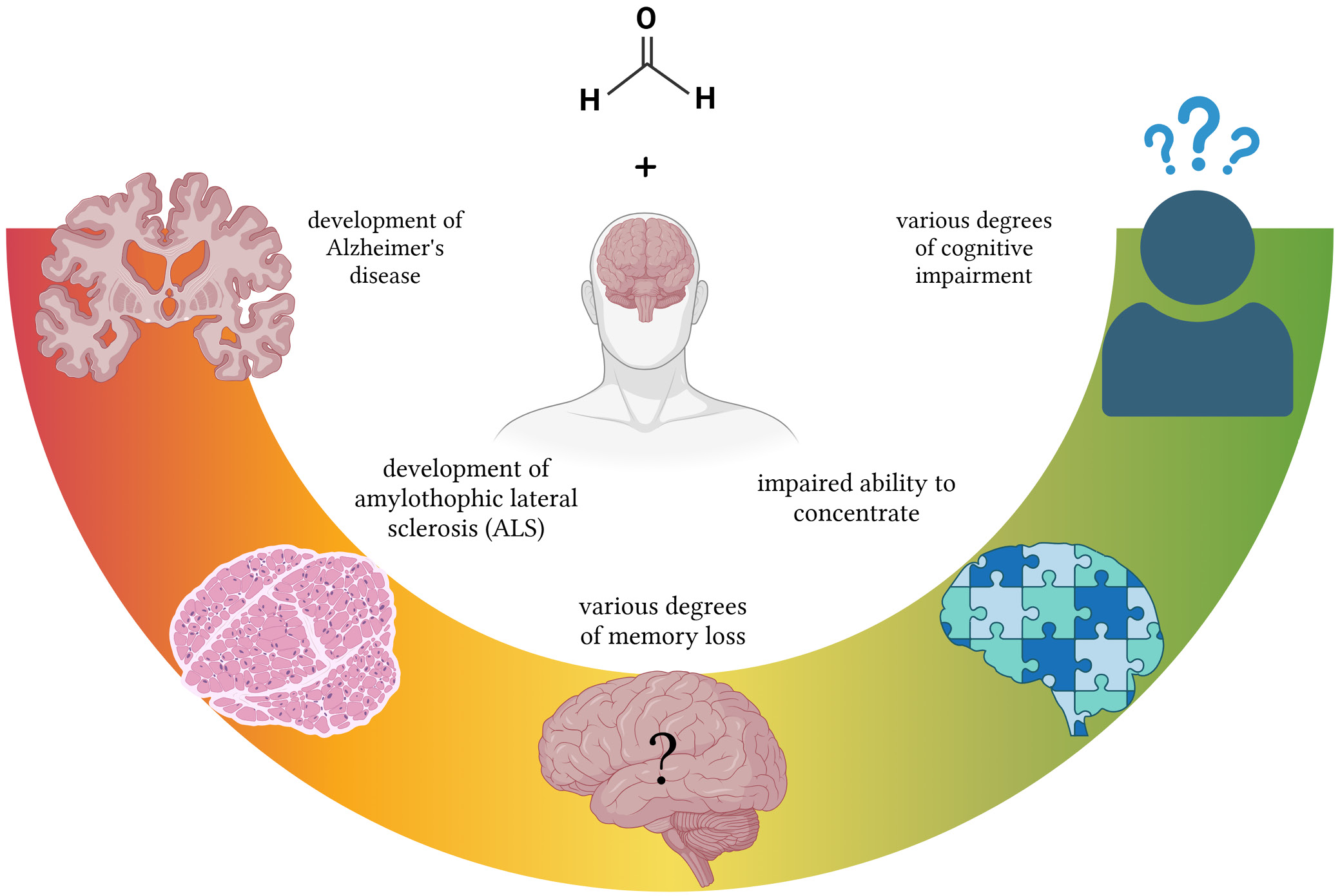
Advances in Clinical and Experimental Medicine

Title abbreviation: Adv Clin Exp Med
Journal Impact Factor (JIF 2025) – 2.5
Journal Citation Indicator (JCI 2025) – 0.40
Scopus CiteScore (2025) – 4.2
Index Copernicus Value (ICV 2024) – 161.00
MNiSW – 70 pts
ISSN 1899–5276 (print), ISSN 2451-2680 (online)
Periodicity – monthly

Download original text (EN)

Advances in Clinical and Experimental Medicine

Ahead of print

doi: 10.17219/acem/209617

Publication type: review

Thematic category: Forensic toxicology; neurology; neuroscience; occupational diseases

Language: English

License: Creative Commons Attribution 3.0 Unported (CC BY 3.0)

Download citation:

  • BIBTEX (JabRef, Mendeley)
  • RIS (Papers, Reference Manager, RefWorks, Zotero)

Cite as:


Drążyk M, Pyc Z, Pietrzyk SJ, et al. Formaldehyde neurotoxicity: Effects on the mammalian brain, cognitive function, and neurodegenerative risk. A scoping review [published online as ahead of print on June 25, 2026]. Adv Clin Exp Med. 2026. doi:10.17219/acem/209617

Formaldehyde neurotoxicity: Effects on the mammalian brain, cognitive function, and neurodegenerative risk. A scoping review

Mateusz Drążyk1,A,B,C,D,F, Zuzanna Pyc2,B,C,D, Szymon J. Pietrzyk1,B,C,D, Antonina Gajda-Janiak1,B,C,D, Filip Godziszewski1,B,C,D, Oliwier Pioterek1,B,E,F, Michał Tulski1,B,E, Mateusz Mazurek1,A,E,F, Zygmunt A. Domagała3,A,E,F

1 Student Scientific Association of Clinical and Dissecting Anatomy, Wroclaw Medical University, Poland

2 Student Scientific Association of Biomedical and Environmental Analyses, Wroclaw Medical University, Poland

3 Division of Anatomy, Wroclaw Medical University, Poland

Graphical abstract


Graphical abstracts

Highlights


• Formaldehyde exposure may contribute to deterioration in mental health.
• Research suggests that formaldehyde exposure may affect mood, cognitive function, and sleep quality.
• Formaldehyde contributes to oxidative stress and apoptosis in neural cells, potentially leading to cognitive decline.
• Reducing or eliminating formaldehyde use in research, particularly in medical settings, should be a priority for occupational safety and health.

Abstract

Aqueous formaldehyde (FA) solution, known as formalin, is currently the primary agent used for preserving tissue samples and anatomical specimens. Formaldehyde is widely used in laboratories and the chemical industry; it also occurs as an air pollutant and endogenous cellular metabolite. The potential carcinogenic effects of formalin on the respiratory tract are well documented. A less recognized consequence of occupational exposure to FA is its detrimental effect on the central nervous system (CNS) and brain function. A literature review was conducted to investigate the effects of FA on the brain. Five databases were searched: PubMed, Web of Science (WoS), Embase, ScienceDirect, and Google Scholar. To describe the effects of FA exposure and endogenous FA generation, 35 relevant publications were collected and analyzed. The literature review demonstrated that inhalation is the most common route of FA exposure. Several studies have shown that FA may cause hippocampal damage, disrupt melatonin secretion, and induce a wide range of cognitive disorders with varying characteristics and severity. These disorders include memory impairment, disturbances in balance and spatial orientation, learning difficulties, sleep disturbances, impaired judgment, and prolonged reaction times to stimuli. Increased endogenous FA concentration has also been associated with a higher risk of neurodegenerative diseases, such as Alzheimer’s disease (AD) and amyotrophic lateral sclerosis. The literature analysis demonstrated the high neurotoxicity of FA, which may lead to numerous neuropsychiatric disorders. We aim to draw attention to the risks associated with the routine use of formalin, particularly among anatomists and pathologists, and to encourage consideration of less harmful alternative preservation agents.

Key words: formaldehyde, occupational exposure, cognitive impairment, neurotoxicity, neurodegenerative diseases

Introduction

Formaldehyde (FA) has been known for more than 150 years. It was first synthesized in gaseous form in 1859 by Alexandr Butlerov. Nearly a decade later, in 1868, German scientist August Wilhelm von Hofmann described its chemical structure.1 Since then, numerous applications of this substance have been identified. Formaldehyde is commonly used in the textile industry and carpentry as a substrate for the production of resins and glues and, in the form of an aqueous solution known as formalin, is widely recognized as the main ingredient of body-preserving solutions used for anatomical and funeral purposes. The use of FA in anatomy began at the end of the 19th century,2 and since then it has continued to be used as a fixative for tissue preservation by pathologists and histologists.3

Formaldehyde is also present in the air we breathe as an environmental pollutant,4, 5 and is a component of cigarette smoke and vehicle exhaust fumes.6, 7, 8 It is widely used in industrial production as a substrate in the chemical industry9 and can also be found in cosmetic products.10, 11 A lesser-known fact is that FA is one of the products of biochemical pathways in the human body.12 These examples demonstrate that the human population is constantly exposed to FA. The level of exposure may vary depending on occupation or place of residence, but exposure is unavoidable.

In 2004, FA was classified as a carcinogen by the International Agency for Research on Cancer (IARC).13 There is sufficient evidence suggesting that it increases the risk of nasopharyngeal cancer and leukemia.14 Several studies have also investigated whether FA exposure may increase the risk of brain tumor development.15 Formaldehyde has been identified as a neurotoxic substance in numerous in vivo and ex vivo studies. Although the carcinogenic properties of FA are widely recognized, it continues to be extensively used. Its low cost and ease of use contribute to its popularity in pathology, histology, and education (e.g., gross anatomy courses). In contrast to other fixatives, FA penetrates tissue easily, acts rapidly, and does not require unusual storage conditions. However, relatively few studies have examined the neurotoxicity of FA and its potential effects on cognition and behavior.

Objectives

In this study, we examined the relationship between neuronal damage and death and reduced brain function associated with exposure to FA and endogenous FA production. In particular, we focused on cognitive impairment, including FA-induced alterations in memory and attention span. We also investigated whether FA may contribute to the development of mood disorders.

Furthermore, we investigated the molecular mechanisms of FA action, its biological and psychological implications, and its potential contribution to the development of neurodegenerative diseases. Considering the contribution of FA to carcinogenesis, we hypothesized that FA may also adversely affect neuronal health and neuropsychological functioning. Our investigation aimed to provide an objective and comprehensive evaluation of the available scientific evidence regarding FA and nervous system health.

Materials and methods

Eligibility criteria

To explore the influence of FA on the central nervous system (CNS), we reviewed the available literature. Only original full-text articles written in English were included. Review articles were excluded. No restrictions regarding publication date were applied. We included studies conducted in humans, animal models, and cell cultures. We searched for information regarding changes in cognitive function, cognitive decline, and behavioral alterations following FA exposure. Additional selected articles described FA metabolism and biochemical pathways in organisms, particularly mechanisms underlying neuronal damage and alterations in neuronal physiology. Finally, we also included articles addressing the influence of exogenous and endogenous FA on neurodegenerative diseases.

Information sources and search strategy

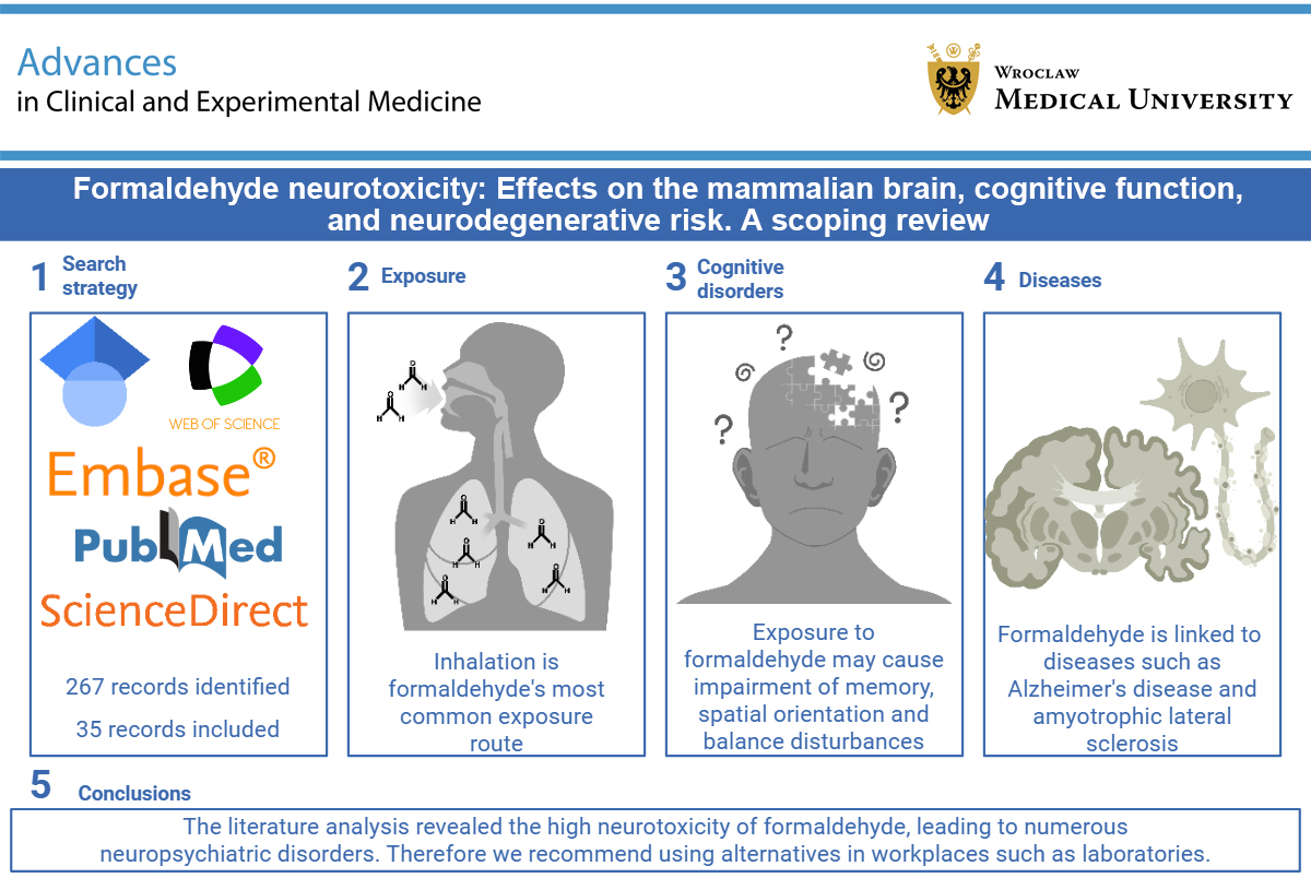
Our search was conducted in 5 databases: PubMed, Web of Science (WoS), Embase, ScienceDirect, and Google Scholar. The search began on November 27, 2023, and ended on December 7, 2023.

We defined search phrases that were subsequently used to identify articles describing FA and the CNS. The search phrases consisted of the constant term “formaldehyde exposure”, combined with the following additional terms: “human”, “brain damage”, “cognition”, “cognitive functions”, “dementia”, “intelligence”, “memory”, “memory loss”, “neurobehavioral impairment”, “neurological disorders”, “neuropsychiatric disorders”, “neurotoxicity”, and “induced neuron apoptosis”.

Data collection and outcomes

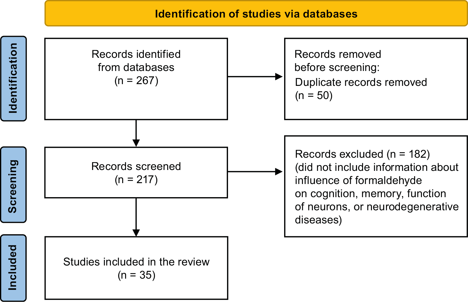
In the initial search, based on title screening conducted in the above-mentioned databases, we identified 267 articles. After removing duplicates, 217 papers remained. Subsequently, abstracts of these articles were screened, which reduced the number of eligible studies to 35. Studies conducted on animal models comprised 18 of the 35 articles included in the final pool of selected studies (Figure 1).

In the next step, data were extracted from the selected articles, which were subsequently reviewed and summarized. Using an extraction form created in Microsoft Word 365 (Microsoft Corp., Redmond, USA), we collected all data related to the above-mentioned terms. We searched for information regarding the effects of FA on neuronal function and the CNS. The collected data also addressed the neurotoxicity of FA. We then examined the impact of neuronal damage or death on cognitive functions, including memory, learning processes, spatial orientation, reaction time, abstract thinking, and judgment, as well as prolonged reaction times to stimuli. We were also interested in attention span, behavioral changes, and affective disorders following exposure. Although the focus was mainly on acute and chronic FA exposure, we also considered endogenous FA formation in the context of cognitive decline and neurodegenerative diseases.

The collected data were subsequently organized into 4 main subject areas: neurotoxicity, the influence of FA on memory, the influence of FA on other cognitive functions, and the development of FA-related neurodegenerative diseases. The summarized data from all studies included in this review are presented in Table 1,16, 17, 18, 19, 20, 21, 22, 23, 24 Table 2,24, 25, 26, 27, 28, 29, 30, 31, 32, 33, 34, 35, 36, 37, 38, 39, 40, 41, 42, 43 and Table 3.4, 5, 44, 45, 46, 47, 48, 49, 50, 51, 52

Results and discussion

Neurotoxicity of formaldehyde

Formaldehyde readily crosses the blood–brain barrier (BBB).53 Once in the brain, it may damage neurons. For example, the methyl donor S-adenosylmethionine (SAM) in PC12 cell cultures derived from patients with Parkinson’s disease induces a cascade of methanol-derived reactions leading to the formation of endogenous FA. Formaldehyde, produced through oxidation of methanol during SAM-induced reactions, showed marked toxicity toward neuron-like PC12 model cells, which are catecholaminergic pheochromocytoma cells. Under the same conditions, methanol and formic acid did not exhibit comparable toxicity. Furthermore, FA induced a considerably weaker toxic effect in C6 glial cell cultures – mainly excessive protein carboxymethylation and inhibition of tyrosine hydroxylase expression – indicating that neurons may be more susceptible to FA toxicity than glial cells. However, prolonged incubation resulted in decreased viability of both PC12 and C6 cells, suggesting that exposure duration plays a crucial role in the toxic effects of FA on human neuronal and glial cells.19

Moreover, numerous studies conducted in laboratory animals have demonstrated visible alterations in brain neuronal tissue following FA exposure. Detected abnormalities included increased reactive oxygen species (ROS) formation,16, 18 other manifestations of oxidative stress (OS),17 neuronal apoptosis,16, 20 and neuronal degeneration (Figure 2, Figure 3).18

Oxidative damage induced by FA

Following FA exposure, numerous biochemical changes indicative of redox imbalance have been observed across different studies. Levels of malondialdehyde (MDA) in the frontal cortex and hippocampus increased after FA exposure.16, 18, 21 Malondialdehyde is a product of lipid peroxidation and is considered one of the most important markers of OS.54 Increased MDA levels have also been observed under controlled conditions simulating occupational exposure.16 In the frontal cortex and hippocampus, catalase and superoxide dismutase (SOD) activity decreased following FA exposure.16, 18 Both enzymes are involved in defense against OS – catalase through decomposition of hydrogen peroxide (H2O2)55 and SOD through dismutation of superoxide anion radicals into H2O2.56 Rats exposed to FA also exhibited elevated levels of 8-hydroxy-deoxyguanosine (8-OHdG), another important marker of OS,57 in both brain tissue and urine.17

Exposure to FA decreases glutathione levels while increasing concentrations of 4-hydroxynonenal.21 Concentrations of lipid peroxides and oxidized proteins also increased,16, 18, 21 as did total ROS levels.21, 22 Increased ROS levels reduced the viability of PC12 cells.22 This overall increase in OS marker levels following exposure suggests substantial intensification of OS induced by FA.

The intensification of OS may result from multiple pathways. Incubation of PC12 cells with FA leads to a significant decrease in the concentration and production of the antioxidant molecule H2S. Exposed cells also displayed a significant increase in inducible nitric oxide synthase (iNOS)16 and neuronal nitric oxide synthase (nNOS) levels and overproduced nitric oxide (NO), which is a major mechanism inhibiting cystathionine β-synthase, the H2S-producing enzyme in PC12 cells.22 Formaldehyde also interacts with glutathione (GSH), thereby depleting the organism’s antioxidant defenses.23 Intensification of OS may additionally result from inhibition of paraoxonase-1, an antioxidant enzyme.58

It is well established that neurons are highly susceptible to damage caused by OS, especially those located in the hippocampus and amygdala.59 Given the information presented above, OS may be considered one of the major mechanisms of cellular damage associated with FA exposure.

Apoptosis induced by formaldehyde

Cells exposed to FA showed changes typical of apoptosis.16, 22 Following exposure, neurons in the frontal cortex and hippocampus of rats exhibited extensive degenerative changes, dark pyknotic nuclei, and shrunken cytoplasm.18, 21 Moreover, their axons retracted, and the cells became separated from one another.21 The cerebellum is also affected by FA exposure, leading to neuronal damage in all its layers, with Purkinje cells being the most severely affected.16 Formaldehyde exposure also results in altered expression of proteins involved in apoptosis and cellular protection. In capillaries supplying Purkinje cells, Bax protein levels increased. Bax is a pro-apoptotic protein and a member of the Bcl-2 gene family involved in p53-mediated apoptosis activation.16

In PC12 cells, FA exposure induced cytotoxic and apoptotic effects, including decreased levels of Bcl-2 protein and increased release of cytochrome C.58 Bcl-2 is a protein responsible for inhibiting apoptosis,60 whereas increased release of cytochrome C into the cytoplasm leads to apoptosome formation and activation of caspase-9, thereby initiating the caspase cascade and inducing apoptosis.61

Formaldehyde may also induce ferroptosis – an iron-dependent form of programmed cell death – in HT22 cells (a mouse hippocampal cell line) by increasing expression of genes involved in the ferroptosis pathway. A possible mechanism underlying ferroptosis in this cell line may involve upregulation of the Warburg effect following FA exposure.21

Other possible mechanisms of formaldehyde neurotoxicity

Formaldehyde can cross-link proteins with nucleic acids62 and form covalent bonds with proteins.19 The presence of covalent bonds between FA and proteins results in accumulation of protein methylation changes typically observed in aging cells.19 Additionally, cross-linking between proteins and nucleic acids may lead to mutations in the cellular genome.62 Reduced dopamine activity has also been observed following FA exposure, possibly as a result of decreased expression of tyrosine hydroxylase, an enzyme involved in dopamine synthesis.38

Workers exposed to FA show a significant correlation between occupational FA exposure and increased blood acetylcholinesterase (AChE) activity,24 which is used as a marker of neurotoxicity.63 Therefore, elevated AChE activity may indicate neuronal damage induced by FA.

Neuronal death in the hippocampus disrupts learning processes and memory.64 Damage to cerebellar neurons is associated with impaired motor coordination, imbalance, and loss of spatial orientation.65 Finally, damage to the frontal lobes may lead to personality changes, reduced attention span, impaired concentration, language dysfunction, and impaired decision-making.66 These examples demonstrate how FA-induced neuronal damage may contribute to cognitive decline.

Formaldehyde exposure leads to cognitive impairment and behavioral changes

The correlation between FA exposure and the occurrence of numerous types of cognitive impairment in humans suggests that FA may be harmful to human health (Figure 4).7, 14 Even moderate but persistent exposure to FA may significantly affect the cognitive functions of exposed individuals.31 People occupationally exposed to FA had a statistically higher risk of cognitive decline. The risk of impairment increased with the duration of exposure. Moreover, FA-induced changes persisted even after exposure had ended.31

Inhalation of airborne FA in the form of “fry joints” or cigarettes immersed in FA solution and laced with phencyclidine (PCP) was associated with decreased language abilities and vocabulary decline.36 In the case of fry joints, however, it remains unclear whether the observed decline resulted solely from FA exposure or whether other components of “fry” also contributed.36 Studies have also reported impaired abstract thinking following FA exposure.36 Additional studies demonstrated similar language impairments after occupational exposure.31 Another important consequence of FA exposure is reduced attention span and, in some cases, impaired concentration.31, 40 Some individuals exposed to FA also exhibited reduced affect and psychomotor slowing.31, 40

Chemical analyses of workers in melamine tableware factories showed that individuals exposed to airborne FA had significantly elevated blood levels of active AChE. Such alterations are thought to contribute to cognitive impairment through reduction of cholinergic signaling in neurons.24 Cases of acute airborne FA exposure leading to neurobehavioral impairment and cognitive decline have also been described, including confusion, impaired concentration, and deficits in sequential processing. Depression and emotional distress accompanied these symptoms, persisting for up to 3 years after exposure.38

Studies in rat models also demonstrated that animals exposed to FA exhibited depression-like behaviors, such as reduced locomotor activity, decreased food and water intake, and anxiety-like behavior.33 Rats exposed to FA for short periods (3 h/day for 2 days in 3 groups receiving 5 ppm, 10 ppm, and 20 ppm FA67; or 2 h/day for 10 days in 3 groups receiving 0.1 ppm, 0.5 ppm, and 5.4 ppm FA35) exhibited reduced motor activity.35 Prolonged exposure led to enhanced aggressive behavior. This change was associated with disruption of dopaminergic and serotonergic pathways in the frontal cortex. Reduced activity of dopamine and serotonin neurons results in impaired concentration and attention deficits. Moreover, FA has been shown to reduce melatonin levels in rats. Rodents examined in the study conducted by Mei et al. demonstrated memory impairment and abnormal behaviors, including remaining motionless in a huddled position and decreased active exploration.37 Lower melatonin levels may be associated with sleep disorders reported after FA exposure.54 Studies have shown that decreased melatonin levels are also associated with depression, anxiety, Alzheimer’s disease (AD), and other types of dementia. Melatonin supplementation has been shown to attenuate the effects of FA-induced OS and structural hippocampal damage, thereby reducing cognitive decline.37

Formaldehyde exposure leads to memory loss

Formaldehyde can have a detrimental impact on memory. Several studies have investigated this issue, mostly using animal models, although some also involved humans. Animal studies investigating memory were conducted in rats and mice. These experiments employed various tests specifically designed to assess cognitive function in animals following exposure to specific factors, in this case FA in gaseous form or in solutions of various concentrations. Animals exposed to FA, either in gaseous form or through intracerebroventricular injection, exhibited memory alterations characterized by impaired learning ability, as demonstrated in the Morris water maze and water labyrinth tests,34, 35, 41 as well as decreased memory performance observed in the step-down test.26 Studies have shown that FA exposure can reduce spatial memory performance, as demonstrated in the Morris water maze and dry food-reward maze tests.20, 32, 39, 42 Impaired novel object recognition may also indicate memory dysfunction and has been observed in mice exposed to FA.20, 32

Microscopic and biochemical studies have also been performed to investigate the possible mechanisms through which FA induces memory impairment. Formaldehyde may disrupt memory-related processes in the hippocampus by inhibiting alcohol dehydrogenase-3 and aldehyde dehydrogenase-2, thereby suppressing long-term potentiation (LTP) formation, or by nonspecifically inhibiting N-methyl-D-aspartate (NMDA) receptors,43 both of which are believed to be associated with memory formation.68 Formaldehyde may also directly impair the brain’s ability to acquire new information by inducing hippocampal neuronal death,18, 37 as demonstrated in rats following intraperitoneal injections of FA at a dose of 10 mg/kg for 10 days.18

Studies investigating the influence of FA on memory in humans included a total of 944 participants. Most focused on occupational exposure.25, 27, 28, 29, 30 One study was based on full-time residents of houses insulated with urea-formaldehyde foam.40 Only 1 study was conducted under experimental conditions, in which participants were placed in a climate chamber and exposed to gaseous FA at concentrations that increased throughout the experiment.3 Individuals exposed to FA appeared to exhibit deficits in short-term25 and long-term memory,28, 29 as well as visual memory, history memory,27 and episodic verbal memory.31 Formaldehyde originating from building materials such as foam insulation may also impair memory processes.40 Finally, FA exposure may cause memory loss in affected individuals.30 Acute and severe exposure to FA may impair both general memory and the ability to acquire new information.38

Neurodegenerative diseases occurrence after formaldehyde exposure

Exposure to FA is not limited to healthcare or laboratory workers; individuals living in large urban areas may also be at risk. In major metropolitan areas, air pollution, including FA, may adversely affect health.4 Elevated levels of antibodies targeting FA-bound protein adducts have been detected in children living in urban environments exposed to air pollution. We would like to highlight a possible link between this heightened immune response and an increased risk of neurodegenerative diseases such as AD or Parkinson’s disease.4 Occupational exposure to FA has also been suggested to influence the development of Parkinson’s disease.38

Alzheimer’s disease in relation to formaldehyde

High tropospheric and indoor FA levels may be associated with an increased risk of AD in humans.5 Mice exposed to high concentrations of FA (1.55 mg/kg/day and 15.5 mg/kg/day) through nasal instillation exhibit brain changes resembling early manifestations of AD. These mice show disruption of the BBB, inflammation, and increased production of ROS, leading to OS. Additionally, degenerative structural changes are observed in the hippocampus, olfactory bulb, cerebral cortex, and prefrontal cortex. Exposed mice also display impaired spatial memory acquisition and retention, as demonstrated in the Morris water maze test.47 These findings have been proposed as potential mechanisms underlying AD.69

Formaldehyde levels in urine and the hippocampus increase with age and cognitive decline in both rats and humans with AD. In the hippocampus, elevated FA levels correlate with reduced expression of DNA methyltransferase-3 (DNMT3), resulting in suppression of its activity, as well as decreased expression of DNA methyltransferase-1 (DNMT1), as demonstrated in studies of human hippocampal tissue and rats treated with FA injections. These enzymes are responsible for DNA methylation; consequently, DNA methylation was also reduced under these conditions.51 DNA methylation is crucial for spatial memory formation and maintenance of long-term memory.70 Inhibition of DNMT activity and expression by FA may therefore contribute to the pathophysiology of AD.51

Elevated FA levels in mouse and rat cells have also been associated with the presence of hyperphosphorylated tau protein analogs linked to AD.47, 48 Formaldehyde particularly affects nuclear tau protein, inducing hyperphosphorylation mediated by glycogen synthase kinase-3β (GSK-3β), as demonstrated in studies of neuroblastoma (N2a) cell nuclei and mouse brains.48 This hyperphosphorylation is accompanied by aggregation of reversible hyperphosphorylated tau protein polymers within the cytoplasm,48 as well as morphological changes in these cells, including shrinkage, disruption of cellular processes,45, 48 and ultimately cell death.45

Formaldehyde also influences tau protein misfolding and aggregation, which may contribute to the development of tauopathies. Tau protein plays a key role in maintaining cytoskeletal structure through its association with microtubules.71 Formaldehyde induces formation of amyloid-like aggregates in rats and human neuroblastoma cells,49 amyloid plaques in mice and rats,47 and amyloid-β accumulation in mice.50

Alzheimer’s disease is the most common cause of dementia.72 Formaldehyde levels in the hippocampi of individuals with dementia are significantly elevated. However, the study sample was small (n = 4); therefore, further studies are needed to confirm or refute this observation. Urine samples demonstrated significantly increased FA levels in patients with AD and slightly increased levels in patients with mild cognitive impairment. Studies using transgenic mice have shown that elevated FA levels accompany amyloid-β deposition.50 Formaldehyde increases both the number and size of amyloid-β oligomers.52 The potential use of urinary FA measurement as a biomarker of dementia cannot be excluded.50

Amyotrophic lateral sclerosis and formaldehyde

Among individuals exposed to FA, the mortality rate from amyotrophic lateral sclerosis (ALS) is more than twice as high as that observed in non-exposed individuals, and the duration of FA exposure appears to have a significant impact.73 Elevated plasma FA levels may increase the risk of ALS or exacerbate disease progression. Plasma FA levels in patients with ALS were higher than those observed in healthy individuals, as demonstrated in the study conducted by Lee et al.46 Increased FA levels induce abnormal folding and aggregation of tau protein in nerve cells, leading to apoptosis, as shown in the study by Nie et al. conducted on SH-SY5Y human neuroblastoma cells.49 Additionally, the presence of FA in cells increases mitochondrial membrane permeability and decreases SOD activity, thereby causing oxidative damage, as observed by Hanna et al.44 These are all mechanisms associated with ALS.

Air pollution, including the presence of FA, has also been suggested to influence the development of multiple system atrophy. This was supported by findings of extensive cell loss, glial alterations such as glial cytoplasmic inclusions, and depigmentation in the substantia nigra (SN) and locus coeruleus (LC), identified post mortem in a previously exposed patient.38

Limitations of the study

However precise and thorough we aimed to be, our review is not without limitations. First, the heterogeneity of the studies available on FA exposure-related diseases made any type of meta-analysis impossible. We also acknowledge that our search strategy, although broad, may have lacked certain details, which could have resulted in omission of some relevant studies. Even though the data included in this review were collected using structured tables with predefined variables of interest, some information may still have been overlooked; e.g., certain important details may not have been fully captured.

Conclusions

The purpose of this study was to gather and analyze the available literature describing the influence of FA on the human brain and cognition. We aimed to highlight potential problems that may result from exposure to this compound. Formaldehyde may negatively affect cognitive function. Reported symptoms include language and vocabulary difficulties, reduced attention span, depression, emotional distress, and anxiety. Sleep disturbances have also been described following FA exposure. Most studies in this area focus on memory and demonstrate substantial impairment of memory function. Formaldehyde also promotes pathological changes associated with neurodegenerative diseases such as AD and ALS. At the cellular level, FA primarily disrupts the function of the frontal cortex, hippocampus, and cerebellum. The main mechanisms underlying neuronal damage and death appear to be OS and apoptosis induction. Based on the available evidence, FA is not neutral to CNS health. However, most studies have been conducted using animal models. Therefore, further research involving human participants is needed and may be feasible due to the widespread use of FA in industry and medicine.

This review provides additional arguments supporting the need to identify safer alternatives to FA for tissue preservation, particularly in medicine. We hope that increasing awareness of the effects of FA on brain function will contribute to the development of improved strategies for minimizing exposure or, where possible, eliminating it entirely. This issue is especially important in anatomy, where FA remains the primary preservative used for cadavers. As a result, students and teachers are routinely exposed to chemical agents that may impair concentration and learning ability, in addition to potentially increasing the risk of serious neurological disorders. It is in our best interest to create an environment in which young people can study safely and develop into highly qualified specialists and physicians.

Use of AI and AI-assisted technologies

Not applicable.

Tables


Table 1. Formaldehyde-induced mechanisms of neurotoxicity

Reference

Model (human/animal/cell line)

Size – control group

Size – experimental group(s)

Type and duration of exposure

Mechanism of neurotoxicity

Results

Askar and Halloull (2018)16

rat

10 – group 1

10 (group 2),

10 (group 3)

Airborne FA exposure;

Group 2: FA exposure for 6 h/day, 5 days/week for 12 weeks,

Group 3: FA exposure for 6 h/day 5 days/week + omega-3-wheat germ oil supplement, for 12 weeks

Increased production of ROS; upregulation of BAX

Damage to neurons;

Intensified OS, apoptosis

Ciftci et al. (2015)17

rat

10

10 (group 2),

10 (group 3)

Group 2: 37% FA injection, 9 mg/kg for 15 days;

Group 3: 37% FA injection, 9 mg/kg paired with 100 mg/kg curcumin every day, for 15 days

Increased levels of 8-OHDG in the brain tissue and urine of rats from group 2

Oxidative stress in rats’ brains

Gurel et al. (2005)18

rat

13

18 – FA,

18 – FA + vitamin E

Intraperitoneal injection

10 mg/kg of FA

for 10 days

Lipid and protein peroxidation; formation of ROS;

Decreased SOD and cat activity in brain tissue

Increased OS;

Degenerative changes in neurons

Lee et al. (2008)19

PC12 and C6 cell lines

untreated cells

cells treated with FA, methanol and formic acid

Incubation of cells in increasing concentrations of FA for

24 h

Excessive protein carboxymethylation;

Inhibition of tyrosine hydroxylase expression

Death of neurons

Li et al. (2020)20

rat

11

11 FA-treated (group 1)

11 FA-NHS-treated (group 2)

Intracerebroventricular injection

10 μmol/day FA

for 7 days

Significant increase in MDA level and decrease in SOS activity in hippocampal cells;

Reduction of expression of BDNF protein;

Significant increase in number of apoptotic neurons

Promotion of OS and apoptosis in hippocampal neurons

Li et al. (2021)21

mouse HT22 cell line

untreated cells

cells treated with FA, concentration of 0.1, 0.5, or 1.0 mm

Cells cultured with FA

for 6 h

Induction of changes leading to ferroptosis;

Upregulation of Warburg effect

Reduced cell viability and increased mortality;

Iron-depended apoptosis in neurons;

Increase in oxidative stress

Tang et al. (2013)22

PC12 cell line

untreated cells

cells treated with FA

FA present in the phosphate-buffered saline (PBS),

5–15 min incubation

Decrease in anti-apoptotic BCL2 protein;

Release of CYT-C leading to apoptosis;

Increase in ROS levels; concentration dependent inhibition of paroxonase-1 expression and activity

Induction of apoptosis in PC12 cells;

Increase in oxidative stress

Tang et al. (2013)22

PC12 cell line

untreated cells

cells treated with FA

120 and 240 mmol/L FA concentrations,

incubation for 24 h 

Significantly decreased NO and H2S production and CBS expression in cells

Reduced cells’ viability; neurotoxicity of FA;

Increased ROS accumulation in treated cells

Umansky et al. (2022)23

NALM6 and

HCT116 cell line

untreated cells

cells treated with FA

Incubation of cell cultures in FA

FA blocks GSH antioxidant capability

Impairment of anti-oxidant defense;

Accumulation of ROS

Zendehdel et al. (2016)24

human

32

35

Occupational exposure to FA vapors,

exposure predates beginning of the study

Higher levels of ache activity in erythrocytes of experimental group

Possible connection with cognitive decline

FA – formaldehyde; ROS – reactive oxygen species; OS – oxidative stress; 8-OHdG – 8-hydroxy-2-deoxyguanosine; SOD – superoxide dismutase; CAT – catalase; PC12 – pheochromocytoma cell line; C6 – rat glial tumor cell line; MDA – malondialdehyde; BDNF – brain-derived neurotrophic factor; HT22 – mouse hippocampal neuronal cell line; Cyt-C – cytochrome C; NO – nitric oxide; H2S – hydrogen sulfide; CBS – cystathionine β-synthase; GSH – glutathione; AChE – acetylcholinesterase; NHS – N-hydroxysuccinimide.
Table 2. Influence of FA on cognitive functions

Reference

Model (human/animal/cell line)

Size (control group)

Size – experimental group(s)

Type and duration of exposure

Results

Bach et al. (1990)25

human

not disclosed

61:
32 – workers occupationally exposed to FA

29 – unexposed occupationally

FA concentrations: 0.0, 0.15, 0.4, or 1.2 mg/m3

climate chamber for 5.5 h for 16 days

possible distractive sensory irritation due to FA exposure

Dou et al. (2012)26

mouse

12

12 – 1 mg/m3 FA concentration,

12 – 3 mg/m3 FA concentration,

12 – 5 mg/m3 FA concentration

airborne FA exposure

impaired memory and learning ability in mice exposed to FA, increasing with FA concentration

Kilburn et al. (1987)27

human

not disclosed

305

airborne FA exposure,

exposure predated beginning of the study, total duration of exposure varying between individuals

reduced performance on story memory, visual memory, digit span, and sharpened-Romberg

Kilburn et al. (1983)28

human

not disclosed

420 – FA concentration ranging from 0.4 ppm to 5 ppm

airborne FA exposure,

exposure predates beginning of the study,

total duration of exposure varying between individuals

impaired short-term memory

impaired ability to concentrate

Kilburn et al. (1985)29

human

26

45 – exposed to FA during fiberglass batt-making,

18 – exposed to FA during fixation of the tissue in histology laboratories

airborne FA exposure,

exposure predates beginning of the study,

total duration of exposure varying between individuals

impaired memory,

loss of concentration,

sleep disturbances,

dizziness,

mood swings

Kilburn et al. (1985)30

human

56 – only women

76 – only women

airborne FA exposure,

exposure predated beginning of the study,

total duration of exposure varying between individuals

impaired memory, mainly short-term;

loss of concentration,

sleep disturbances,

mood swings,

dizziness and headaches,

loss of balance

Letellier et al. (2022)31

human

~69,296

~6,026

occupational exposure,

exposure predates the beginning of the study,

total duration of exposure varying between individuals

higher risk of global cognitive impairment in exposed population;

the severity of cognitive impairment increasing in correlation with level of exposure to FA

Li et al. (2020)32

mouse

10

10–0.5 mg/m3 FA concentration, 10–3.0 mg/m3 FA concentration

airborne FA exposure

impaired memory and learning ability in mice exposed to FA

Li et al. (2016)33

mouse

15

15–1 ppm FA concentration,

15–2 ppm FA concentration

airborne FA exposure,

2 h a day for 7 days

impaired memory and novel objects recognition in mice exposed to higher (5 ppm) doses of FA,

depression- and anxiety-like behavior

Lu et al. (2008)34

mouse

5

5–1 mg/m3 FA concentration,

5–3 mg/m3 FA concentration

airborne FA exposure,

6 h a day for 7 days

impaired memory,

impaired ability to learn

Malek et al. (2003)35

rat

not disclosed, total number of rats in both groups was 120

not disclosed, total number of rats in both groups was 120, animals in experimental group were divided to smaller assemblies with different levels of FA exposure

airborne FA ingestion,

10 h a day for 7 days

impaired memory,

impaired ability to learn

Marceaux et al. (2008)36

human

15

5

airborne FA ingestion through smoking marihuana cigarettes, laced with PCP;

exposure predates the beginning of the study,

total duration of exposure varying between individuals

vocabulary decline,

impairment of abstract thinking

Mei et al. (2016)37

mouse

8

3 groups of n = 8

group 2 – gaseous FA 3.0 mg/m3,

group 3 – melatonin injection,

group 4 – FA + melatonin

airborne FA ingestion,

8 h for 7 days (groups 2 and 4)

lower spatial learning ability,

impaired memory retrieval ability

Perna et al. (2001) 38

human

not disclosed

1

acute occupational exposure

impaired general memory,

impaired ability to acquire new information

Pitten et al. (2000)39

rat

14

13–2.6 ppm FA concentration,

13–4.6 ppm FA concentration

airborne FA ingestion,

10 min a day for 90 days

increasing with level of FA exposure: impaired memory formation,

impaired learning ability

Schenker et al. (1982)40

human

not disclosed

24

airborne FA ingestion,

exposure predates beginning of the study,

exposure duration ranging from 6 months up to 3 years

impaired memory,

impaired attention span

Tang et al. (2013)41

rat

not disclosed

not disclosed;

FA concentration: 0.1, 1, or 10 μmol/2.5 μL

FA injection,

7 days

impaired ability to learn

Tong et al. (2013)42

rat,

rat hippocampal neurons and astrocytes culture, human brain samples (healthy and from those who suffered from AD)

not disclosed

not applicable

not applicable

not disclosed

not applicable

not applicable

rats: intraperitoneally injected solution 60 mg/kg

acute for 7 days

chronic for 30 days

suppression of long-term potentiation;

NMDA receptor inhibition, resulting in memory loss or memory formation disturbance

Tong et al. (2013)43

rat,

human SY5Y cell culture,

rat hippocampal neuron cell line

not disclosed

not applicable

not applicable

not disclosed

not applicable

not applicable

FA injection

impaired memory in rats exposed to FA,

decreased methylation, re-methylation of the DNA in cells from cell cultures

Zendehdel, et al. (2016)24

human

32

35

airborne FA ingestion during preparation of melanin tableware,

exposure predates the beginning of the study,

total duration of exposure varying between individuals

concentration of active AChE particles in the blood increased

FA – formaldehyde; ROS – reactive oxygen species; OS – oxidative stress; 8-OHdG – 8-hydroxy-2-deoxyguanosine; SOD – superoxide dismutase; CAT – catalase; PC12 – pheochromocytoma cell line; C6 – rat glial tumor cell line; MDA – malondialdehyde; BDNF – brain-derived neurotrophic factor; HT22 – mouse hippocampal neuronal cell line; Cyt-C – cytochrome C; NO – nitric oxide; H2S – hydrogen sulfide; CBS – cystathionine β-synthase; GSH – glutathione; AChE – acetylcholinesterase; NHS – N-hydroxysuccinimide; NMDA – N-methyl-D-aspartate; SH-SY5Y – human neuroblastoma cell line; AD – Alzheimer’s disease.
Table 3. Influence of FA on the development of neurodegenerative diseases

Reference

Model (human/animal/cell line)

Size – control group

Size – experimental group(s)

Type and duration of exposure

Symptoms, results of study

Calderon-Garciduenas et al. (2015)4

human

44 – from group 2

95

67 (group 2)

28 (group 1)

airborne FA ingestion correlated with air pollution,

exposure predates beginning of the study

brain imbalance impacting levels of oxidative stress,

immune response and inflammation in neural tissue;

higher prevalence of tight junction antibodies and neural antibodies;

autoimmune response and higher risk of neuroinflammation,

development of AD and Parkinson’s disease

Hanna et al. (1999)44

human

not included in the study

100 with multiple system atrophy, 100 with Parkinson’s disease

airborne FA exposure,

contact with FA during work-related activities,

exposure predates beginning of the study

advanced glial changes, cell loss and depigmentation in substantia nigra,

development of multiple system atrophy;

possible development of Parkinson’s disease

He et al. (2017)45

N2a cell line

cells without any treatment,

cells with DMSO pre-treatment and FA treatment

Cells with FA treatment,

Cells with resveratrol treatment,

Cells with FA and resveratrol treatment

incubation in FA

smaller and atrophied cells treated with FA,

cell borders were irregular and cellular processes were reduced, FA induced neuronal cell death and tau hyperphosphorylation

Lee et al. (2020)46

human

40

50 with ALS

endogenous FA

significantly higher levels of FA detected in plasma of people affected with ALS

Liu et al. (2018)47

mouse

10

30

10 – FA exposure: 0.155 mg/kg/day,

10 – FA exposure: 1.55 mg/kg/day,

10 – FA exposure: 15.5 mg/kg/day

formalin intranasal instillation,

daily for 7 days

impaired spatial memory in mice exposed to FA concentration 1.55 and 15.5 mg/kg/day,

blood–brain barrier (BBB) damage,

swollen and deformed hippocampal cells, with loose and disordered cell arrangement,

disappearing apical dendrites, decrease in Nissl substance.

damaged circular structure of the synaptic glomerulus in the olfactory bulb,

FA-induced decrease in the number of cells in cerebral and prefrontal cortex,

markedly increased expression of Tau-P in cerebral cortex,

increased activation of glia and astrocytes,

increased OS level and inflammation

Lu et al. (2013)48

mouse,

N2a cell,

rat hippocampal neuron culture

untreated cells

not disclosed

mice injected with FA solution,

cells cultured with 0.5 mM FA solution;

hippocampal neuron cultures treated with 0.2 mM FA at the indicated intervals

shrunken cells and atrophied largely processes,

hyperphosphorylation and polymerization of tau protein in cytoplasm and nuclei of observed cells and in mice brains,

possible role in AD development and other tauopathies

Nie et al. (2007)49

SH-SY5Y cell line,

rat hippocampal cells

not applicable

not applicable

not applicable

not applicable

incubation with increasing concentrations of FA from 0.01% to 0.5%

induction of aggregation of human tau protein,

possible role in development of tauopathies

Tong et al. (2011)50

human

38 patients,

autopsy hippocampus tissues from 4 healthy patients,

mice

141 patients with dementia,

49 hypertension or diabetes patients, autopsy hippocampus tissues from 4 patients with AD, age-related

AD-like animal models – mice

not disclosed

in dementia patients and patients with mild cognitive impairment: higher levels of FA in urine,

in the hippocampal tissues from patients with AD and in brains from AD-like mice models: higher levels of FA detected,

in mice treated with FA for 30 days: impaired spatial reference memory function

Tong et al. (2015)51

human

not disclosed

236 – clinical participants

139 – healthy volunteers

aging related-endogenous FA

increased FA levels in urine of clinical patients, formaldehyde levels showed positive correlation with age and levels of cognitive decline in individuals

Tong et al. (2015)51

human hippocampal tissue

8 from healthy patients

9 patients with AD

endogenous FA

increased formaldehyde levels and reduced global methylation in hippocampus of people affected with AD,

significant decrease of DNMT1 and DNMT3a in the samples from older advanced age group;

1000% increase of levels of hippocampal formaldehyde in AD patients

Tong et al. (2015)51

rat

8

8 – recent spatial memory training,

8 – remote spatial memory training

FA injection,

once a day for 7 days

deficits in spatial memory of formaldehyde-injected rats,

decline in DMNT expression,

significant decrease of DNMT1 and DNMT3a in the samples from 30-month-old rats compared to 3-month-old rats

Tong et al. (2015)51

SH-SY5Y cell line

not applicable

not applicable

incubation in FA

reduction in DNMT1 and DNMT3a expression levels

Weisskopf et al. (2009)52

human

not disclosed

1,156 patients passed due to ALS

unspecified environmental exposure, including FA

doubled mortality rate for ALS for individuals with documented duration of formaldehyde exposure

strong dose-response relationship with number of years of exposure

Yuan et al. (2023)5

human

not included in the study

not disclosed

tropospheric FA exposure,

indoor FA pollution

higher levels of tropospheric and indoor formaldehyde are linked to a higher occurrence of AD

FA – formaldehyde; ROS – reactive oxygen species; OS – oxidative stress; 8-OHdG – 8-hydroxy-2-deoxyguanosine; SOD – superoxide dismutase; CAT – catalase; PC12 – pheochromocytoma cell line; C6 – rat glial tumor cell line; MDA – malondialdehyde; BDNF – brain-derived neurotrophic factor; HT22 – mouse hippocampal neuronal cell line; Cyt-C – cytochrome C; NO – nitric oxide; H2S – hydrogen sulfide; CBS – cystathionine β-synthase; GSH – glutathione; AChE – acetylcholinesterase; NMDA – N-methyl-D-aspartate; SH-SY5Y – human neuroblastoma cell line; ALS – amyotrophic lateral sclerosis; Tau-P – hyperphosphorylated tau protein; DNMT1 – DNA (cytosine-5)-methyltransferase 1; DNMT3a – DNA (cytosine-5)-methyltransferase 3 alpha; DMSO – dimethyl sulfoxide; N2a – mouse neuroblastoma cell line; AD – Alzheimer’s disease.

Figures


Fig. 1. Flow diagram depicting the study selection process
Fig. 2. The range of responses of human and rat/mouse neuronal cell lines to formaldehyde (FA) exposure (created using BioRender.com)
Fig. 3. The effects of formaldehyde (FA) on the function and condition of nervous tissue in mice and rats. Letter labels indicate the studied routes of FA exposure in animals. A. Intranasal instillation of FA; B. Inhalation of FA vapors; C. Intravenous administration of FA solution (created using BioRender.com)
Fig. 4. Potential effects of formaldehyde (FA) on human brain function (created using BioRender.com)

References (73)

  1. Musiał A, Gryglewski RW, Kielczewski S, Loukas M, Wajda J. Formalin use in anatomical and histological science in the 19th and 20th centuries. Folia Med Cracov. 2016;56(3):31–40. PMID:28275269.
  2. Brenner E. Human body preservation: Old and new techniques. J Anat. 2014;224(3):316–344. doi:10.1111/joa.12160
  3. Yahyaei E, Majlesi B, Naimi Joubani M, et al. Occupational exposure and risk assessment of formaldehyde in the pathology departments of hospitals. Asian Pac J Cancer Prev. 2020;21(5):1303–1309. doi:10.31557/APJCP.2020.21.5.1303
  4. Calderón-Garcidueñas L, Vojdani A, Blaurock-Busch E, et al. Air pollution and children: Neural and tight junction antibodies and combustion metals, the role of barrier breakdown and brain immunity in neurodegeneration. J Alzheimers Dis. 2014;43(3):1039–1058. doi:10.3233/JAD-141365
  5. Yuan Y, Wu Y, Zhao H, et al. Tropospheric formaldehyde levels infer ambient formaldehyde-induced brain diseases and global burden in China, 2013–2019. Sci Total Environ. 2023;883:163553. doi:10.1016/j.scitotenv.2023.163553
  6. Nelson PF, Tibbett AR, Day SJ. Effects of vehicle type and fuel quality on real world toxic emissions from diesel vehicles. Atmospheric Environment. 2008;42(21):5291–5303. doi:10.1016/j.atmosenv.2008.02.049
  7. Suarez-Bertoa R, Selleri T, Gioria R, et al. Real-time measurements of formaldehyde emissions from modern vehicles. Energies. 2022;15(20):7680. doi:10.3390/en15207680
  8. Baker RR. The generation of formaldehyde in cigarettes: Overview and recent experiments. Food Chem Toxicol. 2006;44(11):1799–1822. doi:10.1016/j.fct.2006.05.017
  9. Soltanpour Z, Mohammadian Y, Fakhri Y. The exposure to formaldehyde in industries and health care centers: A systematic review and probabilistic health risk assessment. Environ Res. 2022;204:112094. doi:10.1016/j.envres.2021.112094
  10. Doi T, Kajimura K, Taguchi S. Survey of formaldehyde (FA) concentration in cosmetics containing FA-donor preservatives. J Health Sci. 2010;56(1):116–122. doi:10.1248/jhs.56.116
  11. Malinauskiene L, Blaziene A, Chomiciene A, Isaksson M. Formaldehyde may be found in cosmetic Productseven when unlabelled. Open Med (Wars). 2015;10(1):323–328. doi:10.1515/med-2015-0047
  12. Pontel LB, Rosado IV, Burgos-Barragan G, et al. Endogenous formaldehyde is a hematopoietic stem cell genotoxin and metabolic carcinogen. Mol Cell. 2015;60(1):177–188. doi:10.1016/j.molcel.2015.08.020
  13. Cogliano V, Grosse Y, Baan R, Straif K, Secretan B, El Ghissassi F; WHO International Agency for Research on Cancer. Advice on formaldehyde and glycol ethers. Lancet Oncol. 2004;5(9):528. doi:10.1016/s1470-2045(04)01562-1
  14. Protano C, Buomprisco G, Cammalleri V, et al. The carcinogenic effects of formaldehyde occupational exposure: A systematic review. Cancers (Basel). 2021;14(1):165. doi:10.3390/cancers14010165
  15. Rana I, Rieswijk L, Steinmaus C, Zhang L. Formaldehyde and brain disorders: A meta-analysis and bioinformatics approach. Neurotox Res. 2021;39(3):924–948. doi:10.1007/s12640-020-00320-y
  16. Askar EM, Halloull NM. Formaldehyde-induced neurotoxicity in rat cerebellar cortex and possible protective effects of fatty acids from omega 3 and wheat germ oil supplement: A histopathological and biochemical study. J Histotechnol. 2018;41(3):79–87. doi:10.1080/01478885.2018.1458176
  17. Ciftci G, Aksoy A, Cenesiz S, et al. Therapeutic role of curcumin in oxidative DNA damage caused by formaldehyde. Microsc Res Tech. 2015;78(5):391–395. doi:10.1002/jemt.22485
  18. Gurel A, Coskun O, Armutcu F, Kanter M, Ozen OA. Vitamin E against oxidative damage caused by formaldehyde in frontal cortex and hippocampus: Biochemical and histological studies. J Chem Neuroanat. 2005;29(3):173–178. doi:10.1016/j.jchemneu.2005.01.001
  19. Lee ES, Chen H, Hardman C, Simm A, Charlton C. Excessive S-adenosyl-l-methionine-dependent methylation increases levels of methanol, formaldehyde and formic acid in rat brain striatal homogenates: Possible role in S-adenosyl-l-methionine-induced Parkinson’s disease-like disorders. Life Sci. 2008;83(25–26):821–827. doi:10.1016/j.lfs.2008.09.020
  20. Li X, Zhuang YY, Wu L, et al. Hydrogen sulfide ameliorates cognitive dysfunction in formaldehyde-exposed rats: Involvement in the upregulation of brain-derived neurotrophic factor. Neuropsychobiology. 2020;79(2):119–130. doi:10.1159/000501294
  21. Li XN, Yang SQ, Li M, et al. Formaldehyde induces ferroptosis in hippocampal neuronal cells by upregulation of the Warburg effect. Toxicology. 2021;448:152650. doi:10.1016/j.tox.2020.152650
  22. Tang XQ, Fang HR, Zhou CF, et al. A novel mechanism of formaldehyde neurotoxicity: Inhibition of hydrogen sulfide generation by promoting overproduction of nitric oxide. PLoS One. 2013;8(1):e54829. doi:10.1371/journal.pone.0054829
  23. Umansky C, Morellato AE, Rieckher M, et al. Endogenous formaldehyde scavenges cellular glutathione resulting in redox disruption and cytotoxicity. Nat Commun. 2022;13(1):745. doi:10.1038/s41467-022-28242-7
  24. Zendehdel R, Fazli Z, Mazinani M. Neurotoxicity effect of formaldehyde on occupational exposure and influence of individual susceptibility to some metabolism parameters. Environ Monit Assess. 2016;188(11):648. doi:10.1007/s10661-016-5662-z
  25. Bach B, Pedersen OF, Molhave L. Human performance during experimental formaldehyde exposure. Environ Int. 1990;16:105–113. doi:10.1016/0160-4120(90)90150-5
  26. Dou XJ, Zhang Y, Wu YH. Study on neurotoxicity of formaldehyde in mice. Toxicol Environ Health Sci. 2012;4(2):115–120. doi:10.1007/s13530-012-0125-6
  27. Kilburn KH, Warshaw R, Thornton JC. Formaldehyde impairs memory, equilibrium, and dexterity in histology technicians: Effects which persist for days after exposure. Arch Environ Health. 1987;42(2):117–120. doi:10.1080/00039896.1987.9935806
  28. Kilburn KH, Warshaw R, Boylen CT, et al. Toxic effects of formaldehyde and solvents in histology technicians: A preliminary report. J Histotechnol. 1983;6(2):73–76. doi:10.1179/his.1983.6.2.73
  29. Kilburn KH, Warshaw R, Boylen CT, et al. Pulmonary and neurobehavioral effects of formaldehyde exposure. Arch Environ Health. 1985;40(5):254–260. doi:10.1080/00039896.1985.10545928
  30. Kilburn KH, Seidman BC, Warshaw R. Neurobehavioral and respiratory symptoms of formaldehyde and xylene exposure in histology technicians. Arch Environ Health. 1985;40(4):229–233. doi:10.1080/00039896.1985.10545924
  31. Letellier N, Gutierrez LA, Pilorget C, et al. Association between occupational exposure to formaldehyde and cognitive impairment. Neurology. 2022;98(6):e633–e640. doi:10.1212/WNL.0000000000013146
  32. Li F, Yujie Qin, Gong S, Zhang H, Ding S. Learning and memory impairment of mice caused by gaseous formaldehyde. Environ Res. 2020;184:109318. doi:10.1016/j.envres.2020.109318
  33. Li Y, Song Z, Ding Y, et al. Effects of formaldehyde exposure on anxiety-like and depression-like behavior, cognition, central levels of glucocorticoid receptor and tyrosine hydroxylase in mice. Chemosphere. 2016;144:2004–2012. doi:10.1016/j.chemosphere.2015.10.102
  34. Lu Z, Li CM, Qiao Y, Yan Y, Yang X. Effect of inhaled formaldehyde on learning and memory of mice. Indoor Air. 2008;18(2):77–83. doi:10.1111/j.1600-0668.2008.00524.x
  35. Malek FA, Möritz KU, Fanghänel J. A study on the effect of inhalative formaldehyde exposure on water labyrinth test performance in rats. Ann Anat. 2003;185(3):277–285. doi:10.1016/S0940-9602(03)80040-7
  36. Marceaux JC, Dilks LS, Hixson S. Neuropsychological effects of formaldehyde use. J Psychoactive Drugs. 2008;40(2):207–210. doi:10.1080/02791072.2008.10400632
  37. Mei Y, Duan C, Li X, et al. Reduction of endogenous melatonin accelerates cognitive decline in mice in a simulated occupational formaldehyde exposure environment. Int J Environ Res Public Health. 2016;13(3):258. doi:10.3390/ijerph13030258
  38. Perna RB, Bordini EJ, Deinzer-Lifrak M. A case of claimed persistent neuropsychological sequelae of chronic formaldehyde exposure: Clinical, psychometric, and functional findings. Arch Clin Neuropsychol. 2001;16(1):33–44. PMID:14590191.
  39. Pitten FA, Kramer A, Herrmann K, Bremer J, Koch S. Formaldehyde neurotoxicity in animal experiments. Pathol Res Pract. 2000;196(3):193–198. doi:10.1016/S0344-0338(00)80100-4
  40. Schenker MB, Weiss ST, Murawski BJ. Health effects of residence in homes with urea formaldehyde foam insulation: A pilot study. Environ Int. 1982;8(1–6):359–363. doi:10.1016/0160-4120(82)90050-2
  41. Tang XQ, Zhuang YY, Zhang P, et al. Formaldehyde impairs learning and memory involving the disturbance of hydrogen sulfide generation in the hippocampus of rats. J Mol Neurosci. 2013;49(1):140–149. doi:10.1007/s12031-012-9912-4
  42. Tong Z, Han C, Luo W, et al. Aging-associated excess formaldehyde leads to spatial memory deficits. Sci Rep. 2013;3(1):1807. doi:10.1038/srep01807
  43. Tong Z, Han C, Luo W, et al. Accumulated hippocampal formaldehyde induces age-dependent memory decline. Age (Dordr). 2013;35(3):583–596. doi:10.1007/s11357-012-9388-8
  44. Hanna PA, Jankovic J, Kirkpatrick JB. Multiple system atrophy: The putative causative role of environmental toxins. Arch Neurol. 1999;56(1):90. doi:10.1001/archneur.56.1.90
  45. He X, Li Z, Rizak JD, et al. Resveratrol attenuates formaldehyde induced hyperphosphorylation of Tau protein and cytotoxicity in N2a cells. Front Neurosci. 2017;10:598. doi:10.3389/fnins.2016.00598
  46. Lee A, Arachchige BJ, Reed S, Henderson R, Aylward J, McCombe PA. Plasma from some patients with amyotrophic lateral sclerosis exhibits elevated formaldehyde levels. J Neurol Sci. 2020;409:116589. doi:10.1016/j.jns.2019.116589
  47. Liu X, Zhang Y, Wu R, et al. Acute formaldehyde exposure induced early Alzheimer-like changes in mouse brain. Toxicol Mech Methods. 2018;28(2):95–104. doi:10.1080/15376516.2017.1368053
  48. Lu J, Miao J, Su T, Liu Y, He R. Formaldehyde induces hyperphosphorylation and polymerization of Tau protein both in vitro and in vivo. Biochim Biophys Acta Gen Subj. 2013;1830(8):4102–4116. doi:10.1016/j.bbagen.2013.04.028
  49. Nie CL, Wang XS, Liu Y, Perrett S, He RQ. Amyloid-like aggregates of neuronal tau induced by formaldehyde promote apoptosis of neuronal cells. BMC Neurosci. 2007;8(1):9. doi:10.1186/1471-2202-8-9
  50. Tong Z, Zhang J, Luo W, et al. Urine formaldehyde level is inversely correlated to mini mental state examination scores in senile dementia. Neurobiol Aging. 2011;32(1):31–41. doi:10.1016/j.neurobiolaging.2009.07.013
  51. Tong Z, Han C, Qiang M, et al. Age-related formaldehyde interferes with DNA methyltransferase function, causing memory loss in Alzheimer’s disease. Neurobiol Aging. 2015;36(1):100–110. doi:10.1016/j.neurobiolaging.2014.07.018
  52. Weisskopf MG, Morozova N, O’Reilly EJ, et al. Prospective study of chemical exposures and amyotrophic lateral sclerosis. J Neurol Neurosurg Psychiatry. 2009;80(5):558–561. doi:10.1136/jnnp.2008.156976
  53. Shcherbakova LN, Tel’pukhov VI, Trenin SO, Bashilov IA, Lapkina TI. Permeability of the blood-brain barrier to intra-arterial formaldehyde [in Russian]. Biull Eksp Biol Med. 1986;102(11):573–575. PMID:3779084.
  54. Janero DR. Malondialdehyde and thiobarbituric acid-reactivity as diagnostic indices of lipid peroxidation and peroxidative tissue injury. Free Radic Biol Med. 1990;9(6):515–540. doi:10.1016/0891-5849(90)90131-2
  55. Nandi A, Yan LJ, Jana CK, Das N. Role of catalase in oxidative stress- and age-associated degenerative diseases. Oxid Med Cell Longev. 2019;2019:9613090. doi:10.1155/2019/9613090
  56. Islam MN, Rauf A, Fahad FI, et al. Superoxide dismutase: An updated review on its health benefits and industrial applications. Crit Rev Food Sci Nutr. 2022;62(26):7282–7300. doi:10.1080/10408398.2021.1913400
  57. Valavanidis A, Vlachogianni T, Fiotakis C. 8-hydroxy-2′-deoxy­guanosine (8-OHdG): A critical biomarker of oxidative stress and carcinogenesis. J Environ Sci Health C. 2009;27(2):120–139. doi:10.1080/10590500902885684
  58. Tang X, Ren Y, Chen R, et al. Formaldehyde induces neurotoxicity to PC12 cells involving inhibition of paraoxonase-1 expression and activity. Clin Exp Pharmacol Physiol. 2011;38(4):208–214. doi:10.1111/j.1440-1681.2011.05485.x
  59. Salim S. Oxidative stress and the central nervous system. J Pharmacol Exp Ther. 2017;360(1):201–205. doi:10.1124/jpet.116.237503
  60. Hockenbery D, Nuñez G, Milliman C, Schreiber RD, Korsmeyer SJ. Bcl-2 is an inner mitochondrial membrane protein that blocks programmed cell death. Nature. 1990;348(6299):334–336. doi:10.1038/348334a0
  61. Ow YLP, Green DR, Hao Z, Mak TW. Cytochrome c: Functions beyond respiration. Nat Rev Mol Cell Biol. 2008;9(7):532–542. doi:10.1038/nrm2434
  62. Craft TR, Bermudez E, Skopek TR. Formaldehyde mutagenesis and formation of DNA-protein crosslinks in human lymphoblasts in vitro. Mutat Res. 1987;176(1):147–155. doi:10.1016/0027-5107(87)90262-4
  63. Kalafatakis K, Gkanti V, Mackenzie-Gray Scott C, Zarros A, Baillie G, Tsakiris S. Acetylcholinesterase activity as a neurotoxicity marker within the context of experimentally-simulated hyperprolinaemia: An in vitro approach. J Nat Sc Biol Med. 2015;6(3):98. doi:10.4103/0976-9668.166099
  64. Anand K, Dhikav V. Hippocampus in health and disease: An overview. Ann Indian Acad Neurol. 2012;15(4):239. doi:10.4103/0972-2327.104323
  65. Therrien AS, Bastian AJ. Cerebellar damage impairs internal predictions for sensory and motor function. Curr Opin Neurobiol. 2015;33:127–133. doi:10.1016/j.conb.2015.03.013
  66. Yu LQ, Kan IP, Kable JW. Beyond a rod through the skull: A systematic review of lesion studies of the human ventromedial frontal lobe. Cogn Neuropsychol. 2020;37(1–2):97–141. doi:10.1080/02643294.2019.1690981
  67. Boja JW, Nielsen JA, Foldvary E, Truitt EB. Acute low-level formaldehyde behavioural and neurochemical toxicity in the rat. Prog Neuropsychopharmacol Biol Psychiatry. 1985;9(5–6):671–674. doi:10.1016/0278-5846(85)90038-7
  68. Lynch MA. Long-term potentiation and memory. Physiol Rev. 2004;84(1):87–136. doi:10.1152/physrev.00014.2003
  69. Khan S, Barve KH, Kumar MS. Recent advancements in pathogenesis, diagnostics and treatment of Alzheimer’s disease. Curr Neuropharmacol. 2020;18(11):1106–1125. doi:10.2174/1570159X18666200528142429
  70. Feng J, Zhou Y, Campbell SL, et al. Dnmt1 and Dnmt3a maintain DNA methylation and regulate synaptic function in adult forebrain neurons. Nat Neurosci. 2010;13(4):423–430. doi:10.1038/nn.2514
  71. Schweers O, Schönbrunn-Hanebeck E, Marx A, Mandelkow E. Structural studies of tau protein and Alzheimer paired helical filaments show no evidence for beta-structure. J Biol Chem. 1994;269(39):24290–24297. doi:10.1016/S0021-9258(19)51080-8
  72. Chen K, Maley J, Yu PH. Potential implications of endogenous aldehydes in β-amyloid misfolding, oligomerization and fibrillogenesis. J Neurochem. 2006;99(5):1413–1424. doi:10.1111/j.1471-4159.2006.04181.x
  73. Roberts AL, Johnson NJ, Cudkowicz ME, Eum KD, Weisskopf MG. Job-related formaldehyde exposure and ALS mortality in the USA. J Neurol Neurosurg Psychiatry. 2016;87(7):786–788. doi:10.1136/jnnp-2015-310750