


Abstract
Background. Total cholesterol (TC) levels represent the comprehensive level of human cholesterol metabolism, which is closely related to the nutritional status, metabolic level, disease development, and aging of the human body. Total cholesterol plays an important role in the maintenance of bodily functions, regulation of sexual function, immune regulation, and in the development of organisms. Abnormal TC levels are an important risk factor for cardiovascular disease (CVD), and TC is closely related to the development of many diseases, and is used as an important indicator of human blood lipid levels and overall health status. However, the relationship between serum TC levels and the prognosis of patients with hip fractures remains unclear.
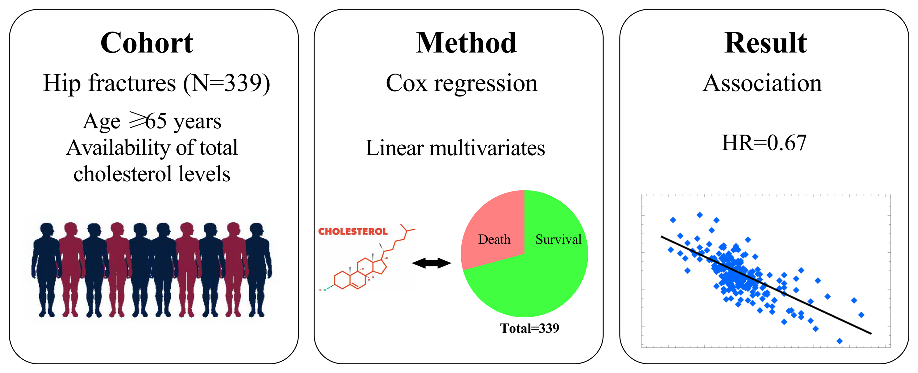
Objectives. To evaluate the association between TC levels and all-cause mortality in patients with geriatric hip fractures.
Materials and methods. Elderly patients with hip fractures were screened between January 2015 and September 2019. Patient demographic and clinical characteristics were recorded. Linear multivariate Cox regression models were used to identify the association between TC levels and all-cause mortality. Analyses were performed using Empower Stats and R software.
Results. Three hundred and thirty-nine patients were enrolled. The mean follow-up period was 34.18 months. There were 99 (29.20%) cases of all-cause mortality. Total cholesterol levels after hip fracture were linearly associated with all-cause mortality in the elderly. Linear multivariate Cox regression models showed that TC levels were associated with mortality (hazard ratio (HR) = 0.67; 95% confidence interval (95% CI): 0.53–0.85; p = 0.001 after adjusting for confounding factors). Each 1 mmol/L increase in TC levels was associated with a 33% reduction in morbidity and mortality. Compared with the low-TC group, mortality was significantly lower in the middle-TC group (HR = 0.58; 95% CI: 0.35–0.94; p = 0.027) and high-TC group (HR = 0.45; 95% CI: 0.27–0.75; p = 0.002).
Conclusions. Total cholesterol levels were associated with mortality in geriatric hip fracture patients and could be considered a protective factor for all-cause mortality.
Key words: all-cause mortality, hip fractures, total cholesterol
Background
Hip fractures in elderly patients are one of the most common and severe types of osteoporotic fractures, occurring overwhelmingly in adults over the age of 70.1 The incidence of hip fractures in the elderly increases with age, and is as high as 7% for those occurring within 10 years in the aging population aged 75–84 years.2 The risk of death in older people with hip fractures is 6.8 times higher than that in people of the same age without fractures.3 It is predicted that by 2050, approx. 52% (3.3 million) of hip fracture cases worldwide will occur in Asian countries, particularly in China.4, 5 The burden of hip fractures is expected to rise owing to the increase in the elderly population,6 and the resulting medical costs in China will reach 85 billion USD.3 Hip fractures pose a serious threat to the quality of life and life expectancy of the elderly population owing to their high prevalence and disability and mortality rates.7
In recent years, several scholars have analyzed the factors affecting the prognosis of hip fractures in elderly patients and concluded that sex, age, American Society of Anesthesiologists (ASA) classification, number of underlying medical conditions, preoperative waiting time, preoperative hemoglobin values, and albumin values are independent risk factors affecting the postoperative prognosis of elderly hip fracture patients.8
Total cholesterol (TC) levels represent the comprehensive level of human cholesterol metabolism, which is closely related to the nutritional status, metabolic level, disease development, and aging of the human body.9 Total cholesterol plays an important role in the maintenance of bodily functions, regulation of sexual functions, immune regulation, and in the development of organisms. Abnormal TC levels are an important risk factor for cardiovascular disease (CVD); therefore, TC is closely related to the development of many diseases,10, 11 and is used as an important indicator of human blood lipid levels and overall health status. Current research suggests that lipids and other nutrient metabolites are associated with the incidences of osteoporosis and osteoporotic fractures.12 Tankó et al. found that changes in hip bone mineral density (BMD) were closely related to TC levels and that these changes were a good predictor of atherosclerosis incidence.13 Related studies have shown that serum TC levels are positively correlated with overall bone density in the intertrochanteric space, greater trochanter, femoral neck, and hip.14 Other studies support the association between BMD in various parts of the hip and blood lipids.15, 16 Few studies on lipid levels and hip BMD levels suggest that the conversion of nutrient metabolites in vivo may have an impact on the metabolism and deposition of bone mineral salts. In a study on independent risk factors for osteoporotic fractures of the hip, Yamaguchi et al. concluded that decreased TC levels were an independent risk factor for fracture.17 Dyslipidemia occurs when TC levels are elevated, possibly contributing to the development of osteoporosis by inhibiting the expression of the osteogenic gene zinc finger structural transcription factor (Osterix) and dwarf-related transcription factor 2 (Runx2) through the BMP-Smad signaling pathway, and this mechanism reduces osteoblastic activity.18 Moreover, abnormal TC levels may differentiate stem cells in the bone marrow into lipogenic precursor cells, reduce the number of osteogenic precursor cells, and aggravate osteoporosis.19
Objectives
The relationship between serum TC levels and the prognosis of patients with hip fractures remains unclear. Therefore, the present study assessed the influence of serum TC levels on patient mortality over a long-term follow-up period. We hypothesized that there would be a linear association between TC levels and mortality. This prospective cohort study aimed to identify the role of TC levels in the incidence of hip fractures.
Materials and methods
Study design
We recruited elderly patients who had a hip fracture between January 1, 2015, and September 30, 2019, at the largest trauma center in Northwest China (Honghui Hospital in X’ian). This prospective study was approved by the Ethics Committee of Honghui Hospital (approval No. 202201009). All procedures involving human participants were performed in accordance with the 1964 Declaration of Helsinki and its later amendments. The study flow diagram is shown in Figure 1.
Participants
The demographic and clinical data of patients were obtained from their original medical records. The inclusion criteria were as follows: 1) age ≥65 years, 2) radiographic or computed tomography (CT) diagnosis of a femoral neck, intertrochanteric or subtrochanteric fracture, 3) receiver of surgical or conservative treatment in a hospital, 4) availability of clinical data in the hospital, and 5) availability and ability to be contacted by telephone. Patients who could not be contacted were excluded.
Setting
Patients were examined using blood tests and ultrasonography during preparation for surgery. Intertrochanteric fractures are often managed with closed/open reduction and internal fixation (ORIF) using a proximal femoral nail anti-rotation implant. Femoral neck fractures were often treated with hemiarthroplasty or total hip arthroplasty, depending on the patient’s age. Prophylaxis for deep vein thrombosis was initiated on admission. Upon discharge, the patients were asked to return monthly to assess the fracture union and orthopedic function.
Variables
Variables in our study were as follows: age, sex, occupation, history of allergy, injury mechanism, fracture classification, presence of hypertension, diabetes, coronary heart disease (CHD), arrhythmia, hemorrhagic stroke, ischemic stroke, cancer, multiple injuries, dementia, chronic obstructive pulmonary disease (COPD), hepatitis or gastritis, age-adjusted Charlson comorbidity index (aCCI), time from injury to admission, time from admission to operation, TC levels, operation time, blood loss, infusion, transfusion, treatment strategy, length in hospital stay, and length of follow-up.
Data sources and measurement
After discharge, the patient’s family members were contacted by telephone from January 2022 to March 2022 to record data on the patient’s survival, survival time and activities of daily life.
Bias
Follow-up was conducted by 2 medical professionals with 2 weeks of training and 1 year of experience. Patients who could not be contacted initially were attempted to be contacted 2 times more. When the family members of the patients did not respond, the patients were considered lost to follow-up.
Study size
We recruited elderly patients who had a hip fracture between January 1, 2015, and September 30, 2019.
Quantitative variables
Total cholesterol levels were defined using blood test results at admission. The dependent variable was all-cause mortality, whereas the independent variable was the TC level. The other variables were potential confounding factors. The endpoint event in this study was all-cause mortality after treatment. All-cause mortality was defined as mortality reported by the patient’s family members.
Statistics analyses
Continuous variables were reported as mean ± standard deviation (M ±SD, Gaussian distribution) or median (range, skewed distribution). Categorical variables are presented as numbers with proportions. A χ2 (categorical variables), one-way analysis of variance (ANOVA, normal distribution) or Kruskal–Wallis H test (skewed distribution) were used to detect the differences between TC levels. Proportional hazard assumptions were checked graphically and with the Schoenfeld residual test. Multicollinearity was checked using the variance inflation factor (VIF). Univariate and multivariate Cox proportional hazards regression models (3 models) were used to test the association between TC levels and mortality. Model 1 was not adjusted for covariates, model 2 was a minimally adjusted model, with adjusted sociodemographic variables, and model 3 was fully adjusted for all covariates (Table 1). For the Cox regression model, we used the concordance index to assess the goodness-of-fit. To test the robustness of our results, a sensitivity analysis was performed. We converted the TC levels into categorical variables according to the anemia criteria and calculated the p-value for the trend to verify the results. Since Cox proportional hazards regression model-based methods are often unable to study nonlinear models, the nonlinearity between TC levels and mortality was accounted for using a Cox proportional hazards regression model with cubic spline functions and smooth curve fitting (penalized spline method). In the spline analysis, a restricted cubic spline transformation of TC with 4 knots (25th, 50th, 75th, and 95th percentiles in all participants) was used to evaluate nonlinear associations. If nonlinearity was detected, we first calculated the inflection point using a recursive algorithm, and subsequently constructed a 2-piecewise Cox proportional hazards regression model on both sides of the inflection point.
All analyses were performed using statistical software packages R (R Foundation for Statistical Computing, Vienna, Austria), SAS 9.4 (SAS Institute Inc., Cary, USA) and EmpowerStats (X&Y Solutions Inc., Boston, USA). Hazard ratios (HRs) with 95% confidence intervals (95% CIs) were calculated. A 2-sided p < 0.05 was considered statistically significant.
Results
Patient characteristics
Three hundred and thirty-nine patients were enrolled in our study. The mean follow-up period was 34.18 months. There were 99 (29.20%) cases of all-cause mortality. The patients were divided into 3 groups according to TC levels: low-TC group, middle-TC group and high-TC group. Results of normality tests are presented in Supplementary Table 1; continuous variables were non-normally distributed according to the results of Shapiro–Wilk test. Table 1 lists the demographic and clinical characteristics of the patients, including their comorbidities, factors associated with injuries, and treatment.
Univariate analysis of the association between variates and mortality
We performed a univariate analysis to identify the potential confounding factors and the relationship between variables and mortality (Table 2). Owing to p < 0.1, the following variables were considered in the multivariate Cox regression analysis: age, CHD, arrhythmia, dementia, treatment strategy, and aCCI.
Multivariate analysis between TC and mortality
We used 3 models (Table 3) to correlate the TC levels and mortality. When the concentration of TC was variable, a linear regression was observed. The fully adjusted model showed a 33% decrease in the mortality risk when the TC concentration increased by 1 mmol/L after controlling for confounding factors (HR = 0.67; 95% CI: 0.53–0.85; p = 0.001). The concordance index was 0.70 (95% CI: 0.65–0.75), indicating that the Cox regression model was adequate. When the TC concentration was used as a categorical variable, there were statistically significant differences in TC levels among the 3 models (p < 0.001). Moreover, the p-value for the trend also showed a linear correlation in the 3 models (p < 0.001). The restricted cubic spline regression revealed no significant nonlinear associations between TC and mortality (Supplementary Fig. 1), and none of the predictors violated the proportional hazards assumption (Supplementary Table 2). The Kaplan–Meier survival curves in the 3 groups are depicted in Figure 2.
Discussion
We found a linear correlation between TC levels and all-cause mortality after hip fracture in older adults. A higher TC was associated with lower mortality (HR = 0.67; 95% CI: 0.53–0.85; p = 0.0012), suggesting that each 1 mmol/L increase in TC was associated with a 33% reduction in the mortality rate. Compared to the low-TC group, the middle-TC group had a significantly lower mortality rate (HR = 0.58; 95% CI: 0.35–0.94; p = 0.027), and the high-TC group had a significantly lower mortality rate (HR = 0.45; 95% CI: 0.27–0.75; p = 0.002). Moreover, the results were stable in the trend test (p-value for trend). In clinical practice, the patient’s TC levels at admission could be used as a predictor of all-cause mortality in the mid-clinical course of hip fractures in elderly patients, and a higher TC level is associated with lower mortality.
Serum TC has been shown to be a protective factor not only for vascular-related diseases but also for osteoporotic fractures. An indirect association between TC and BMD has been demonstrated, and BMD is considered an important determinant of osteoporotic fractures.20, 21, 22, 23, 24 Total cholesterol concentration can be identified as an independent risk factor for osteoporotic fractures, and its predictive power increases over time.25 Hypercholesterolemia has been reported to be a risk factor for atherosclerosis in patients with CHD.26 It also increases bone loss in postmenopausal women.27 Several studies have confirmed that elevated TC levels leading to hyperlipidemia may also induce bone loss and osteoporosis by affecting the function and differentiation of osteoblasts and osteoclasts.28, 29 However, the interrelationship between serum cholesterol and osteoporosis fractures is still controversial.30 This study is the first to prospectively examine the relationship between TC levels at admission and hip fracture prognosis.
Being a lipid-like substance, TC is an important component of the cell and organelle membranes, comprising approx. 30% of the cell membrane lipids, maintains the fluidity of the cell membrane, and facilitates the exchange of substances and information inside and outside the cell membrane. Simultaneously, TC is involved in the synthesis of bile acids and steroid hormones, and plays an important role in the formation and development of the nervous system. Importantly, the results of several studies have shown that low TC levels may cause mental and neurological symptoms such as irritability, cognitive impairment, memory impairment, dementia,31, 32, 33 and ultimately an increased risk of death.34 In other words, the risk of death decreases with elevated TC levels. The relationship between abnormal serum lipid levels and cognition, memory and other functions in the elderly may result from amyloid deposition in the brain and the development of cerebrovascular disease.35, 36, 37 In contrast, high-density lipoprotein is a component of TC. A study by Yang et al. established a multivariate Cox regression model to analyze the 90-day postoperative all-cause mortality and reported that high-density lipoprotein (HDL) was an independent protective factor for postoperative death in elderly patients with intertrochanteric fractures (HR = 0.41).38
In addition to the linear relationship, we also speculated about the possibility of a curvilinear relationship based on a subgroup analysis and curve fitting. However, no inflection point on the curve was found in this study. Currently, in the context of this field, a linear relationship is more appropriate for explaining the relationship between TC and mortality.
To identify the confounding factors and draw reliable conclusions from this study, we first identified factors affecting the prognosis and TC levels. Age, sex, CHD, arrhythmia, dementia, and treatment strategy have been reported in previous studies as risk factors for hip fracture prognosis.39, 40, 41, 42, 43 Thus, we controlled for the vast majority of these confounding factors.
Limitations of the study
This study has a few limitations. First, because our study was prospectively designed, a certain amount of loss to follow-up was unavoidable. To determine the prognosis of patients, we conducted several telephone follow-ups with patients or their family members when the patients did not respond the first time. Second, we were unable to determine the causal relationship between TC levels and prognosis, which needs to be confirmed in future studies. Third, our study population was from Western China, which has geographical and ethnic limitations. The generalizability of the conclusions of this study should be treated carefully.
Conclusions
In summary, TC levels were associated with all-cause mortality in patients with geriatric hip fractures and could be considered a protective factor for all-cause mortality.
Supplementary data
The Supplementary materials are available at https://doi.org/10.5281/zenodo.8227882. The package contains the following files:
Supplementary Fig. 1. Multivariable-adjusted HRs (black solid lines) and 95% Cls (dotted and dashed lines) for risk of all-cause mortality according to TC levels.
Supplementary Table 1. Results for Shapiro–Wilk test.
Supplementary Table 2. Test of the proportional hazards assumption for all covariates and mortality used in the Cox regression model.

