Advances in Clinical and Experimental Medicine

Title abbreviation: Adv Clin Exp Med
JCR Impact Factor (IF) – 2.1
5-Year Impact Factor – 2.2
Scopus CiteScore – 3.4
Index Copernicus  – 168.52; MEiN – 140 pts

ISSN 1899–5276 (print)
ISSN 2451-2680 (online)
Periodicity – monthly

Download original text (EN)

Advances in Clinical and Experimental Medicine

Ahead of print

doi: 10.17219/acem/162537

Publication type: review

Language: English

License: Creative Commons Attribution 3.0 Unported (CC BY 3.0)

Download citation:

  • BIBTEX (JabRef, Mendeley)
  • RIS (Papers, Reference Manager, RefWorks, Zotero)

Cite as:


Nishi R, Fukumoto T, Asakawa A. Possible effect of natural light on emotion recognition and the prefrontal cortex: A scoping review of near-infrared (NIR) spectroscopy [published online as ahead of print on April 24, 2023]. Adv Clin Exp Med. 2023. doi:10.17219/acem/162537

Possible effect of natural light on emotion recognition and the prefrontal cortex: A scoping review of near-infrared (NIR) spectroscopy

Ryusei Nishi1,A,B,C,D,E,F, Takamasa Fukumoto1,B,C,E,F, Akihiro Asakawa1,A,E,F

1 Department of Psychosomatic Internal Medicine, Graduate School of Medical and Dental Sciences, Kagoshima University, Japan

Graphical abstract


Graphical abstracts

Abstract

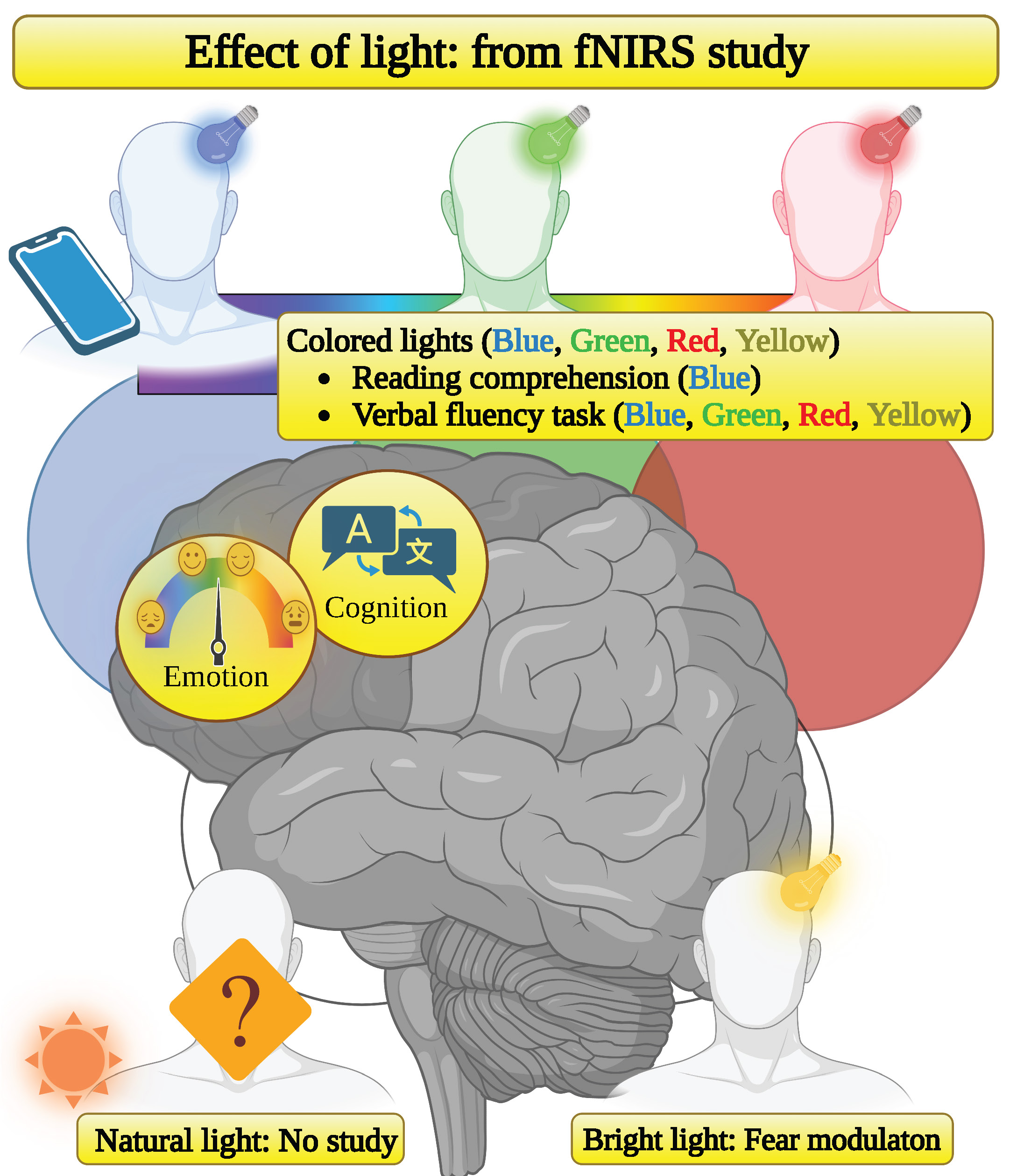
Near-infrared (NIR) spectroscopy, also known as functional NIR spectroscopy (fNIRS), is a tool for measuring the hemodynamic response of the prefrontal cortex (PFC) by using NIR light, enabling a noninvasive indirect neural activity assessment. The application of fNIRS in emotion recognition or the differential diagnosis of psychiatric disorders of depressive patients, including major depressive disorder, bipolar disorder and schizophrenia, has previously been reported. Although the use of fNIRS has gradually expanded in cognitive neuroscience studies, few researchers have focused on the effects of light exposure in fNIRS studies. In addition, to the best of our knowledge, there are no scoping reviews of fNIRS studies on light exposure. Because light is an important topic in cognitive neuroscience and psychiatry, we evaluated fNIRS studies on light exposure in humans. We reviewed 10 papers in their entirety. Bright light (BR) modulates fear, and the color differences showed no significance in 1 study, whereas other studies delved extensively into the effects of colored light, finding some individual hemodynamic responses. In our study, we highlighted that the effects of natural light have not been studied using fNIRS. Light is becoming a critical topic in cognitive neuroscience and psychiatry, and fNIRS is critical for improving public health and managing psychiatric disorders.

Key words: prefrontal cortex, emotion recognition, light, NIR spectroscopy

Introduction

Functional near-infrared spectroscopy (fNIRS) is a tool for measuring cerebral hemodynamic responses, enabling a noninvasive assessment of indirect neural activity. This technique has developed rapidly over the 30 years since its first application.1 The fNIRS tolerates moderate movements, which is an advantage over other tools, such as functional magnetic resonance imaging (fMRI).

The first demonstration of measuring cerebral oxygen level was performed by Jöbsis in 1977,2 while the first commercial near-infrared (NIR) spectroscopy with a single channel was developed by Hamamatsu Photonics K.K. (Hamamatsu, Japan) in 1989. The original fNIRS system was expensive and cumbersome, and the many wires required rendered subjects immobile, making its application in clinical settings challenging. In 1998, a 10-channel system developed by Hitachi (Tokyo, Japan) was used on intractable epilepsy patients in a clinical setting.3 Recently, researchers have elaborated on developing more miniaturized, wireless and low-cost fNIRS systems.4, 5, 6

The principles of the mainstream continuous wave (CW) fNIRS technique are outlined in Figure 1. When NIR light travels through cerebral capillaries, the light is absorbed, scattered and transmitted. This light absorption can be calculated using the Beer–Lambert law. However, it is inappropriate to apply the original Beer–Lambert law. First, scattering light should be considered because the human brain includes blood vessels and other highly scattering tissues, such as the skin and skull. Second, the trajectory of light is not linear but banana-shaped (Figure 1) when applying NIRS to the human brain. Therefore, the law needed to be modified.7 When measuring light absorption of 2 different wavelengths, the common factor of scattering is negligible. Because the molar extinction coefficient for oxygenated hemoglobin (oxy-Hb) and deoxygenated hemoglobin (H-Hb) can be obtained experimentally, the relative concentrations of both oxy-Hb and H-Hb result from solving the 2 equations simultaneously.

Functional near-infrared spectroscopy can be a useful tool for measuring brain activity in various settings besides limited laboratory situations. However, there are critical disadvantages of fNIRS to consider. Although fNIRS measures hemodynamic response noninvasively,8, 9, 10 the spatial resolution is not as high compared to an electroencephalogram. In addition, the deep brain of adults cannot be measured since light penetration does not reach it. Therefore, fNIRS can only target the lateral surface of the brain hemispheres, mainly the cortex.11 The forehead is easy to approach regions of interest in fNIRS. These regions includes motor functionality (the primary sensorimotor cortex and the premotor cortex12) and higher-order functionality (the frontopolar cortex and the prefrontal cortex (PFC)13).

The PFC contributes to both fear acquisition and extinction, as PFC has the ability to learn from a precedent fear.14 Recent studies have suggested that the medial PFC is impaired by the basolateral complex of the amygdala (BLA), which is activated by the locus coeruleus (LC). In addition, the posterior ventromedial PFC (BA11) activates powerfully during late fear conditioning.15 In contrast, the LC activates the medial PFC, and the medial PFC inhibits the BLA.16 Therefore, the LC, medial PFC and BLA are vital for noradrenergic fear modulation, but the mechanisms differ between fear acquisition and fear extinction. In functional brain imaging studies, the fear and anxiety regions of the frontal cortex are not only in the medial prefrontal but also the dorsal, ventrolateral and prefrontal orbitofrontal cortex.17 In patients with post-traumatic stress disorder (PTSD), impairment of recalling fear extinction from the hypoactivity of the hippocampus was reported, whereas patients were conditioned to both fear acquisition and extinction normally.14 Also, the hippocampus projection to the PFC is aberrant in patients with PTSD, depression and schizophrenia,18 suggesting that hippocampal function affects fear, anxiety and cognition.

The fNIRS application in the differential diagnosis of depressive patients, including major depressive disorder, bipolar disorder and schizophrenia, has been reported.19 In Japan, fNIRS, as an optical topography test, has been covered by the healthcare insurance system for the differential diagnosis of the depressive state. However, clinical applications in neuroimaging are still controversial. In 2018, the American Psychiatric Association (APA) concluded that neuroimaging should remain a research tool only. There may be 2 main reasons for this. Firstly, most studies have a small number of participants and identify a relatively tiny effect size. Moreover, although neuroimaging is noninvasive, it is challenging to draw an identical assessment between clinicians, which makes it difficult to support its application for clinical diagnosis.

The use of fNIRS has expanded gradually in cognitive neuroscience studies. Cognitive neuroscience is a broad multidisciplinary field focusing mainly on the neural mechanisms behind cognitive function by linking together disciplines such as physiology, anatomy, psychology, psychiatry, and computational biology.20 Table 1 shows the main subjects of cognitive neuroscience with reported topics for fNIRS application. It should be noted that the mentioned studies were almost but not entirely separate, and each field will overlap with other fields. For example, dyslexia is in the reading field but can also be categorized in the decision or language fields. In terms of the attention field, attention deficit hyperactivity disorder (ADHD) during the developmental ages is a critical topic in fNIRS studies.21 Studies regarding traumatic brain injury,22, 23 autism spectrum disorders,24, 25 drowsy driving,26 and meditation27 are also recent clinical applications of fNIRS. Studies on consciousness, anesthesia management,28 disorders of consciousness,29 and locked-in syndrome30 have been reported. Decision-making is one of the essential applications of cognitive neuroscience. The application of the brain–computer interface31 to examine group decision,32 purchase decision33, 34 and risk-taking35, 36 yielded some pieces of evidence. In terms of executive control, obesity,37 exercise38 and older adults39 have been researched. In the intelligence field, problem-solving40 and work performance41 have been studied. The language field42, 43 is the most extensively studied topic in cognitive neuroscience, and the verbal fluency task (VFT) has recently demonstrated its ability to measure depressive disorder.44 We perceive almost all stimuli under any circumstances; therefore, it is challenging to aggregate perception studies. To the best of our knowledge, perception of depth,45 emotion,46, 47, 48 face,49 reward,50, 51 self-agency,52 and time53 have been reported. Finally, reading studies include dyslexia54 and overt reading55 (reading aloud).

Objectives

Light is essential not only for visualization but also for sleep, mood and cognition. Meta-analyses and systematic reviews support the impact of light on alertness.56 In addition, light therapy is an effective treatment for depression in Parkinson’s disease57 and premenstrual dysphoric disorder,58 and the effects of light therapy on sleep disturbance have also been reported.57, 59, 60, 61 In cognitive neuroscience and psychiatry, light is an important topic. However, few researchers have focused on the effects of light exposure measured using fNIRS. In addition, to the best of our knowledge, there is no scoping review of fNIRS studies on light exposure. Therefore, in the present review, we evaluate fNIRS studies on light exposure in humans.

Review protocol

We delineated 4 inclusion criteria. First, studies must be performed on humans. Second, studies should focus on light exposure. Therefore, papers that only mentioned light exposure for the technical principles of fNIRS or light effect on fNIRS experiments itself62 (ambient light) were excluded from our review. Third, not only direct but also indirect light exposure measurements were included, as was smartphone use63 (effect of blue light (BL)). However, studies were excluded if there were other non-negligible effects, such as the natural light effect on garden therapy64 with green plants. Fourth, since the first fNIRS study was published in 1991, studies published only after that year were included in the analysis.1 Following an initial Internet search, we concluded the literature on light exposure encompassed various fields such as architecture, education, religion, and complementary medicine. Therefore, from the standpoint of scientific rigor, we required papers to have a PubMed Identifier (PMID) in PubMed and did not take into account information available from other websites, dissertations and books. Our review presents the whole PubMed-indexed research on the topic.

We started the literature search on November 15, 2022, and finished it on January 20, 2023. The process of selecting literature is shown in Figure 2. We used Google Scholar, Scopus and PubMed databases to identify relevant studies, and carefully selected queries in order to identify fNIRS research on light exposure. The queries that were used are listed in Table 2. Google Scholar facilitates literature identification,65 but many irrelevant results appear because of its low precision.66 In addition, search results are not reproducible.67 Therefore, using Google Scholar solely is challenging in identifying literature68; thus, Scopus and PubMed were used to improve coverage and maintain search consistency. On January 20, 2023, 2 authors (RN and TF) independently searched the databases and found that all results were consistent. In addition, we confirmed that the results had not been updated since the previous search. After literature identification, we deduplicated the references using Zotero v. 6.0.18 (https://www.zotero.org/). After deduplication, articles with unavailable full-text articles were excluded, abstracts were screened and 10 papers remained for full-text review. The main highlight of the fNIRS studies was the oxy-Hb concentration.

The bias risk assessment was performed based on the characteristics and number of participants and study protocols. Moreover, the study protocol was assessed. We used robvis tool to create risk-of-bias plots.69

Results

A brief overview of the 10 reviewed studies is presented in Table 3. In summary, bright light (BR) modulates fear. Regarding the color differences, no significance was indicated in 1 study, whereas other studies suggested that colored light produced some individual hemodynamic responses.

Bias risk assessment

Figure 3 summarizes the risk of bias. All studies included both male and female participants. Although the number of participants ranged from 10 to 34 in most studies, 1 study had 757 participants. Due to the inhomogeneous signal processing methods, comparing oxy-Hb concentrations between studies would be inappropriate. In 4 studies,70, 71, 72, 73 the reported number, male/female proportion, standard deviation (SD), mean (M), and age ranges of participants were identical. In 1 study, a machine learning technique was used.71 However, although the machine learning technique is overused in various fields, artificial intelligence (AI) does not apply to small biased datasets.74 Therefore, even if machine learning methods successfully find plausible clustering, these models are not always suitable for all datasets, and sometimes the clustering is a result of sheer luck.

Detailed description
of the reviewed literature

Hori et al. stimulated 757 subjects with various colored lights and waterfall sounds using different frequencies to study the emotional effects on the PFC.75 Their feedback system enabled them to alter the stimuli in response to increased oxy-Hb concentration in the PFC, resulting from pleasantness or unpleasantness. Pleasantness or unpleasantness was determined according to a previous study.11 After the stimulation, 298 participants answered a questionnaire to assess their comfort with the stimuli, ranging from −5 (unpleasant) to +5 (pleasant). Results indicated that sound frequency rather than light color affects how pleasant or unpleasant the feelings of the participants were. High-frequency sounds accounted for approx. 43% of both pleasantness and unpleasantness responses.75

Yoshiike et al. performed an fNIRS study to show the effect of BR on PFC activity during human fear conditioning and fear extinction. They conducted a single-blinded study by exposing 29 participants to either bright (9000 lx) or regular (<500 lx) light, and presented visual conditioned stimuli (CS) randomly, followed by unconditioned stimuli (US) using an unpleasant but not painful electric shock. For CS, each sign represented a learning process using triangles, squares and circles to represent fear extinction, fear acquisition and a safety state, respectively. In addition to the PFC activity on fNIRS, skin conductance was recorded with each response to stimuli.76 The results were intriguing. Bright light decreases skin conductance during the recall session of any learning process. However, fNIRS showed that BR modulates fear immediately and differently, promoting fear extinction and safe learning while inhibiting fear acquisition. Moreover, BR modulates fear in any type of learning, which generally lasts at least 24 h.

Wolf’s research group extensively focused on the color of light and conducted 8 fNIRS studies in the years 2011–2022.70, 71, 72, 73, 77, 78, 82, 83 In 2011, they first investigated the effect of red and blue light on oxygen consumption in the brain and muscles of 10 participants.77 Then, they exposed the 12 volunteers to these 2-colored lights sequentially to examine the hypothesis stating that red light enhances the effects of BL.78 The source of light was not a light-emitting diode (LED) but a white light bulb with colored filters. The constant hemoglobin concentration and high tissue oxygen saturation indicated that BL, but not red light, exposure decreases oxygen demand in the brain.77, 78 However, this contradicts the activation of the PFC in both long (30 min) and short (1 min) BL exposures.79, 80, 81 Interestingly, blood oxygenation was independent of the sequence, and the oxygenation in both sequences was equivalent to a single red light exposure, suggesting that red light is preponderant to BL.78

Metz et al. in 2017 proposed physiology-augmented fNIRS (SPA-fNIRS) and conducted the first 2 SPA-fNIRS studies. As the name suggests, SPA-fNIRS associates fNIRS with physiological parameters, including skin conductance, end-tidal carbon dioxide (PETCO2) and heart rate (HR).82, 83 Both studies exposed the participants to colored light, but the major difference was whether the exposure was continuous82 or intermittent.83

Metz et al. continuously exposed the 12 participants to blue, green, red, and yellow LED lights for 10 min while recording their physiological and neurological activities using SPA-fNIRS. Skin conductance increased for all colors except green, and only yellow light significantly elevated oxy-Hb concentration in the left but not the right PFC. Additionally, as in their previous study, oxygen demand decreases were only observed when exposed to blue, but not red light. Except for the green light, HR increased while the partial pressure of CO2 (PaCO2, equivalent to PETCO284) decreased. The HR elevation and PETCO2 reduction indicate that participants hyperventilated during fNIRS. They described that this hyperventilation occurred due to weak pain in the recovery phase, but not from the light itself.

Scholkmann et al. used blue, green and red LEDs to beam light for 10 min, on and off every 20 s, but did not use yellow LEDs. In addition to PFC activity, visual cortex (VC) activity was measured using the SPA-fNIRS. The results indicated that BL and red light significantly increased respiratory rate (RR), whereas green light decreased it. Regarding HR and PETCO2, only BL elevated HR, and PETCO2 showed no significant alteration. In addition, only BL elicited an elevation in PFC oxy-Hb, while VC activity-related oxy-Hb elevation was observed with any color.83

In 2021, followed by another SPA-fNIRS study to investigate the colored light effect (CLE) during a VFT,73 Wolf’s research group delved into the interindividual differences of hemodynamic responses and consecutively published 4 studies in January 2021, April 2021, May 2022, and October 2022.70, 71, 72, 73 Briefly, followed by performing a VFT under CLE, they patterned or clustered hemodynamic responses using various techniques, including unsupervised machine learning,71 namely k-means.

In 2022, Honma et al. enrolled 34 healthy individuals to compare reading comprehension on a smartphone compared to paper by recording the number of sighs the subjects made while reading and bilateral PFC activity obtained using fNIRS. The 10-point comprehension test used 10 multiple-choice questions. This crossover study revealed that reading on paper achieves at least 1 point higher score than reading on a smartphone. In addition, sighs decreased by approx. 70% while reading on a smartphone, and the left PFC was simultaneously overactivated. These findings indicated that BL decreases sighs and increases left PFC overactivation, resulting in poor comprehension due to excessive cognitive loads.63

Discussion

Results show that the CLEs on verbal fluency have been extensively researched, and the hemodynamic responses may differ individually. In addition, BL overly activates the PFC. Moreover, BR modulates fear immediately and differently between learning processes, promoting fear extinction and safe learning, as well as inhibiting fear acquisition.

Although our review provides some evidence of the effects of colored light, some concerns should be mentioned. First, according to previously mentioned findings,75 light color does not affect emotions. On the other hand, Battaglia et al. suggested that both positive and negative emotions equally control our behaviors.85 Therefore, light color differences may not affect our behaviors. However, it is not surprising that numerous studies suggest that color underlies mood, quality of life (QOL) and cognition. In 2010, Carruthers et al. developed the Manchester Color Wheel to assess patients by the color of light they were exposed to using 38 colors representing positive, neutral or negative conditions that were rigorously reproduced on the participants.86 Color-using art therapy can increase purpose in the life of stroke patients and their caregivers, resulting in QOL improvements.87 In addition, as Berndt et al. indicated in 2020, color plays a vital role in language comprehension, facilitating anagram solving by presenting the referent color of the solution words.88

Second, as previously mentioned, Metz et al. reported that exposure to green light did not increase HR and decrease PaCO2,82 suggesting that green light did not induce hyperventilation (in turn caused by weak pain) during fNIRS. However, the light color should not be the only difference studied, and hyperventilation during fNIRS needs to be elucidated more complex. Although previously mentioned, it should be noted again that noninvasiveness is a core feature of fNIRS. Moreover, hyperventilation suggesting HR elevation and concurrent PETCO2 reduction were not observed with intermittent light exposure.83 Research on the effect of green light is scarce because exposure to green light in the natural environment is rare; nonetheless, the effects of green light need to be elucidated.

Finally, there are some discrepancies to be noted when comparing continuous light82 and intermittent light83 exposure. Regarding the BL effect, continuous light does not induce activation,77, 82 whereas intermittent light induces an oxy-Hb concentration increase in the PFC.83 However, interindividual differences can be the main plausible cause of varying results, as mentioned.70, 71, 72

Blue light activates intrinsically photosensitive retinal ganglion cells (ipRGCs), and the signal is projected to several brain regions, including the ventral tegmental area, raphe and LC via the suprachiasmatic nucleus, to modulate circadian rhythms.89 The ipRGCs do not form a vision, suggesting that even in blind people exposure to colored light may result in circadian modulation.80, 90, 91 A LED display on smartphones is an example of BL emitter. Blue light harms photoreceptors and pigmented epithelium cells,92 and the reviewed fNIRS study suggests the negative impact of BL on reading comprehension.63

In the reviewed studies, BL increased total hemoglobin concentrations when the participants were exposed to light emitted by a color-filtered white lightbulb, but it remained constant under LED illumination. However, the difference between incandescent light and LED can be applied to this result. Recently, Farghly et al. reared rabbits under various light conditions and found that rearing animals under LED conditions resulted in the highest glucose concentrations.93 In addition, Niemierzycka et al. showed that LED light with a color temperature of 4200 K achieves higher speeds with maintained accuracy on the Kraepelin test.94 As the Kraepelin test induces PFC activity95 and glucose is primarily present in the brain, it can be inferred that LED light plays a different role than incandescent light on the PFC.

The present review indicated that there are only 10 studies on the effects of light exposure measured using fNIRS measurements in PubMed. In addition, we should carefully interpret these results. For example, since colored light effects have been studied only by a single research group, there are some non-negligible biases, such as confirmation bias. Thereby, the present review identified the sparsity of light exposure studies with fNIRS usage.

The effect of natural light has not been studied using fNIRS. As previously mentioned, garden therapy was studied,64 but the effect of green plants is not negligible. However, studying solely natural light is challenging because natural light is a component of the environment. For example, when downtown, there can be additional stimuli like noise from cars or rising temperatures caused by the heat island effect.

We hypothesize that fNIRS can yield critical findings in combination with other biomarkers. For example, because fNIRS measures the oxidation of hemoglobin in the cerebral capillaries, cerebral oxidative stress can be noninvasively measured in combination with other oxidative indicators, such as the kynurenine-3-monooxygenase, which is activated by oxygen.96, 97, 98, 99, 100, 101 Moreover, fNIRS might facilitate the understanding of fear learning, in combination with cardiac autonomic dynamics of heart rate variability.102

Another important hypothesis is that fNIRS can bring critical findings for psychiatric disorders in terms of light effects. For example, since light therapy was reported to be effective in seasonal affective disorder (SAD) in 1984,103 only the atypical pattern of rod electroretinogram has been reported as a possible biomarker.104 Due to a much stronger invasiveness of electroretinogram than fNIRS, the present review suggests the need for developing a biomarker in patients with SAD. The fNIRS can also be used in combination with inflammatory cytokines in dementia105, 106 or the severe acute respiratory syndrome coronavirus 2 (SARS-CoV-2) in various gut microbiota-related psychiatric illnesses.107 Although MRI is essential for diseases such as cerebral small vessel disease,108 there might be a possibility of using fNIRS to predict the critical need for an MRI, which would significantly spare the clinician’s time and effort.

As previously mentioned, fNIRS is an effective tool for cognitive neuroscience due to its non-invasiveness, tolerance of movements and high resolution. On the other hand, most fNIRS studies corroborate PFC activity by associating oxy-Hb concentration increase with brain activity.109, 110, 111, 112, 113 The idea of such association is derived from the neurovascular coupling theory. Although the mechanism by which neural tissue activation leads to increased cerebral blood flow remains unclear,114, 115 in terms of neurovascular coupling, the integration of fNIRS and functional MRI can be helpful in understanding brain hemodynamics.116

Limitations

Although this scoping review presents the whole PubMed-indexed research on fNIRS and the effects of light exposure, it has some limitations. We focused only on PubMed-indexed literature, which excludes information available from websites, dissertations and books. In addition, fNIRS-related fields, such as engineering, computer science and physics, were not incorporated because PubMed does not include biomedical journals. Finally, we did not include studies published after January 2023.

Conclusions

The fNIRS techniques enable real-time monitoring of human brain activity with tolerance to motion artifacts to some extent. Humans live with light, but its quality changes from natural to artificial light exposure. Notwithstanding that natural light is still essential, its effect on the brain’s hemodynamic response has not been thoroughly researched. Delving into light effects under commonplace environments using fNIRS is a reasonable approach. In conclusion, light will be a more critical topic in cognitive neuroscience and psychiatry, and fNIRS can be a critical tool for improving public health and managing psychiatric disorders.

Tables


Table 1. Major topics of cognitive neuroscience and reported application of functional near-infrared spectroscopy (fNIRS)

Topic

Application

Attention

attention deficit hyperactivity disorder (ADHD)

impairments after traumatic brain injury

autism spectrum disorder

drowsy driving

meditation practice

Consciousness

anesthesia monitoring

disorders of consciousness

locked-in syndrome

Decision

brain–computer interface

group decision

purchasing decisions

risk behavior in financial decisions

risky decision-making in social interactions

Executive control

central obesity

exercise

older adults

Intelligence

problem solving

work performance

Language

aphasia

bilinguality

developments

presurgical assessment

second language

unknown language

verbal fluency

Perception

depth

emotion

light

face

reward

self-agency

time

Reading

dyslexia

overt reading

Table 2. Queries used for database search

Database

Queries

Matches

Google Scholar

“functional near-infrared spectroscopy” AND “light exposure”

212

“functional near-infrared spectroscopy” in title: “light”

231

PubMed

“functional near-infrared spectroscopy (title)” AND “light (title/abstract)”

83

Scopus

TITLE-ABS-KEY (“functional near-infrared spectroscopy” OR “functional near infrared spectroscopy” OR “fNIRS”) AND TITLE-ABS-KEY (“light W/3 exposure” AND (LIMIT-TO (DOCTYPE, “ar”) OR LIMIT-TO (DOCTYPE, “re”)

10

Table 3. Summary of fully reviewed references

Year

Author and reference

Types of light

Participants, n (male/female)

Age [years]

fNIRS findings

range

M

SD

2011

Weinzirl

et al.78

C

13 (7/6)

18–44

30.0

N/A

indifferent to color sequence

2012

Weinzirl

et al.77

C

10 (5/5)

23–44

27

N/A

BL decreases oxygen demand

2017

Hori

et al.75

C

fNIRS measurement: 757 (294/463)

questionnaire: 299 (118/181)

6–78

32.0

14.0

light itself does not change an emotion

Metz

et al.82

C

17 (11/6)

25–65

N/A*

N/A

yellow light elevates oxy-Hb concentration in the left PFC

Scholkmann

et al.83

C

14 (9/5)

24–57

33.4

10.5

only BL elicits PFC oxy-Hb concentration elevation

2018

Yoshiike

et al.76

BR

29 (10/19)

20–25

21.7

N/A

BR decreases oxy-Hb concentration in the PFC

2021

Zohdi

et al.72, 73

C

32 (17/15)

N/A

25.5

4.3

indicates inter-participant hemodynamic variability

C

32 (17/15)

19–45

25.5

4.3

various deviations in cerebral hemodynamics

2022

Honma et al.63

BL

34 (14/20)

N/A

20.4

0.8

BL overly activates the PFC

Zohdi et al.71

C

32 (17/15)

N/A

25.5

4.3

Scholkmann et al.70

C

32 (17/15)

19–45

25.5

4.3

oxy-Hb and deoxy-Hb concentration change is color-independent;

BL rather than red light response is stronger in the VC

BL – blue light; BR – bright light; C – colored light; N/A – not available; oxy-Hb – oxygenated hemoglobin; PFC – prefrontal cortex; fNIRS – functional near-infrared spectroscopy; M – mean; SD – standard deviation; VC – visual cortex; * median age was 29 years.

Figures


Fig. 1. The principle of the continuous wave (CW) functional near-infrared spectroscopy (fNIRS) system. Two different wavelengths of light are emitted from the emitter and then detected with 2 detectors. Detection is performed under the principles of the modified Beer–Lambert law; therefore, 2 equations are formulated. Finally, these equations are solved to calculate the oxygenated hemoglobin (oxy-Hb) concentration
Fig. 2. Selection of target literature
fNIRS – functional near-infrared spectroscopy.
Fig. 3. Summary of bias risk assessment. A. Risk of bias for each study and domain; B. Summarized bias risk assessment for all 10 studies

References (116)

  1. Ferrari M, Quaresima V. A brief review on the history of human functional near-infrared spectroscopy (fNIRS) development and fields of application. Neuroimage. 2012;63(2):921–935. doi:10.1016/j.neuroimage.2012.03.049
  2. Jöbsis FF. Noninvasive, infrared monitoring of cerebral and myocardial oxygen sufficiency and circulatory parameters. Science. 1977;198(4323):1264–1267. doi:10.1126/science.929199
  3. Watanabe E, Maki A, Kawaguchi F, et al. Non-invasive assessment of language dominance with near-infrared spectroscopic mapping. Neurosci Lett. 1998;256(1):49–52. doi:10.1016/S0304-3940(98)00754-X
  4. Liu G, Cui W, Hu X, et al. Development of a miniaturized and modular probe for fNIRS instrument. Lasers Med Sci. 2022;37(4):2269–2277. doi:10.1007/s10103-021-03493-w
  5. Bolton FJ, Bernat AS, Bar-Am K, Levitz D, Jacques S. Portable, low-cost multispectral imaging system: Design, development, validation, and utilization. J Biomed Opt. 2018;23(12):1–11. doi:10.1117/1.JBO.23.12.121612
  6. Farzam P, Starkweather Z, Franceschini MA. Validation of a novel wearable, wireless technology to estimate oxygen levels and lactate threshold power in the exercising muscle. Physiol Rep. 2018;6(7):e13664. doi:10.14814/phy2.13664
  7. Delpy DT, Cope M, van der Zee P, Arridge S, Wray S, Wyatt J. Estimation of optical pathlength through tissue from direct time of flight measurement. Phys Med Biol. 1988;33(12):1433–1442. doi:10.1088/0031-9155/33/12/008
  8. Perrey S. Non-invasive NIR spectroscopy of human brain function during exercise. Methods. 2008;45(4):289–299. doi:10.1016/j.ymeth.2008.04.005
  9. Strangman G, Boas DA, Sutton JP. Non-invasive neuroimaging using near-infrared light. Biol Psychiatry. 2002;52(7):679–693. doi:10.1016/S0006-3223(02)01550-0
  10. Villringer A, Chance B. Non-invasive optical spectroscopy and imaging of human brain function. Trends Neurosci. 1997;20(10):435–442. doi:10.1016/S0166-2236(97)01132-6
  11. Hoshi Y, Huang J, Kohri S, et al. Recognition of human emotions from cerebral blood flow changes in the frontal region: A study with event-related near-infrared spectroscopy. J Neuroimaging. 2011;21(2):e94–e101. doi:10.1111/j.1552-6569.2009.00454.x
  12. Leff DR, Orihuela-Espina F, Elwell CE, et al. Assessment of the cerebral cortex during motor task behaviours in adults: A systematic review of functional near infrared spectroscopy (fNIRS) studies. Neuroimage. 2011;54(4):2922–2936. doi:10.1016/j.neuroimage.2010.10.058
  13. Cazzell M, Li L, Lin ZJ, Patel SJ, Liu H. Comparison of neural correlates of risk decision making between genders: An exploratory fNIRS study of the Balloon Analogue Risk Task (BART). Neuroimage. 2012;62(3):1896–1911. doi:10.1016/j.neuroimage.2012.05.030
  14. Battaglia S. Neurobiological advances of learned fear in humans. Adv Clin Exp Med. 2022;31(3):217–221. doi:10.17219/acem/146756
  15. Fullana MA, Harrison BJ, Soriano-Mas C, et al. Neural signatures of human fear conditioning: An updated and extended meta-analysis of fMRI studies. Mol Psychiatry. 2016;21(4):500–508. doi:10.1038/mp.2015.88
  16. Giustino TF, Maren S. Noradrenergic modulation of fear conditioning and extinction. Front Behav Neurosci. 2018;12:43. doi:10.3389/fnbeh.2018.00043
  17. Shiba Y, Santangelo AM, Roberts AC. Beyond the medial regions of prefrontal cortex in the regulation of fear and anxiety. Front Syst Neurosci. 2016;10:12. doi:10.3389/fnsys.2016.00012
  18. Godsil BP, Kiss JP, Spedding M, Jay TM. The hippocampal–prefrontal pathway: The weak link in psychiatric disorders? Eur Neuro­psychopharmacol. 2013;23(10):1165–1181. doi:10.1016/j.euroneuro.2012.10.018
  19. Lai CYY, Ho CSH, Lim CR, Ho RCM. Functional near-infrared spectroscopy in psychiatry. BJPsych Adv. 2017;23(5):324–330. doi:10.1192/apt.bp.115.015610
  20. Cutini S, Moro SB, Bisconti S. Functional near infrared optical imaging in cognitive neuroscience: An introductory review. JNIRS. 2012;20(1):75–92. doi:10.1255/jnirs.969
  21. Mauri M, Nobile M, Bellina M, Crippa A, Brambilla P. Light up ADHD: I. Cortical hemodynamic responses measured by functional Near Infrared Spectroscopy (fNIRS). J Affect Disord. 2018;234:358–364. doi:10.1016/j.jad.2017.11.087
  22. Chang F, Li H, Li N, et al. Functional near-infrared spectroscopy as a potential objective evaluation technique in neurocognitive disorders after traumatic brain injury. Front Psychiatry. 2022;13:903756. doi:10.3389/fpsyt.2022.903756
  23. Wu Z, Mazzola CA, Catania L, et al. Altered cortical activation and connectivity patterns for visual attention processing in young adults post-traumatic brain injury: A functional near infrared spectroscopy study. CNS Neurosci Ther. 2018;24(6):539–548. doi:10.1111/cns.12811
  24. Pecukonis M, Perdue KL, Wong J, Tager-Flusberg H, Nelson CA. Exploring the relation between brain response to speech at 6-months and language outcomes at 24-months in infants at high and low risk for autism spectrum disorder: A preliminary functional near-infrared spectroscopy study. Dev Cogn Neurosci. 2021;47:100897. doi:10.1016/j.dcn.2020.100897
  25. Xu L, Liu Y, Yu J, et al. Characterizing autism spectrum disorder by deep learning spontaneous brain activity from functional near-infrared spectroscopy. J Neurosci Methods. 2020;331:108538. doi:10.1016/j.jneumeth.2019.108538
  26. Li T, Lin Y, Gao Y, Zhong F. Longtime driving induced cerebral hemodynamic elevation and behavior degradation as assessed by functional near-infrared spectroscopy and a voluntary attention test. J Biophotonics. 2018;11(12):e201800160. doi:10.1002/jbio.201800160
  27. Yamaya N, Tsuchiya K, Takizawa I, Shimoda K, Kitazawa K, Tozato F. Effect of one‐session focused attention meditation on the working memory capacity of meditation novices: A functional near‐infrared spectroscopy study. Brain Behav. 2021;11(8):e2288. doi:10.1002/brb3.2288
  28. Liang Z, Gu Y, Duan X, et al. Design of multichannel functional near-infrared spectroscopy system with application to propofol and sevoflurane anesthesia monitoring. Neurophotonics. 2016;3(4):045001. doi:10.1117/1.NPh.3.4.045001
  29. Zhang Y, Yang Y, Si J, Xia X, He J, Jiang T. Influence of inter-stimulus interval of spinal cord stimulation in patients with disorders of consciousness: A preliminary functional near-infrared spectroscopy study. Neuroimage Clin. 2017;17:1–9. doi:10.1016/j.nicl.2017.09.017
  30. Abdalmalak A, Milej D, Norton L, et al. Single-session communication with a locked-in patient by functional near-infrared spectroscopy. Neurophotonics. 2017;4(4):040501. doi:10.1117/1.NPh.4.4.040501
  31. Zephaniah PV, Kim JG. Recent functional near infrared spectroscopy based brain computer interface systems: Developments, applications and challenges. Biomed Eng Lett. 2014;4(3):223–230. doi:10.1007/s13534-014-0156-9
  32. Zhao H, Li Y, Wang Y, et al. Acute stress makes women’s group decisions more rational: A functional near-infrared spectroscopy (fNIRS)-based hyperscanning study. J Neurosci Psychol Econ. 2021;14(1):20–35. doi:10.1037/npe0000138
  33. Bak S, Yeu M, Jeong J. Forecasting unplanned purchase behavior under buy-one get-one-free promotions using functional near-infrared spectroscopy. Comput Intell Neurosci. 2022;2022:1034983. doi:10.1155/2022/1034983
  34. Ding Z, Zhang Z, Chen W. The influence of media in purchasing decisions for recycled construction and demolition waste products: An functional near infrared spectroscopy study. Front Neurosci. 2022;16:881537. doi:10.3389/fnins.2022.881537
  35. Zhang M, Liu T, Pelowski M, Jia H, Yu D. Social risky decision-making reveals gender differences in the TPJ: A hyperscanning study using functional near-infrared spectroscopy. Brain Cogn. 2017;119:54–63. doi:10.1016/j.bandc.2017.08.008
  36. Holper L, Wolf M, Tobler PN. Comparison of functional near-infrared spectroscopy and electrodermal activity in assessing objective versus subjective risk during risky financial decisions. Neuroimage. 2014;84:833–842. doi:10.1016/j.neuroimage.2013.09.047
  37. Deng Z, Huang Q, Huang J, Zhang W, Qi C, Xu X. Association between central obesity and executive function as assessed by stroop task performance: A functional near-infrared spectroscopy study. J Innov Opt Health Sci. 2018;11(1):1750010. doi:10.1142/S1793545817500109
  38. Herold F, Wiegel P, Scholkmann F, Müller N. Applications of functional near-infrared spectroscopy (fNIRS) neuroimaging in exercise−cognition science: A systematic, methodology-focused review. J Clin Med. 2018;7(12):466. doi:10.3390/jcm7120466
  39. Bishnoi A, Holtzer R, Hernandez ME. Brain activation changes while walking in adults with and without neurological disease: Systematic review and meta-analysis of functional near-infrared spectroscopy studies. Brain Sci. 2021;11(3):291. doi:10.3390/brainsci11030291
  40. Jin L, Jia H, Li H, Yu D. Differences in brain signal complexity between experts and novices when solving conceptual science problem: A functional near-infrared spectroscopy study. Neurosci Lett. 2019;699:172–176. doi:10.1016/j.neulet.2019.02.015
  41. Hiwa S, Katayama T, Hiroyasu T. Functional near-infrared spectroscopy study of the neural correlates between auditory environments and intellectual work performance. Brain Behav. 2018;8(10):e01104. doi:10.1002/brb3.1104
  42. Soltanlou M, Sitnikova MA, Nuerk HC, Dresler T. Applications of functional near-infrared spectroscopy (fNIRS) in studying cognitive development: The case of mathematics and language. Front Psychol. 2018;9:277. doi:10.3389/fpsyg.2018.00277
  43. Butler LK, Kiran S, Tager-Flusberg H. Functional near-infrared spectroscopy in the study of speech and language impairment across the life span: A systematic review. Am J Speech Lang Pathol. 2020;29(3):1674–1701. doi:10.1044/2020_AJSLP-19-00050
  44. Husain SF, Yu R, Tang TB, et al. Validating a functional near-infrared spectroscopy diagnostic paradigm for Major Depressive Disorder. Sci Rep. 2020;10(1):9740. doi:10.1038/s41598-020-66784-2
  45. Ward LM, Morison G, Simpson WA, Simmers AJ, Shahani U. Using functional near infrared spectroscopy (fNIRS) to study dynamic stereoscopic depth perception. Brain Topogr. 2016;29(4):515–523. doi:10.1007/s10548-016-0476-4
  46. Gruber T, Debracque C, Ceravolo L, et al. Human discrimination and categorization of emotions in voices: A functional near-infrared spectroscopy (fNIRS) study. Front Neurosci. 2020;14:570. doi:10.3389/fnins.2020.00570
  47. Schneider S, Christensen A, Häußinger FB, Fallgatter AJ, Giese MA, Ehlis AC. Show me how you walk and I tell you how you feel: Functional near-infrared spectroscopy study on emotion perception based on human gait. Neuroimage. 2014;85(Pt 1):380–390. doi:10.1016/j.neuroimage.2013.07.078
  48. Bandara D, Velipasalar S, Bratt S, Hirshfield L. Building predictive models of emotion with functional near-infrared spectroscopy. Int J Hum Comput. 2018;110:75–85. doi:10.1016/j.ijhcs.2017.10.001
  49. Yamanaka N, Kanazawa S, Yamaguchi MK. Infants’ brain activity to cartoon face using functional near-infrared spectroscopy. PLoS One. 2022;17(2):e0262679. doi:10.1371/journal.pone.0262679
  50. Kaimal G, Ayaz H, Herres J, et al. Functional near-infrared spectroscopy assessment of reward perception based on visual self-expression: Coloring, doodling, and free drawing. Arts Psychother. 2017;55:85–92. doi:10.1016/j.aip.2017.05.004
  51. Liang Y, Fu G, Yu R, Bi Y, Ding XP. The role of reward system in dishonest behavior: A functional near-infrared spectroscopy study. Brain Topogr. 2021;34(1):64–77. doi:10.1007/s10548-020-00804-2
  52. Wakata S, Morioka S. Brain activity and the perception of self-agency while viewing a video of hand grasping: A functional near-infrared spectroscopy study. Neuroreport. 2015;26(7):394–398. doi:10.1097/WNR.0000000000000355
  53. Soshi T, Kuriyama K, Aritake S, et al. Sleep deprivation influences diurnal variation of human time perception with prefrontal activity change: A functional near-infrared spectroscopy study. PLoS One. 2010;5(1):e8395. doi:10.1371/journal.pone.0008395
  54. Sela I, Izzetoglu M, Izzetoglu K, Onaral B. A functional near-infrared spectroscopy study of lexical decision task supports the dual route model and the phonological deficit theory of dyslexia. J Learn Disabil. 2014;47(3):279–288. doi:10.1177/0022219412451998
  55. Safi D, Lassonde M, Nguyen DK, et al. Functional near‐infrared spectroscopy for the assessment of overt reading. Brain Behav. 2012;2(6):825–837. doi:10.1002/brb3.100
  56. Mu YM, Huang XD, Zhu S, et al. Alerting effects of light in healthy individuals: A systematic review and meta-analysis. Neural Regen Res. 2022;17(9):1929–1936. doi:10.4103/1673-5374.335141
  57. Lin F, Su Y, Weng Y, et al. The effects of bright light therapy on depression and sleep disturbances in patients with Parkinson’s disease: A systematic review and meta-analysis of randomized controlled trials. Sleep Med. 2021;83:280–289. doi:10.1016/j.sleep.2021.03.035
  58. Krasnik C, Montori VM, Guyatt GH, Heels-Ansdell D, Busse JW. The effect of bright light therapy on depression associated with premenstrual dysphoric disorder. Am J Obstet Gynecol. 2005;193(3 Pt 1):658–661. doi:10.1016/j.ajog.2005.01.055
  59. Lam C, Chung MH. Dose–response effects of light therapy on sleepiness and circadian phase shift in shift workers: A meta-analysis and moderator analysis. Sci Rep. 2021;11(1):11976. doi:10.1038/s41598-021-89321-1
  60. van Maanen A, Meijer AM, van der Heijden KB, Oort FJ. The effects of light therapy on sleep problems: A systematic review and meta-analysis. Sleep Med Rev. 2016;29:52–62. doi:10.1016/j.smrv.2015.08.009
  61. Mitolo M, Tonon C, La Morgia C, Testa C, Carelli V, Lodi R. Effects of light treatment on sleep, cognition, mood, and behavior in Alzheimer’s disease: A systematic review. Dement Geriatr Cogn Disord. 2018;46(5–6):371–384. doi:10.1159/000494921
  62. Orihuela-Espina F, Leff DR, James DRC, Darzi AW, Yang GZ. Quality control and assurance in functional near infrared spectroscopy (fNIRS) experimentation. Phys Med Biol. 2010;55(13):3701–3724. doi:10.1088/0031-9155/55/13/009
  63. Honma M, Masaoka Y, Iizuka N, et al. Reading on a smartphone affects sigh generation, brain activity, and comprehension. Sci Rep. 2022;12(1):1589. doi:10.1038/s41598-022-05605-0
  64. Olszewska-Guizzo A, Fogel A, Escoffier N, et al. Therapeutic garden with contemplative features induces desirable changes in mood and brain activity in depressed adults. Front Psychiatry. 2022;13:757056. doi:10.3389/fpsyt.2022.757056
  65. Gottlieb M, Heinrich S, Gore K. Leveraging Google Scholar to facilitate resident research reporting. Cureus. 2021;13(10):e19022. doi:10.7759/cureus.19022
  66. Boeker M, Vach W, Motschall E. Google Scholar as replacement for systematic literature searches: Good relative recall and precision are not enough. BMC Med Res Methodol. 2013;13(1):131. doi:10.1186/1471-2288-13-131
  67. Pozsgai G, Lövei GL, Vasseur L, et al. Irreproducibility in searches of scientific literature: A comparative analysis. Ecol Evol. 2021;11(21):14658–14668. doi:10.1002/ece3.8154
  68. Martín-Martín A, Thelwall M, Orduna-Malea E, Delgado López-Cózar E. Google Scholar, Microsoft Academic, Scopus, Dimensions, Web of Science, and OpenCitations’ COCI: A multidisciplinary comparison of coverage via citations. Scientometrics. 2021;126(1):871–906. doi:10.1007/s11192-020-03690-4
  69. McGuinness LA, Higgins JPT. Risk‐of‐bias VISualization (robvis): An R package and Shiny web app for visualizing risk‐of‐bias assessments. Res Synth Methods. 2021;12(1):55–61. doi:10.1002/jrsm.1411
  70. Scholkmann F, Zohdi H, Wolf U. The role of systemic physiology in individual hemodynamic responses measured on the head due to long-term stimulation involving colored light exposure and a cognitive task: An SPA-fNIRS study. Brain Sci. 2022;12(5):597. doi:10.3390/brainsci12050597
  71. Zohdi H, Natale L, Scholkmann F, Wolf U. Intersubject variability in cerebrovascular hemodynamics and systemic physiology during a verbal fluency task under colored light exposure: Clustering of subjects by unsupervised machine learning. Brain Sci. 2022;12(11):1449. doi:10.3390/brainsci12111449
  72. Zohdi H, Scholkmann F, Wolf U. Individual differences in hemodynamic responses measured on the head due to a long-term stimulation involving colored light exposure and a cognitive task: A SPA-fNIRS study. Brain Sci. 2021;11(1):54. doi:10.3390/brainsci11010054
  73. Zohdi H, Egli R, Guthruf D, Scholkmann F, Wolf U. Color-dependent changes in humans during a verbal fluency task under colored light exposure assessed by SPA-fNIRS. Sci Rep. 2021;11(1):9654. doi:10.1038/s41598-021-88059-0
  74. Volovici V, Syn NL, Ercole A, Zhao JJ, Liu N. Steps to avoid overuse and misuse of machine learning in clinical research. Nat Med. 2022;28(10):1996–1999. doi:10.1038/s41591-022-01961-6
  75. Hori S, Mori K, Mashimo T, Seiyama A. Effects of light and sound on the prefrontal cortex activation and emotional function: A functional near-infrared spectroscopy study. Front Neurosci. 2017;11:321. doi:10.3389/fnins.2017.00321
  76. Yoshiike T, Honma M, Yamada N, Kim Y, Kuriyama K. Effects of bright light exposure on human fear conditioning, extinction, and associated prefrontal activation. Physiol Behav. 2018;194:268–276. doi:10.1016/j.physbeh.2018.06.015
  77. Weinzirl J, Wolf M, Nelle M, Heusser P, Wolf U. Colored light and brain and muscle oxygenation. Adv Exp Med Biol. 2012;737:33–36. doi:10.1007/978-1-4614-1566-4_5
  78. Weinzirl J, Wolf M, Heusser P, Nelle M, Wolf U. Effects of changes in colored light on brain and calf muscle blood concentration and oxygenation. ScientificWorldJournal. 2011;11:1216–1225. doi:10.1100/tsw.2011.118
  79. Alkozei A, Smith R, Pisner DA, et al. Exposure to blue light increases subsequent functional activation of the prefrontal cortex during performance of a working memory task. Sleep. 2016;39(9):1671–1680. doi:10.5665/sleep.6090
  80. Vandewalle G, Collignon O, Hull JT, et al. Blue light stimulates cognitive brain activity in visually blind individuals. J Cogn Neurosci. 2013;25(12):2072–2085. doi:10.1162/jocn_a_00450
  81. Vandewalle G, Archer SN, Wuillaume C, et al. Effects of light on cognitive brain responses depend on circadian phase and sleep homeostasis. J Biol Rhythms. 2011;26(3):249–259. doi:10.1177/0748730411401736
  82. Metz AJ, Klein SD, Scholkmann F, Wolf U. Continuous coloured light altered human brain haemodynamics and oxygenation assessed by systemic physiology augmented functional near-infrared spectroscopy. Sci Rep. 2017;7(1):10027. doi:10.1038/s41598-017-09970-z
  83. Scholkmann F, Hafner T, Metz AJ, Wolf M, Wolf U. Effect of short-term colored-light exposure on cerebral hemodynamics and oxygenation, and systemic physiological activity. Neurophotonics. 2017;4(4):045005. doi:10.1117/1.NPh.4.4.045005
  84. Nunn JF, Hill DW. Respiratory dead space and arterial to end-tidal CO2 tension difference in anesthetized man. J Appl Physiol. 1960;15(3):383–389. doi:10.1152/jappl.1960.15.3.383
  85. Battaglia S, Cardellicchio P, Di Fazio C, Nazzi C, Fracasso A, Borgomaneri S. Stopping in (e)motion: Reactive action inhibition when facing valence-independent emotional stimuli. Front Behav Neurosci. 2022;16:998714. doi:10.3389/fnbeh.2022.998714
  86. Carruthers HR, Morris J, Tarrier N, Whorwell PJ. The Manchester Color Wheel: Development of a novel way of identifying color choice and its validation in healthy, anxious and depressed individuals. BMC Med Res Methodol. 2010;10(1):12. doi:10.1186/1471-2288-10-12
  87. Kim MK, Kang SD. Effects of art therapy using color on purpose in life in patients with stroke and their caregivers. Yonsei Med J. 2013;54(1):15–20. doi:10.3349/ymj.2013.54.1.15
  88. Berndt E, Dudschig C, Kaup B. Green as a cbemcuru: Modal as well as amodal color cues can help to solve anagrams. Psychol Res. 2020;84(2):491–501. doi:10.1007/s00426-018-1055-y
  89. LeGates TA, Fernandez DC, Hattar S. Light as a central modulator of circadian rhythms, sleep and affect. Nat Rev Neurosci. 2014;15(7):443–454. doi:10.1038/nrn3743
  90. Hull JT, Czeisler CA, Lockley SW. Suppression of melatonin secretion in totally visually blind people by ocular exposure to white light: Clinical characteristics. Ophthalmology. 2018;125(8):1160–1171. doi:10.1016/j.ophtha.2018.01.036
  91. Vandewalle G, van Ackeren MJ, Daneault V, et al. Light modulates oscillatory alpha activity in the occipital cortex of totally visually blind individuals with intact non-image-forming photoreception. Sci Rep. 2018;8(1):16968. doi:10.1038/s41598-018-35400-9
  92. Algvere PV, Marshall J, Seregard S. Age-related maculopathy and the impact of blue light hazard. Acta Ophthalmol Scand. 2006;84(1):4–15. doi:10.1111/j.1600-0420.2005.00627.x
  93. Farghly MFA, Mahrose KM, Peris SI, et al. Effects of lighting source as an environmental strategy for heat stress amelioration in growing Californian rabbits during summer season. Anim Biotechnol. 2022;33(1):159–166. doi:10.1080/10495398.2021.1895186
  94. Niemierzycka A, Tomczuk K, Mikicin M, Zdrodowska A, Orzechowski G, Kowalczyk M. Examinations of the methods used to power supply of different light sources and their effect on bioelectrical brain activity. Neurol Neurochir Pol. 2018;52(4):505–513. doi:10.1016/j.pjnns.2018.02.007
  95. Hamazaki-Fujita N, Hamazaki K, Tohno H, et al. Polyunsaturated fatty acids and blood circulation in the forebrain during a mental arithmetic task. Brain Res. 2011;1397:38–45. doi:10.1016/j.brainres.2011.04.044
  96. Tanaka M, Szabó Á, Spekker E, Polyák H, Tóth F, Vécsei L. Mitochondrial impairment: A common motif in neuropsychiatric presentation? The link to the tryptophan–kynurenine metabolic system. Cells. 2022;11(16):2607. doi:10.3390/cells11162607
  97. Tanaka M, Toldi J, Vécsei L. Exploring the etiological links behind neurodegenerative diseases: Inflammatory cytokines and bioactive kynurenines. Int J Mol Sci. 2020;21(7):2431. doi:10.3390/ijms21072431
  98. Tanaka M, Tóth F, Polyák H, Szabó Á, Mándi Y, Vécsei L. Immune influencers in action: Metabolites and enzymes of the tryptophan-kynurenine metabolic pathway. Biomedicines. 2021;9(7):734. doi:10.3390/biomedicines9070734
  99. Tanaka M, Bohár Z, Vécsei L. Are kynurenines accomplices or principal villains in dementia? Maintenance of kynurenine metabolism. Molecules. 2020;25(3):564. doi:10.3390/molecules25030564
  100. Török N, Maszlag-Török R, Molnár K, et al. Single nucleotide polymorphisms of indoleamine 2,3-dioxygenase 1 influenced the age onset of Parkinson’s disease. Front Biosci (Landmark Ed). 2022;27(9):265. doi:10.31083/j.fbl2709265
  101. Martos D, Tuka B, Tanaka M, Vécsei L, Telegdy G. Memory enhancement with kynurenic acid and its mechanisms in neurotransmission. Biomedicines. 2022;10(4):849. doi:10.3390/biomedicines10040849
  102. Battaglia S, Orsolini S, Borgomaneri S, Barbieri R, Diciotti S, di Pellegrino G. Characterizing cardiac autonomic dynamics of fear learning in humans. Psychophysiology. 2022;59(12):e14122. doi:10.1111/psyp.14122
  103. Rosenthal NE, Sack D, Gillin J, et al. Seasonal affective disorder: A description of the syndrome and preliminary findings with light therapy. Arch Gen Psychiatry. 1984;41(1):72–80. doi:10.1001/archpsyc.1984.01790120076010
  104. Gagné AM, Hébert M. Atypical pattern of rod electroretinogram modulation by recent light history: A possible biomarker of seasonal affective disorder. Psychiatry Res. 2011;187(3):370–374. doi:10.1016/j.psychres.2010.08.010
  105. Carrera-González M del P, Cantón-Habas V, Rich-Ruiz M. Aging, depression and dementia: The inflammatory process. Adv Clin Exp Med. 2022;31(5):469–473. doi:10.17219/acem/149897
  106. Lee EC, Hong DY, Lee DH, et al. Inflammation and rho-associated protein kinase-induced brain changes in vascular dementia. Biomedicines. 2022;10(2):446. doi:10.3390/biomedicines10020446
  107. Sfera A, Hazan S, Kozlakidis Z, Klein C. Microbiota-derived psychedelics: Lessons from COVID-19 [published as ahead of print on February 8, 2023]. Adv Clin Exp Med. 2023. doi:10.17219/acem/159477
  108. Chojdak-Łukasiewicz J, Dziadkowiak E, Zimny A, Paradowski B. Cerebral small vessel disease: A review. Adv Clin Exp Med. 2021;30(3):349–356. doi:10.17219/acem/131216
  109. Deepeshwar S, Vinchurkar SA, Visweswaraiah NK, Nagendra HR. Hemodynamic responses on prefrontal cortex related to meditation and attentional task. Front Syst Neurosci. 2015;8:252. doi:10.3389/fnsys.2014.00252
  110. Hoshi Y, Tsou BH, Billock VA, et al. Spatiotemporal characteristics of hemodynamic changes in the human lateral prefrontal cortex during working memory tasks. Neuroimage. 2003;20(3):1493–1504. doi:10.1016/S1053-8119(03)00412-9
  111. Leon-Carrion J, Martín-Rodríguez JF, Damas-López J, et al. Does dorsolateral prefrontal cortex (DLPFC) activation return to baseline when sexual stimuli cease? Neurosci Lett. 2007;416(1):55–60. doi:10.1016/j.neulet.2007.01.058
  112. Numata N, Hirano Y, Sutoh C, et al. Hemodynamic responses in prefrontal cortex and personality characteristics in patients with bulimic disorders: A near-infrared spectroscopy study. Eat Weight Disord. 2020;25(1):59–67. doi:10.1007/s40519-018-0500-7
  113. Sakatani K, Lichty W, Xie Y, Li S, Zuo H. Effects of aging on language-activated cerebral blood oxygenation changes of the left prefrontal cortex: Near infrared spectroscopy study. J Stroke Cerebrovasc Dis. 1999;8(6):398–403. doi:10.1016/S1052-3057(99)80047-0
  114. Hillman EMC. Coupling mechanism and significance of the BOLD signal: A status report. Annu Rev Neurosci. 2014;37(1):161–181. doi:10.1146/annurev-neuro-071013-014111
  115. Nair DG. About being BOLD. Brain Res Rev. 2005;50(2):229–243. doi:10.1016/j.brainresrev.2005.07.001
  116. Steinbrink J, Villringer A, Kempf F, Haux D, Boden S, Obrig H. Illuminating the BOLD signal: Combined fMRI–fNIRS studies. Magn Reson Imaging. 2006;24(4):495–505. doi:10.1016/j.mri.2005.12.034