



Abstract
Pancytopenia is a significant hematological complication in patients with systemic lupus erythematosus (SLE), with an estimated prevalence of 10–40%. It is defined as a simultaneous decrease in red blood cells, white blood cells, and platelets. Although isolated cytopenias are more common, pancytopenia may indicate severe disease activity or secondary complications, such as bone marrow suppression or hemophagocytic syndromes. This review aims to summarize and synthesize current knowledge on the etiology, pathophysiology, diagnostic approach, and treatment strategies for pancytopenia in SLE, as well as its differentiation from other causes of bone marrow failure. The pathogenesis of pancytopenia in SLE is multifactorial and includes drug-induced bone marrow suppression, hypersplenism, myelofibrosis, macrophage activation syndrome (MAS), and autoimmune bone marrow failure. The diagnostic evaluation includes hematologic assessment, bone marrow examination, and exclusion of alternative diagnoses, such as aplastic anemia and paroxysmal nocturnal hemoglobinuria. Therapeutic management depends on the underlying cause. We also summarize published case reports comparing treatment approaches and clinical outcomes. The role of rituximab (RTX) in the management of pancytopenia and isolated cytopenias associated with SLE is discussed in detail. Particular attention is given to its mechanisms of action, safety profile and mixed clinical outcomes, documented in multicenter retrospective cohort studies, meta-analyses, and case series. Early recognition of pancytopenia remains a clinical challenge and requires well-structured diagnostic and therapeutic strategies. A thorough understanding of its underlying mechanisms and clinical manifestations is essential to avoid delays in treatment and prevent complications.
Key words: systemic lupus erythematosus, pancytopenia, rituximab, leukopenia, thrombocytopenia
Introduction
Systemic lupus erythematosus (SLE) is a chronic autoimmune disorder characterized by the production of autoantibodies against nuclear antigens and the formation of immune complexes that contribute to multiorgan tissue damage. Hematological manifestations are among the most common clinical features of SLE, with pancytopenia – defined as a concurrent reduction in red blood cells, white blood cells, and platelets – representing a particularly important clinical challenge. Its clinical presentation ranges from asymptomatic or mild, incidentally detected cases to severe manifestations associated with, e.g., sepsis.1
Pancytopenia in the context of SLE may result from several pathogenic mechanisms, including autoimmune destruction of hematopoietic cells, hypersplenism, bone marrow suppression, and adverse effects of immunosuppressive therapy. Coexisting conditions, such as macrophage activation syndrome (MAS) or secondary myelofibrosis, may further complicate the clinical picture and pose significant diagnostic challenges.2 Differentiating primary autoimmune cytopenias from other causes often requires a comprehensive diagnostic workup, including bone marrow biopsy, immunologic studies, and infectious disease screening.1 The search for optimal diagnostic algorithms, supported by multicenter retrospective cohort studies, has led to the adoption of tools such as the Hemophagocytic Syndrome Diagnostic Score (HScore), which facilitates the diagnosis of hemophagocytic lymphohistiocytosis (HLH) and reduces the risk of overlooking the underlying cause of the patient’s condition.3
Pancytopenia is considered not only a hematological manifestation of SLE but also a marker of disease activity and severity. Although isolated leukopenia, lymphopenia, and thrombocytopenia are relatively common, their coexistence as pancytopenia warrants a thorough etiological investigation. Effective treatment depends on identifying the underlying cause – whether uncontrolled autoimmunity, iatrogenic effects, or overlapping syndromes – and determining whether the pathology involves peripheral destruction of blood cells, intrinsic bone marrow dysfunction, or both.1
Moreover, the 2nd part of this review focuses on the role of rituximab (RTX) in the treatment of pancytopenia and isolated cytopenias associated with SLE. Particular emphasis is placed on the mechanisms of action of RTX, with consideration given to the pathophysiological basis of the autoimmune process and its molecular targets. The efficacy of RTX is discussed in the context of evidence from multicenter retrospective cohort studies, meta-analyses, and case series. Its safety profile is also reviewed, with particular attention to patients with immune-mediated cytopenias.
Objectives
This review synthesizes current knowledge on the pathophysiology, differential diagnosis, and treatment of pancytopenia in SLE.4 Based on clinical studies, retrospective analyses, and current therapeutic guidelines, it aims to provide a comprehensive overview of this multifaceted complication while emphasizing the importance of an individualized, multidisciplinary approach.
Materials and methods
This review aimed to synthesize current knowledge on pancytopenia and other hematological complications associated with SLE. To achieve this, a comprehensive literature search was conducted using major scientific databases, focusing primarily on studies published within the past 15 years. The search keywords included “systemic lupus erythematosus,” “pancytopenia,” “cytopenia,” “autoimmune myelofibrosis,” “hemophagocytic syndrome,” “rituximab in SLE,” and “iatrogenic cytopenia.” We included studies addressing the pathophysiology, clinical presentation, differential diagnosis, and treatment of pancytopenia, as well as isolated cytopenias in the context of SLE. Eligible publications included original clinical studies, case series, and systematic reviews.
A total of more than 90 articles were screened, of which 71 peer-reviewed publications and case reports were selected for in-depth analysis. These publications covered topics such as autoimmune mechanisms, drug-induced toxicity (e.g., iberdomide-induced degradation of the transcription factors Ikaros and Aiolos), laboratory biomarkers, differential diagnosis, and various immunosuppressive treatment protocols, including those described in case series as practical references for clinicians. In addition, the role of RTX in the treatment of immune-mediated cytopenias associated with SLE was thoroughly analyzed. Population-based studies were also included, providing broader insights into the true prevalence of hematological complications in this patient population. The data were organized thematically to illustrate the relationship between SLE and pancytopenia, leukopenia – with particular emphasis on neutropenia – and thrombocytopenia, followed by a section on differential diagnosis, including aplastic anemia, and concluding with evidence-based treatment strategies.
Pancytopenia in the context of SLE
Pancytopenia, defined as a reduction in all 3 major blood cell lineages, is a potential hematological complication in patients with SLE. This condition is diagnosed when hemoglobin (Hb) levels are below 10 g/dL, the total white blood cell count is ≤4 × 10⁹/L, the neutrophil count is <1.5 × 10⁹/L, and the platelet count is <100 × 109/L.5 Pancytopenia occurs less frequently than isolated cytopenias in patients with SLE. It is estimated to affect 10–40% of patients with SLE, whereas neutropenia and thrombocytopenia are observed more frequently than aplastic anemia.5, 6
Pathogenesis of pancytopenia in SLE
Potential causes of pancytopenia in patients with SLE include medications, peripheral blood cell destruction, autoimmune myelofibrosis, and rare conditions such as MAS.7 In addition, bone marrow aspiration typically reveals hypocellularity and necrosis, which are attributed to autoimmune mechanisms. Various triggering factors, including immune activation and infections, may lead to excessive macrophage activation in the bone marrow and other tissues.8 Moreover, hemophagocytosis is frequently observed in the bone marrow but is neither pathognomonic nor sufficient for establishing the diagnosis.9
Autoimmune diseases such as SLE, rheumatoid arthritis, and Felty’s syndrome contribute to pancytopenia through hypersplenism, which results in peripheral destruction of blood cells by the reticuloendothelial system. Furthermore, hypersplenism occurs in 9–46% of patients with SLE,10 particularly during periods of active disease.5
Myelofibrosis is another important cause of pancytopenia and is characterized by clonal proliferation of myeloid stem cells and the production of a fibrotic bone marrow stroma.6 In addition, life-threatening conditions such as HLH, disseminated intravascular coagulation (DIC), and paroxysmal nocturnal hemoglobinuria (PNH) should also be considered in the differential diagnosis of pancytopenia in critically ill patients.5
Iatrogenic pancytopenia in SLE
Pancytopenia may be induced by various iatrogenic factors, including chemotherapy, radiotherapy, and antiepileptic drugs such as carbamazepine and valproate. Drug interactions, including those between allopurinol and azathioprine (AZA) or methotrexate (MTX) and trimethoprim, may also contribute to the development of pancytopenia. Additional causes include sepsis and certain antibiotics, particularly chloramphenicol, trimethoprim, and linezolid.5
In addition, immunosuppressive drugs used in the treatment of SLE, including AZA, MTX, and cyclophosphamide (CYC), may cause bone marrow suppression.5 The mechanisms underlying drug-induced cytopenia include immune-mediated reactions. These reactions may impair hematopoiesis within the bone marrow or enhance peripheral destruction of blood cells. They may also be associated with the patient’s immune profile or coexisting diseases.6 Moreover, pancytopenia in SLE may be induced by interferon alpha (IFN-α) in the context of HLH.11 Cases of pancytopenia developing during sulfasalazine therapy have also been reported. Furthermore, resolution of pancytopenia following discontinuation of sulfasalazine has been described.12
Anemia
Studies consistently identify anemia as the most common hematological manifestation in patients with SLE, with a reported prevalence exceeding 50%.6 Another review reported rates as high as 98%, reflecting the diverse etiologies of anemia in SLE, including anemia of chronic disease, autoimmune hemolytic anemia, and microangiopathic hemolytic anemia. Anemia of chronic disease was the predominant subtype, underscoring the impact of chronic inflammation and hematologic dysregulation associated with SLE.13
Anemia of chronic disease is considered the most common type of anemia in SLE, accounting for approx. 33% of cases. In asymptomatic patients, treatment beyond adequate control of the underlying disease is usually unnecessary; however, in severe cases, erythropoiesis-stimulating agents may be considered.6
`Autoimmune hemolytic anemia (AIHA) occurs in approx. 10% of patients with SLE, may precede the diagnosis of SLE by several years, and may represent the predominant clinical manifestation. Warm AIHA is the most common subtype, and its treatment is based primarily on glucocorticosteroids (GCs). However, increasing attention has been paid to the use of RTX in patients with SLE-associated AIHA, as it may avoid the need for splenectomy and its associated complications.6 Aplastic anemia may also occur in patients with SLE, and careful monitoring while awaiting the response to treatment is essential because blood transfusions may be required.6
Microangiopathic hemolytic anemia leading to thrombotic microangiopathy (TMA) occurs in approx. 0.5–10% of patients with SLE and is associated with a poor clinical prognosis. Although this is a rare complication, its coexistence with SLE is associated with high mortality, ranging from 33.9% to 62.5%, despite the use of plasmapheresis, which remains the cornerstone of treatment in this setting.6
Regardless of the underlying etiology of anemia in SLE, it is essential to determine whether the patient requires urgent intervention, including blood transfusion. It should be emphasized that transfusion is a supportive measure aimed at stabilizing the patient’s condition until immunosuppressive or hematological treatment becomes effective.
Transfusion is generally considered when the Hb concentration is <7 g/dL in hemodynamically stable patients, or <9 g/dL in patients with cardiovascular disease (CVD), impaired tissue perfusion, acute hemolytic anemia, or active AIHA. Transfusion is also indicated in symptomatic AIHA accompanied by signs of circulatory failure and in anemia resulting from aplastic anemia or myelofibrosis when causal treatment is ineffective.1
In patients with SLE, the use of leukoreduced blood components is recommended because it reduces the risk of febrile nonhemolytic transfusion reactions, alloimmunization, and transmission of leukocyte-associated viruses. In addition, irradiated blood components are recommended for patients with profound immunosuppression, particularly those receiving RTX, CYC, mycophenolate mofetil, or those who have undergone hematopoietic stem cell transplantation (HSCT). This approach aims to prevent transfusion-associated graft-versus-host disease (TA-GVHD), a potentially life-threatening complication in this patient population.1
Leukopenia
Leukopenia, defined as a reduced white blood cell count, is a common hematological manifestation of SLE and often correlates with disease activity. Proposed mechanisms include increased peripheral destruction of granulocytes, alterations in the marginal granulocyte pool and spleen, and reduced bone marrow production.6 The most common form of lymphopenia in SLE is T-cell lymphopenia, which is strongly associated with disease activity, particularly when accompanied by thrombocytopenia. Notably, both the American College of Rheumatology (ACR) and the Systemic Lupus International Collaborating Clinics (SLICC) recognize leukopenia as an important classification criterion for SLE.6
In addition, lymphopenia has been reported to be significantly associated with overall SLE disease activity, erythrocyte sedimentation rate (ESR), serological findings, and the use of various medications, including prednisolone, AZA, MTX, CYC, and RTX.14 In contrast, MTX and cyclosporine have been reported to be associated with a lower risk of neutropenia. Lower SLE disease activity has likewise been associated with a lower prevalence of both lymphopenia and neutropenia.15
Neutropenia
Neutropenia is the most common neutrophil abnormality in SLE, occurring in 20–47% of patients. It is classified as mild when the neutrophil count is 1.0–1.5 × 10⁹/L, moderate at 0.5–1.0 × 10⁹/L, and severe at <0.5 × 10⁹/L.16 Neutrophils have long been recognized as a potential source of autoantigens and inflammatory cytokines in SLE.17 Neutrophil populations in patients with SLE differ from those in healthy individuals, with impaired phagocytic function, altered metabolism, and increased apoptosis representing the principal abnormalities.18 Consequently, these neutrophils exhibit an increased capacity to form neutrophil extracellular traps (NETs) and contain autoantigens as well as elevated levels of other immunostimulatory molecules.19 Excessive NET formation may contribute to disease progression by stimulating type I interferon production by plasmacytoid dendritic cells and promoting endothelial dysfunction and prothrombotic changes.17
In addition to reduced bone marrow production and immune-mediated destruction, transient reductions in circulating neutrophil counts in SLE may also result from increased margination of neutrophils along the vascular endothelium.20 Margination reflects the redistribution of neutrophils from the freely circulating pool into marginated intravascular reservoirs (e.g., the spleen and the pulmonary and hepatic microvasculature) and may result in apparent peripheral neutropenia without a true reduction in neutrophil mass.21 Moreover, iberdomide, a novel therapeutic agent under investigation for SLE, promotes degradation of the transcription factors Ikaros and Aiolos, thereby influencing leukocyte development and autoimmune processes. However, its use is associated with adverse effects, including neutropenia.22
A study comparing 208 patients with neutropenia and 779 patients with SLE without neutropenia found that neutropenia was significantly associated with thrombocytopenia, lymphopenia, and reduced complement C3 levels.17 Chronic neutropenia was also associated with the presence of anti-Ro/SSA antibodies. Kurien et al. proposed that anti-Ro/SSA antibodies may cross-react with neutrophil surface proteins, triggering complement activation and subsequent cell damage.23 Furthermore, Beyan et al.24 reported a 20% prevalence of neutropenia among 115 patients with SLE (<1.8 × 10⁹/L), whereas Dias et al.25 documented a prevalence of 40.3% among 124 patients with SLE, with severe and persistant neutropenia occurring in 0.8% and 4.8% of patients respectively.
Thrombocytopenia
Mild thrombocytopenia (platelet count: 100–150 × 109/L) is observed in 25–50% of patients, whereas severe thrombocytopenia (platelet count <50 × 109/L) occurs in approx. 10%.26 Immune thrombocytopenia (ITP), the primary cause of SLE-associated thrombocytopenia,6 is defined as a platelet count <100 × 109/L and affects 10–40% of patients with SLE.27 Immune thrombocytopenia may precede the clinical manifestations of SLE in 5–16% of patients.28 Moreover, severe thrombocytopenia is associated with a poor prognosis and increased mortality.29 The underlying mechanisms include impaired platelet production by megakaryocytes and increased peripheral platelet destruction mediated by autoimmune processes.7
Treatment of SLE-associated immune thrombocytopenia
Currently, the treatment of SLE-associated ITP (SLE-ITP) is based on immunosuppressive therapy, similarly to the treatment of primary ITP. Approximately 75% of patients with SLE-ITP respond to GCs and antimalarial drugs.29 In cases in which ITP occurs secondary to SLE, GCs play a central role in treatment, resulting in clinical improvement in approx. 2/3 of patients. Moreover, GC monotherapy achieves remission in nearly 20% of patients. Dexamethasone is the preferred GC because of its more rapid onset of action and lower risk of toxicity compared with prolonged prednisolone therapy.30 Furthermore, intravenous immunoglobulin (IVIG) can temporarily increase platelet counts; however, it is used primarily in patients who are intolerant of corticosteroids. Immunoglobulin G (IgG) is administered intravenously at a dose of 1 g/kg/day for 1 or 2 days.6
The latest therapeutic options include thrombopoietin receptor agonists (TPO-RAs), platelet desialylation inhibitors (e.g., oseltamivir),31 neonatal Fc receptor (FcRn) inhibitors, spleen tyrosine kinase (Syk) inhibitors, Bruton’s tyrosine kinase (BTK) inhibitors,32 and B-cell-targeted therapies (e.g., RTX and belimumab).33
The introduction of RTX into the treatment of ITP has resulted in response rates of up to 60%, providing an alternative to splenectomy. However, observational studies indicate that only 20–30% of patients maintain long-term remission, with treatment being more effective in younger women before the disease enters the chronic phase.34
Differentiation of pancytopenia in SLE from aplastic anemia and other types of anemia
The diagnosis of aplastic anemia (AA), a rare complication in patients with SLE, is often delayed because of the presence of other potential causes of pancytopenia.35 In addition, several other types of anemia may occur in patients with SLE, including microangiopathic hemolytic anemia, iron deficiency anemia, autoimmune hemolytic anemia (with a positive Coombs test), pure red cell aplasia, and anemia secondary to chronic kidney disease (CKD). Autoimmune cytopenias in SLE are associated with the presence of autoantibodies directed against blood cell antigens, leading to immune-mediated destruction of hematopoietic cells.4
Differential diagnosis of pancytopenia in SLE
Once pancytopenia has been confirmed based on the initial complete blood count, preliminary noninvasive investigations are recommended, including reticulocyte count and peripheral blood smear analysis. The reticulocyte count serves as a reliable indicator of bone marrow function.14
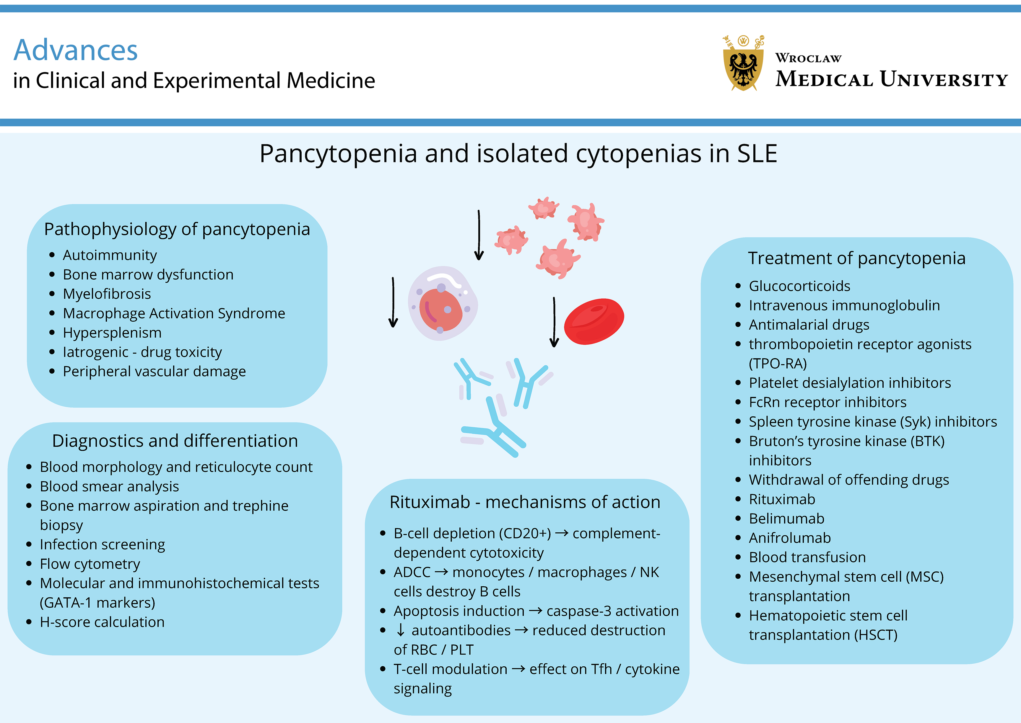
In recent years, several biomarkers of primary myelofibrosis have been identified, including circulating YKL-40 and GATA-1; however, they are not useful for diagnosing myelofibrosis associated with SLE.36 Further hematological evaluation is typically recommended, including bone marrow aspiration and trephine biopsy, which may reveal a hypercellular bone marrow. Trephine biopsy is also used to confirm hypocellularity or bone marrow necrosis5 (Figure 1).
Chronic SLE and long-term immunosuppressive therapy increase the risk of lymphoid malignancies. Therefore, in patients with persistent cytopenia or new clinical features, such as progressive lymphadenopathy, fever, night sweats, or weight loss, lymphoma should be considered in the differential diagnosis.5
In the differential diagnosis, it is important to distinguish between bone marrow suppression caused by medications or active autoimmune disease and malignant bone marrow infiltration. The final diagnosis is based on bone marrow aspiration and trephine biopsy with histopathological evaluation, flow cytometry, and cytogenetic and molecular analyses for monoclonal lymphocyte populations, allowing lymphoma to be confirmed or excluded as the cause of pancytopenia. In diagnostically challenging cases, additional molecular and immunohistochemical analyses, including GATA-1 assessment, may be considered to help distinguish myeloproliferative neoplasms from lymphoproliferative disorders.5
Moreover, infectious causes should also be considered in the differential diagnosis of pancytopenia in patients with SLE. Viral infections, including parvovirus B19, HIV, Epstein–Barr virus (EBV), cytomegalovirus (CMV), and hepatitis viruses (HAV, HBV, and HCV), may impair bone marrow hematopoiesis because of their tropism for hematopoietic progenitor cells and their ability to induce aplastic anemia.5 In endemic regions, visceral leishmaniasis should also be considered because it may closely mimic SLE flares by presenting with fever, pancytopenia, splenomegaly, hypergammaglobulinemia, and autoantibody production.37
Furthermore, additional investigations, including flow cytometry for PNH and measurements of aspartate aminotransferase (AST), ferritin, triglycerides, and fibrinogen, are important for calculating the HScore in patients with suspected HLH.5
Treatment of pancytopenia is primarily directed at identifying and eliminating the underlying cause (Table 1). In addition, antibiotics are indicated in cases of severe neutropenia, and blood transfusions may be administered when clinically indicated to manage severe anemia.5 Prompt recognition and comprehensive diagnostic evaluation are essential to distinguish SLE-related pancytopenia from other potentially life-threatening conditions. A thorough understanding of the pathophysiology and clinical implications of pancytopenia in SLE is crucial for guiding appropriate therapeutic strategies and improving patient outcomes. Further research is warranted to clarify the underlying mechanisms and optimize management strategies for this complex and heterogeneous patient population.
The role of rituximab in the treatment of cytopenias associated with SLE
Given the central role of B lymphocytes in the pathogenesis of SLE, RTX, a chimeric mouse/human monoclonal antibody targeting the CD20 antigen, has been evaluated in numerous randomized controlled trials for the treatment of SLE, including studies assessing its B-cell specificity and efficacy in SLE as well as in other rheumatic diseases.48 Rituximab induces depletion of mature B lymphocytes through complement-dependent cytotoxicity (CDC), antibody-dependent cellular cytotoxicity (ADCC), and induction of apoptosis, providing a biological rationale for its use in autoimmune cytopenias associated with SLE.49
In many countries, RTX is used off-label for the treatment of primary immune cytopenias in adults. Prospective randomized studies have demonstrated response rates of approx. 50–60% in ITP50 and up to 75% in warm AIHA.51 However, studies evaluating RTX in SLE-related cytopenias have not demonstrated a significant clinical benefit; therefore, its use in this setting remains off-label.48
Mechanisms of action of RTX
Direct depletion of CD20-positive B lymphocytes through CDC and ADCC prevents the generation of autoantibody-producing plasma cells directed against blood cell antigens (e.g., antiplatelet antibodies in ITP and antierythrocyte antibodies in AIHA), thereby reducing opsonization and peripheral destruction of blood cells.4 In cases of severe thrombocytopenia, RTX treatment stabilized platelet counts above 100 × 109/µL for more than 6 months and was accompanied by the disappearance of anti-DNA antibodies4 (Figure 2).
Importantly, RTX targets both immature pre-B cells in the bone marrow and mature circulating B lymphocytes. As B cells differentiate into immunoglobulin-producing plasma cells, they lose CD20 expression. Consequently, plasma cells are generally not depleted during RTX therapy, and serum immunoglobulin levels usually remain within the normal range. In addition, RTX does not affect hematopoietic stem cells (HSCs).52
B-cell depletion also influences the number and function of helper T lymphocytes, including T follicular helper (Tfh) cells, as well as cytokine production, which is relevant in immune-mediated bone marrow suppression.33 Furthermore, RTX exerts complement-dependent cytotoxicity by binding C1q, leading to the formation of the membrane attack complex and subsequent lysis of CD20-positive B cells. In addition, RTX induces apoptosis through activation of the caspase-3 signaling pathway and via Fab-dependent mechanisms.53 Antibody-dependent cellular cytotoxicity represents another important mechanism of RTX action. This process involves binding of Fc receptors expressed on monocytes, macrophages, and natural killer (NK) cells to the Fc region of RTX, resulting in activation of effector cells and lysis of B lymphocytes.54 Rituximab may also modulate germinal center responses and the activity of follicular regulatory T (Tfr) cells, which suppress B-cell activation within germinal centers.53
In cases associated with MAS or HLH, the therapeutic effect of RTX may result from disruption of the pathological feedback loop between autoreactive B cells and macrophage overactivation through modulation of the cytokine network. Rituximab may be used both as monotherapy and as an adjunct to the HLH-2004 treatment protocol. Compared with etoposide, RTX has a more favorable safety profile and is generally better tolerated.55
Efficacy of RTX in the treatment of hematological manifestations of SLE
Evidence supporting the efficacy of RTX in the treatment of hematological manifestations of SLE is derived primarily from case series, large observational cohorts, and meta-analyses. In a multicenter retrospective study by Serris et al., RTX produced a high rate of rapid responses in immune cytopenias associated with SLE. The overall initial response rate was 82%, including a complete response (CR) rate of 59%. The initial response rates were 87% for ITP, 86% for AIHA, and 50% for Evans syndrome. Although relapse occurred in 41% of patients despite an initial response, re-treatment with RTX was effective in 94% of these cases.56
In the study by Sans-Pola et al., RTX was administered to 33 patients with extrarenal SLE, resulting in partial or complete responses. The median SLEDAI-2K score decreased from 9 to 1.5, and patients with thrombocytopenia experienced significant increases in platelet counts. Anti-double-stranded DNA (anti-dsDNA) antibody levels also declined, from a median of 64.3 to 32.7. However, the median time to relapse after the first treatment cycle was 1.6 years, and all patients required ongoing therapy to maintain remission.57
In a multicenter retrospective cohort study including 71 adults with SLE-associated cytopenias, 86% of patients responded initially to RTX (91% with ITP, 87.5% with AIHA, and 60% with Evans syndrome), and 60.5% achieved complete remission. No cases of RTX-induced neutropenia were reported, and only 3 patients developed severe infections. Among the 24 patients (39.3%) who relapsed after an initial response, 18 underwent re-treatment with RTX, which was successful in 88.8% of cases.49
Pediatric case reports have also described severe, treatment-refractory pancytopenia that achieved remission following RTX therapy without significant adverse events.58
Although the placebo-controlled randomized EXPLORER (extrarenal lupus) and LUNAR (lupus nephritis) trials did not demonstrate statistically significant superiority of RTX combined with standard therapy, observational evidence suggests that carefully selected patients with hematological manifestations of SLE, including ITP, AIHA, and pancytopenia, may benefit from RTX treatment.59
In an interventional study by Zhang et al., 8 patients with treatment-refractory SLE-associated ITP received low-dose RTX (2 infusions of 200 mg administered at 2-week intervals) together with tapering corticosteroid therapy. Overall response rates (ORRs) at 1, 3, 6, and 12 months were 25.0%, 87.5%, 75.0%, and 75.0%, respectively, suggesting that this regimen may represent a promising therapeutic approach.60
In a retrospective cohort study by Harris et al., the ORR to RTX (56%) did not differ significantly between children (58%, n = 24) and adults (55%, n = 40). Response rates were also similar in primary and secondary ITP (53% and 62%, respectively). Evans syndrome was more common in children (42%) than in adults (18%) treated with RTX.61
Safety profile of RTX, with emphasis on patients with immune cytopenias
Rituximab is not without adverse effects. It may induce secondary hypogammaglobulinemia, thereby increasing the risk of severe infections, particularly in patients with low IgG levels before treatment or after repeated treatment courses. This complication may require IVIG replacement therapy. The risk is particularly relevant in patients with baseline pancytopenia, as this condition itself predisposes them to infections and necessitates regular monitoring of serum immunoglobulin levels.62
In addition, late-onset neutropenia (LON) may occur weeks to months after RTX administration, although it is usually transient. Concomitant use of other immunosuppressive agents or previous treatment courses may increase this risk.63
Infusion-related reactions may also occur; however, they are generally mild to moderate.53 There are also isolated reports suggesting that RTX may exacerbate bone marrow suppression.64
Other treatment methods in SLE patients with cytopenias
Clinical studies have demonstrated that mesenchymal stem cell (MSC) transplantation can improve outcomes in patients with SLE by enhancing regulatory T-cell (Treg) and T-helper 2 (Th2) responses while suppressing pro-inflammatory Th1, Th17, and B-cell responses, with an overall favorable safety profile. However, therapeutic efficacy remains variable, which may reflect differences in donor cell quality and recipient immune characteristics.65
An alternative treatment for severe SLE is hematopoietic stem cell transplantation (HSCT), which may induce long-term remission but is associated with risks such as infections and conditioning-related toxicity.66
Belimumab, a B-lymphocyte stimulator (BLyS/BAFF) inhibitor, has beneficial effects on the course of SLE, including its hematological manifestations, by suppressing excessive activation of autoreactive B lymphocytes.29 Analyses of published case reports suggest that belimumab may improve platelet counts and Hb concentrations in patients with SLE-ITP and autoimmune hemolytic anemia (AIHA).67 In addition, belimumab therapy may allow reduction of GC doses.68 Its safety profile has been shown to be favorable across different clinical phenotypes of SLE. However, it should be noted that patients with severe, life-threatening cytopenias were excluded from the pivotal registration trials. Therefore, belimumab may be considered an adjunctive treatment option for chronic, refractory cytopenias associated with SLE, although the decision to initiate therapy should be individualized.69
Another biologic agent that may be useful in the treatment of cytopenias in patients with SLE is anifrolumab. It is a monoclonal antibody that blocks the type I interferon receptor (IFNAR1), thereby targeting a key pathogenic pathway in SLE and reducing disease activity, including improvement of hematological parameters in some patients.70 Post hoc analyses of clinical trials have shown that inhibition of type I interferon signaling is associated with improvements across multiple domains of disease activity, including hematological manifestations. However, no clinical studies have specifically evaluated anifrolumab for the treatment of severe cytopenias in patients with SLE.71
Limitations of the study
This review has several limitations, primarily related to the heterogeneity and limited availability of large, high-quality studies investigating pancytopenia in SLE. Much of the available evidence is derived from small case series, introducing the potential for selection and reporting bias. In addition, the retrospective design of many studies and the overlap between iatrogenic causes and disease manifestations limit causal inference. There is a clear need for prospective cohort studies and randomized clinical trials to support the development of robust diagnostic and therapeutic guidelines for the management of hematological disorders in patients with SLE.
Conclusions
Pancytopenia in SLE is a multifactorial complication with a diverse etiology. It may reflect primary autoimmune activity or arise secondary to infections, drug toxicity, bone marrow suppression, or concomitant hematological disorders such as MAS. Early recognition and a systematic diagnostic evaluation are essential for identifying the underlying cause and selecting appropriate treatment. Bone marrow examination, serological testing, and immunologic profiling are key components of the diagnostic workup and help exclude conditions that may mimic SLE-associated pancytopenia, such as hematological malignancies or aplastic anemia.
Therapeutic interventions should be directed at the underlying cause. In autoimmune-mediated pancytopenia, immunosuppressive therapy – including GCs, IVIG, and biologic agents such as RTX – may be effective. In cases related to drug toxicity or infection, supportive care and modification of the immunosuppressive regimen are required. Novel therapeutic approaches, including neonatal Fc receptor (FcRn) inhibitors and immunomodulatory agents such as iberdomide, are currently under clinical investigation and may offer promising treatment options for refractory cases.
Importantly, pancytopenia should not be regarded merely as a laboratory abnormality; it often reflects increased disease activity and an elevated risk of systemic complications. Its presence should prompt comprehensive clinical reassessment and multidisciplinary collaboration involving rheumatologists, hematologists, and infectious disease specialists.
Further research is needed to clarify the immunopathogenesis of pancytopenia in SLE and to optimize evidence-based treatment algorithms. Given its potential for rapid progression and its prognostic significance, pancytopenia should be recognized as a serious clinical manifestation requiring prompt evaluation and appropriate management within the broader context of systemic autoimmunity.
Use of AI and AI-assisted technologies
Not applicable.
.jpg)
.jpg)