Abstract
Background. Sevoflurane is a widely used inhalation anesthetic during surgical procedures. Experimental animal models have demonstrated that exposure to sevoflurane leads to cognitive dysfunction. The molecular mechanisms underlying sevoflurane-induced neurotoxicity, as well as the potential neuroprotective mechanisms of berberine, have not yet been fully elucidated.
Objectives. The current investigation sought to evaluate the effects of berberine on memory and learning in aged rats undergoing sevoflurane anesthesia.
Materials and methods. Fifty-six male Sprague Dawley rats aged 20–24 weeks were assigned to 4 groups (n = 14/group): control, berberine, sevoflurane + berberine, and sevoflurane. The control group was exposed to 50% O2 and 50% air at a flow rate of 2 L/min for 6 h. The sevoflurane group underwent inhalation anesthesia with 3% sevoflurane mixed with 50% O2 and 50% air at 2 L/min for 6 h. In the sevoflurane + berberine group, berberine was administered intraperitoneally at a dose of 50 mg/kg/day, starting 1 week before sevoflurane anesthesia. Following these procedures, the Morris water maze (MWM), elevated plus maze (EPM), and novel object recognition tests were conducted. Brain tissues obtained from the remaining 7 rats were preserved for TUNEL staining. The expression levels of the investigated microRNAs and BDNF (brain-derived neurotrophic factor) in the frontal lobe, hippocampus, and plasma were determined using real-time quantitative polymerase chain reaction (qPCR), while BDNF levels were measured using enzyme-linked immunosorbent assay (ELISA).
Results. In the berberine group, compared with the sevoflurane group, plasma, frontal lobe, and hippocampal levels of microRNAs 9, 124, and 132 and BDNF were significantly increased, and TUNEL staining revealed a statistically significant reduction in apoptotic bodies.
Conclusions. This study indicates that berberine exerts beneficial effects on memory and learning at the molecular level in the geriatric population.
Key words: microRNA, berberine, sevoflurane, geriatric anesthesia
Background
Postoperative cognitive dysfunction (POCD) is a form of cognitive impairment that may develop following anesthesia administration and surgical procedures. It is characterized by deficits in multiple cognitive domains, including information processing, visuospatial ability, executive function, attention, concentration, memory, and psychomotor speed.1 The prevalence of cognitive dysfunction depends on several factors, including age, education level, sex, comorbidities, type of surgery, and assessment methods.2 Postoperative cognitive dysfunction represents a common postoperative complication that affects the central nervous system in older adult patients after surgery and is associated with increased mortality, greater pain and distress, prolonged hospital stays, and higher healthcare costs.3 Investigating the pathophysiology of POCD may help identify new therapeutic targets, improve postoperative quality of life in older adults, and reduce clinical and social burdens. However, the pathophysiological mechanisms underpinning POCD remain unclear, effective preventive or therapeutic interventions are currently lacking, and no effective therapies are currently available.
Sevoflurane is a widely used volatile anesthetic agent owing to its rapid onset, fast induction, and precise controllability. Nevertheless, accumulating evidence indicates that exposure to sevoflurane-based anesthesia, particularly when repeated, may induce neuropathological alterations and contribute to long-term cognitive impairment in humans and animals.4 Experimental studies have demonstrated that sevoflurane increases hippocampal neuronal inflammation and apoptosis in aged rats.5 Proposed mechanisms underlying sevoflurane-induced POCD include neuroinflammation, alterations in neurotransmitter systems, reductions in brain-derived neurotrophic factor (BDNF) levels, mitochondrial oxidative stress, and dysregulation of amyloid-β metabolism.6
Berberine is a naturally occurring benzylisoquinoline alkaloid present in several medicinal plants and exhibits a broad spectrum of pharmacological activities.7 In addition to its well-documented anti-inflammatory, antioxidant, antitumor, and cholesterol-lowering properties,8, 9, 10, 11, 12, 13 berberine has been shown to exert neuroprotective effects and to ameliorate diabetes-associated cognitive decline in mice.14, 15 Moreover, its protective effects in neurodegenerative disorders and aging-related cognitive impairment have attracted increasing attention.16
MicroRNAs (miRNAs) are small endogenous non-coding RNA molecules, approx. 21 nucleotides in length, that act as key epigenetic regulators of gene expression.17 Among them, microRNA-9 (miR-9) is highly expressed in the mammalian brain and plays a critical role in neuronal development and function by modulating the expression of ion channels, enzymes, transcription factors, and other essential molecular targets.18 MicroRNA-124 (miR-124) is essential for neuronal differentiation and maturation and facilitates neural reprogramming and functional integration of newly generated neurons.19 MicroRNAs miR-132 and miR-212 are co-transcribed from a shared genomic locus and are closely linked to BDNF signaling through the cyclic AMP response element-binding protein (CREB), a key transcriptional regulator of genes involved in synaptic plasticity and memory formation.20
MicroRNA-134 (miR-134), a brain-specific miRNA, has been implicated in the regulation of synaptic activity and dendritic spine morphology.21 Together with miR-132 and miR-138, miR-134 contributes to the fine-tuning of dendritic spine structure, which represents the primary site of excitatory synaptic transmission. In hippocampal circuits – widely used as experimental models for learning and memory – miR-138 is abundantly expressed and serves as a critical regulator of excitatory synaptic activity in pyramidal neurons.22 We hypothesize that berberine administration prior to sevoflurane exposure would attenuate anesthesia-induced cognitive dysfunction by modulating key molecular pathways, particularly through the upregulation of BDNF, alteration of microRNA expression profiles, and reduction of neuronal apoptosis.
Objectives
In this study, we aimed to investigate the roles of miR-9, miR-124, miR-132, miR-134, miR-138, and BDNF and examine the neuroprotective effects of berberine in sevoflurane-
induced neurotoxicity in aged rats. Specifically, we sought to evaluate the impact of berberine on memory and learning in aged rats exposed to sevoflurane-based anesthesia.
Materials and methods
Fifty-six male Sprague Dawley rats (20–24 weeks old, weighing 350–500 g) were obtained from the Eskişehir Osmangazi University Saki Yenilli Experimental Animal Production and Application Laboratory (Eskişehir, Turkey). All experimental procedures were approved by the Animal Experiments Local Ethics Committee of Eskişehir Osmangazi University, Turkey (decision No. 893-1, issued on June 16, 2022). Animals were housed under controlled conditions (19–21°C, 12-h light/dark cycle) with ad libitum access to food and water. Rats were randomly assigned to 4 groups (n = 14 per group): control, sevoflurane, berberine, and sevoflurane + berberine. Berberine (50 mg/kg, intraperitoneally) was administered once daily for 7 days in the berberine and sevoflurane + berberine groups. The selected dose was below the reported LD50 of berberine (57.61 mg/kg).23 On the final day, rats in the sevoflurane and sevoflurane + berberine groups received inhalation anesthesia with 3% sevoflurane in a 50% O2/air mixture for 6 h (2 L/min). Control animals were exposed to the same gas mixture without sevoflurane. Inclusion of a berberine-only group allowed evaluation of the independent effects of berberine in the absence of anesthesia. Twenty-four hours after anesthesia was designated as day 0. Behavioral assessments were conducted between days 1 and 5, including the elevated plus maze (EPM), novel object recognition, and Morris water maze (MWM), using standard protocols. After completion of behavioral testing, blood samples were collected, and the rats were euthanized. The hippocampus and frontal lobes were dissected for molecular and biochemical analyses. Tissues from 7 rats per group were used for histological evaluation, and the remaining samples were allocated to real-time quantitative polymerase chain reaction (qPCR) and enzyme-linked immunosorbent assay (ELISA) analyses.
Anxiety-like behavior was assessed using the EPM by recording the time spent in the open and closed arms and the number of arm entries. Spatial learning and memory were evaluated using the MWM by measuring escape latency, time spent in the target quadrant, swimming speed, and total distance traveled. Recognition memory was assessed using the novel object recognition test by calculating the relative exploration index and novelty preference ratio. Total RNA was isolated from hippocampal, frontal lobe, and serum samples, followed by cDNA synthesis. Expression levels of miR-9, miR-124, miR-132, miR-134, miR-138, and BDNF were quantified using SYBR Green-based quantitative PCR, and relative expression was calculated using the 2–ΔΔCt method. Primer sequences are provided in Table 1. Brain-derived neurotrophic factor protein concentrations were measured using a commercially available ELISA kit according to the manufacturer’s instructions (cat. No. 201-11-0477-96T; Shanghai Sunred Biological Technology Co., Ltd., Shanghai, China). Brain tissues were fixed, paraffin-embedded, and sectioned for histopathological evaluation. Apoptosis in the hippocampus was assessed using terminal deoxynucleotidyl transferase dUTP nick-end labeling (TUNEL), and the apoptotic index was calculated as the percentage of TUNEL-positive cells. Immunohistochemical staining was performed, and staining intensity was quantified using the H-score method by blinded observers. Morphometric damage in the cortical and hippocampal regions was graded using a standardized semi-quantitative scale.
Statistical analyses
All analyses were conducted on 4 independent groups using IBM SPSS Statistics v. 28 (IBM Corp., Armonk, USA). Continuous outcomes were summarized using mean ± standard deviation (SD) and median (interquartile range (IQR)). Before inferential testing, distributional assumptions were assessed with the Shapiro–Wilk test and visual inspection of Q–Q plots; homogeneity of variances was examined using the Levene or Brown–Forsythe test. When assumptions were met, between-group differences were evaluated using one-way analysis of variance (ANOVA); otherwise, the Kruskal–Wallis test was applied. For outcomes with a significant omnibus result, pairwise post hoc comparisons employed Tukey’s honestly significant difference (HSD) test (after ANOVA) or Dunn’s test (after Kruskal–Wallis), with family-wise error controlled using the Holm or Bonferroni adjustment. All tests were two-tailed with α = 0.05, and exact p-values are reported (very small values denoted as p < 0.001); significance levels are indicated as p < 0.05, p < 0.01, and p < 0.001.
Results
Results of the object recognition test are summarized in Table 2, and assumption checks are provided in Supplementary Table 1. As the assumptions of normality and/or homogeneity of variance were violated, between-group comparisons were performed using the Kruskal–Wallis test followed by Dunn–Bonferroni post hoc test.
Exploration times during the familiarization phases (phases I and II) did not differ among groups (p = 0.716 and p = 0.670), indicating comparable baseline exploratory behavior. For the familiar object, a significant group effect was observed (p = 0.019), with reduced exploration time in the berberine group compared with the sevoflurane + berberine group (adjusted p = 0.014). No other pairwise differences were significant.
Exploration of the novel object differed significantly among groups (p = 0.001). The sevoflurane group showed markedly reduced exploration compared with the control and berberine groups (both adjusted p = 0.001). In addition, the berberine group exhibited greater novel object exploration than the sevoflurane + berberine group (adjusted p = 0.019), suggesting preservation of recognition performance with berberine treatment.
Novel object preference results are presented in Table 3, with assumption checks shown in Supplementary Table 2. Relative exploration time differed significantly among groups (p < 0.001); the control and berberine groups demonstrated higher values than both sevoflurane-exposed groups (all adjusted p ≤ 0.005), while no difference was observed between the control and berberine groups. Preference ratio also varied significantly (p < 0.001), with lower values in the sevoflurane group compared with the control and berberine groups (adjusted p < 0.01).
The ELISA results for hippocampal tissue, frontal lobe tissue, and blood samples are summarized in Table 4. Hippocampal biomarker levels differed significantly among groups (p = 0.02), with higher concentrations in the sevoflurane + berberine group than in the control group (adjusted p = 0.014). Frontal lobe concentrations also showed significant group differences (p = 0.003), with higher levels in the control and berberine groups compared with the sevoflurane and sevoflurane + berberine groups. Blood biomarker levels differed at the group level (p = 0.027); however, no pairwise comparisons remained significant after correction.
Hippocampal expression levels of all examined miRNAs and BDNF differed significantly among groups (all p < 0.001; Table 5). Sevoflurane exposure was associated with pronounced downregulation of miRNAs and BDNF, whereas berberine treatment significantly attenuated these molecular alterations.
Significant group-dependent differences were observed for all frontal lobe miRNAs and BDNF (all p < 0.001; Table 6). Sevoflurane produced the lowest expression levels, while berberine administration resulted in higher expression of miR-9, miR-124, miR-132, miR-134, miR-138, and BDNF.
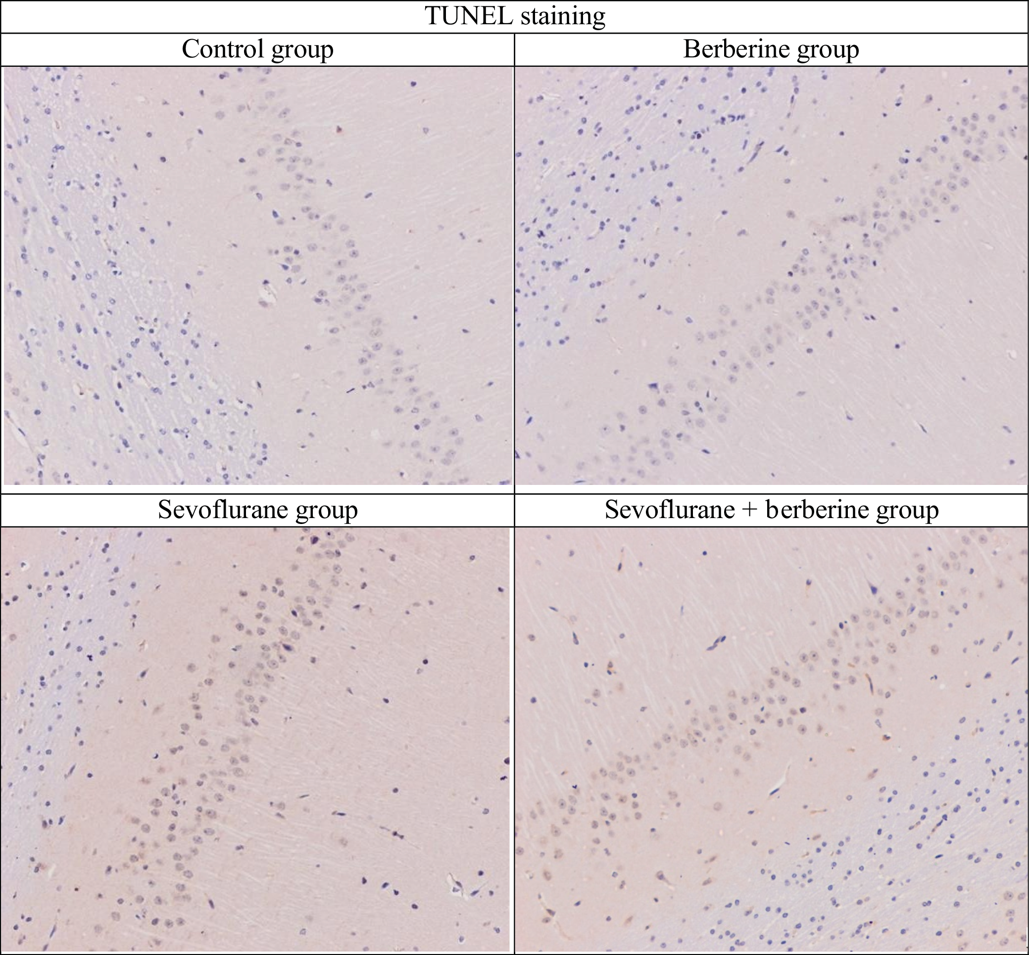
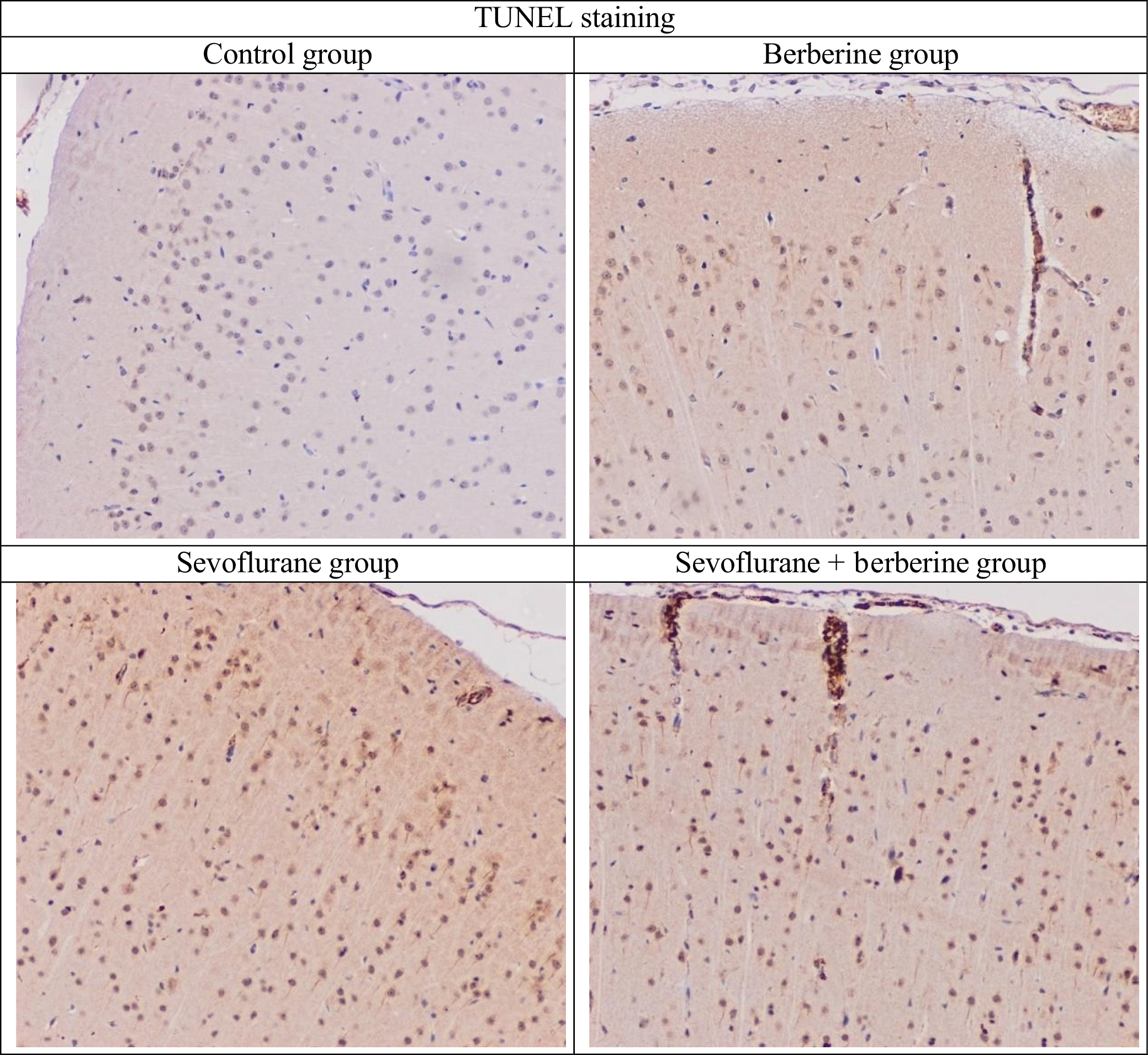
Serum miRNA and BDNF results are shown in Table 7. Significant group differences were detected for all markers except miR-9 (p = 0.086). Sevoflurane exposure significantly reduced serum miR-124, miR-132, miR-134, miR-138, and BDNF levels, whereas berberine treatment reversed these reductions and yielded the highest overall expression levels. Representative TUNEL-stained histopathological images of the hippocampus and cortex are presented in Figure 1 and Figure 2, respectively.
Across behavioral, biochemical, and molecular analyses, sevoflurane consistently impaired recognition-related performance and suppressed miRNA and BDNF expression in brain tissue and serum. Berberine treatment mitigated these effects, preserving cognitive performance and restoring molecular profiles.
Discussion
This study measured hippocampal, frontal lobe, and serum levels of BDNF and miR-9, miR-124, miR-132, miR-134, and miR-138, which are regulators of dendritic outgrowth, synaptic plasticity, long-term potentiation, memory, and learning, and demonstrated that berberine attenuated the adverse effects of sevoflurane. TUNEL staining also demonstrated a statistically significant reduction in the apoptotic index and damage scores in the hippocampus and cortex of the berberine-treated groups.
Behavioral assessments showed that sevoflurane exposure impaired cognitive performance, as reflected by reduced exploration of novel objects in the object recognition test and decreased time spent in the open arms of the EPM. The MWM further confirmed spatial memory deficits, with prolonged escape latency and reduced time spent in the target quadrant. In contrast, berberine treatment ameliorated these impairments, improving exploration indices, memory retention, and anxiety-related behaviors. At the molecular level, hippocampal analyses revealed that berberine markedly upregulated miR-9, miR-132, miR-134, and BDNF expression, while sevoflurane significantly suppressed miR-9, miR-124, miR-132, and BDNF levels. Frontal lobe qPCR data showed a similar pattern, with miR-9 and miR-124 downregulated and miR-138 markedly reduced in the sevoflurane group, whereas berberine preserved expression levels closer to those of the control group. Serum analyses supported these findings, demonstrating significantly reduced miR-124 and BDNF levels in the sevoflurane group and a marked elevation of BDNF in the berberine group. Collectively, these results indicate that berberine mitigates sevoflurane-induced cognitive and molecular impairments by preserving memory performance and normalizing miRNA and BDNF expression.
Previous studies have examined the effects of sevoflurane on memory. In one study, 60 male Wistar rats (200–250 g) were divided into 6 groups: control, diabetic control, sevoflurane anesthesia, isoflurane anesthesia, diabetic sevoflurane anesthesia, and diabetic isoflurane anesthesia. Animals received 2.5% sevoflurane or 1.5% isoflurane for 2 h, and the MWM test was conducted 1 week later. While the diabetic control and sevoflurane anesthesia groups showed no significant changes, the isoflurane group had a longer escape latency than the controls, suggesting impaired learning and memory. Diabetic rats exposed to either anesthetic also spent less time in the target quadrant than the controls.24 Unlike that study, which initiated MWM testing 1 week after exposure, we began testing 1 day after sevoflurane exposure and found a statistically significant increase in escape latency in the sevoflurane group.
Brain-derived neurotrophic factor, a member of the neurotrophin family, activates intracellular signaling pathways such as mitogen-activated protein kinase/extracellular signal-regulated kinase and phosphoinositide 3-kinase through stimulation of tropomyosin receptor kinase B. It has been shown to upregulate miR-132 expression in cortical neuron cultures.25 MicroRNA-132 promotes primary neuron growth. Consistent with these findings, our study demonstrated significantly higher BDNF and miR-132 levels in the frontal cortex and hippocampus of the berberine groups compared with the sevoflurane group. The relationship between BDNF and miR-132 underscores the significance of our results.
Our study also revealed higher miR-9 expression, increased active microglia, and reduced neuronal cell counts in rats, similar to findings from a study in mice with late-stage Alzheimer’s disease.26 In the present study, hippocampal miR-9 expression was markedly suppressed following sevoflurane exposure, while berberine administration significantly increased miR-9 levels compared with the sevoflurane group, suggesting a potential protective role against anesthesia-induced neurodegeneration.
MicroRNA-124 plays a central role in neuronal cell proliferation, differentiation, migration, memory formation, apoptosis, and neurodegenerative diseases. Experimental studies have shown that reduced miR-124 expression is associated with increased neuronal apoptosis and impaired axonal growth.27, 28 In line with these observations, the present study demonstrated significantly reduced miR-124 expression and increased hippocampal apoptosis in the sevoflurane group, as confirmed by TUNEL staining, whereas berberine treatment attenuated these effects.
MicroRNA-134, a brain-specific microRNA primarily localized to the synapto-dendritic region, is thought to negatively regulate dendritic spine development and plasticity.29 Recent findings indicate that miR-134 inhibits hippocampal synaptic plasticity by targeting plasticity-related proteins, such as LIM kinase 1, resulting in cognitive deficits. In contrast, our study found increased hippocampal miR-134 expression in the berberine group.
MicroRNA-138 has been shown to reduce dendritic spine size and suppress excitatory synaptic transmission in hippocampal neurons.30 Elevated miR-138 levels inhibit SIRT1 signaling, impair axonal growth, and are associated with stress-related behavioral alterations.31, 32 Consistent with these findings, hippocampal miR-138 expression was significantly higher in the sevoflurane group and accompanied by reduced BDNF levels, whereas berberine treatment normalized both miR-138 and BDNF expression.
Saha et al.33 established an epilepsy model in male Wistar rats by intraperitoneal administration of pentylenetetrazol (30 mg/kg) every other day for 6 weeks. Rats with pentylenetetrazol-induced epilepsy were treated with 50, 100, and 200 mg/kg of berberine and 25, 50, and 100 mg/kg of nano-berberine. Outcomes included seizure scores, the percentage of rats experiencing seizures, histopathological scores, oxidative stress, apoptosis, and inflammation. None of the animals in the nano-berberine group exhibited seizures. This effect was attributed to significantly increased levels of antioxidants, such as superoxide dismutase and glutathione, in the nano-berberine group, whereas standard berberine did not produce a comparable antioxidant response. These results suggest that the superior antioxidant properties of nano-berberine may be due to its higher plasma concentration compared with standard berberine. Nano-berberine exhibited greater antioxidant properties than berberine.
In the present study, berberine hydrochloride was freshly prepared in 0.9% isotonic saline immediately before administration. Histological TUNEL staining showed significantly reduced apoptosis in the berberine-treated group compared with the sevoflurane group; however, the absence of significant differences in behavioral tests may be attributed to the rapid metabolism, slow elimination, and short half-life of berberine, which may limit plasma levels. This limitation could potentially be addressed by developing novel nano-formulations with improved bioavailability. Future studies using nano-berberine are expected to yield more pronounced effects than both sevoflurane and the control group.
Previous studies have demonstrated that miR-124, miR-132, miR-134, and miR-138 play critical roles in neuronal differentiation, neurite outgrowth, dendritic spine remodeling, synaptic plasticity, and axonal regeneration, and that dysregulation of these microRNAs is associated with stress-related, anesthesia-induced, and age-related cognitive impairment and affective behavioral changes.28, 29, 30, 31, 32 In this context, our findings suggest that berberine exerts neuroprotective effects, potentially through modulation of miRNA expression, BDNF levels, and reduced apoptosis. It is important to acknowledge that berberine has a broad pharmacological profile. In addition to its effects on neuronal signaling, berberine is known to exert anti-inflammatory, antioxidant, and metabolic effects, all of which could contribute to the observed behavioral improvements.34, 35 Furthermore, although rodent models provide valuable mechanistic insight, interspecies differences in brain structure, pharmacokinetics, and immune responses limit direct translational applicability to postoperative cognitive dysfunction in humans.36 Additionally, our study was conducted exclusively in male animals, which may overlook potential sex-based differences in response to berberine, as previous studies have shown that pharmacokinetic and pharmacodynamic profiles can vary significantly by sex.37
Finally, the observed miRNA alterations are consistent with emerging evidence that integrative bioinformatics and machine learning approaches can identify miRNA-driven regulatory networks and protective biomarkers in complex diseases, including neurocognitive and oncological conditions.38 Future studies should therefore incorporate both sexes, explore longer-term outcomes, and evaluate alternative dosing strategies to better inform clinical applications.
Limitations of the study
Our study had several limitations. First, isotonic fluid maintenance was not administered for 6 h during sevoflurane anesthesia, and neither the minimum alveolar concentration of sevoflurane nor mean arterial blood pressure were continuously monitored. Second, the anti-inflammatory effects of berberine were not specifically evaluated, and the 1-week pre-anesthesia administration of berberine may be insufficient to assess its potential for long-term neuroprotection. In this study, our primary objective was to assess the prophylactic effects of berberine in the early phase following anesthesia; therefore, berberine administration was not continued post-anesthesia, and we recognize that this may have limited our ability to assess the sustainability of its effects. In addition, we acknowledge that the short interval between anesthesia, behavioral testing, and tissue collection may not fully capture delayed or persistent cognitive deficits, which are indeed critical in the context of POCD. Finally, the lack of nano-berberine administration in our study is a limitation.
Conclusions
This study evaluated the hippocampal, frontal lobe, and serum levels of BDNF and miRNAs 9, 124, 132, 134, and 138, which regulate dendritic outgrowth, synaptic plasticity, long-term potentiation, memory, and learning, and showed that berberine ameliorated the detrimental effects of sevoflurane. TUNEL staining also demonstrated a significant reduction in the apoptotic index and damage scores in the hippocampus and cortex of the berberine-treated groups. However, the absence of significant differences among groups in behavioral tests such as the MWM and EPM suggests that these effects may not translate into observable clinical improvement. To enhance clinical relevance, further research should examine berberine in nano-formulations, evaluate longer treatment durations, and assess continued administration after sevoflurane exposure. These findings may help optimize the neuroprotective potential and clinical relevance of berberine.
Supplementary data
The supplementary materials are available at https://doi.org/10.5281/zenodo.17822503. The package contains the following files:
Supplementary Table 1. Results of assumption checks for variables in Table 2 (with p-values).
Supplementary Table 2. Results of assumption checks for variables in Table 3 (with p-values).
Supplementary Table 3. Results of assumption checks for variables in Table 4 (with p-values).
Supplementary Table 4. Results of assumption checks for variables in Table 5 (with p-values).
Supplementary Table 5. Results of assumption checks for variables in Table 6 (with p-values).
Supplementary Table 6. Results of assumption checks for variables in Table 7 (with p-values).
Data Availability Statement
The datasets supporting the findings of the current study are openly available in Zenodo at https://doi.org/10.5281/zenodo.17831138.
Consent for publication of personal information
Not applicable.
Use of AI and AI-assisted technologies
Not applicable.