



Abstract
Background. Psoriasis is a chronic, immune-mediated skin disease characterized by keratinocyte hyperproliferation and persistent inflammation. Recent studies suggest that long non-coding RNAs (lncRNAs), such as HOX transcript antisense RNA (HOTAIR), may play critical roles in regulating inflammatory pathways involved in psoriasis pathogenesis.
Objectives. This study aimed to investigate the association between HOTAIR gene polymorphisms (rs12826786 and rs4759314) and psoriasis susceptibility, and to assess their potential regulatory effects on gene expression using expression quantitative trait loci (eQTL) data.
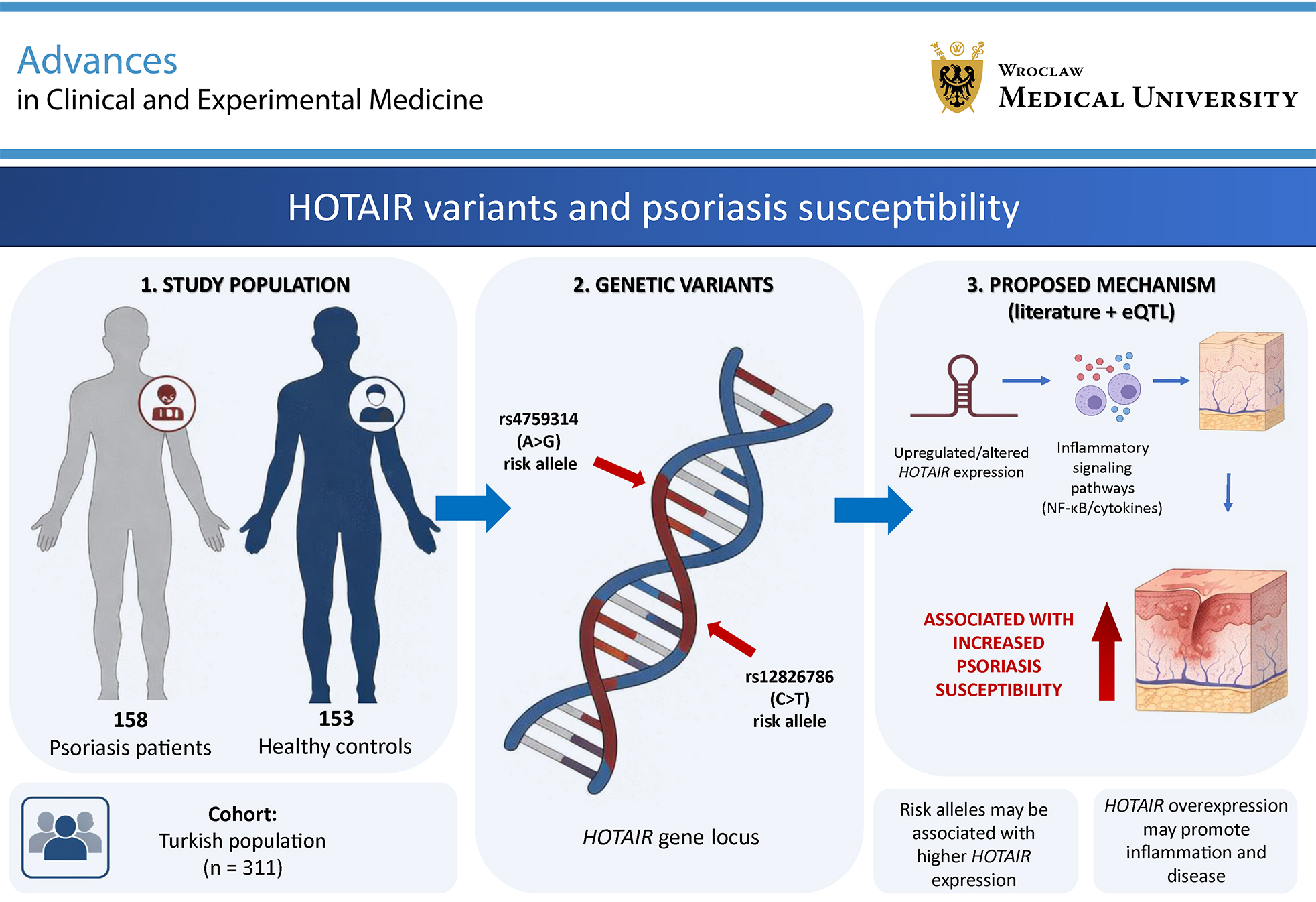
Materials and methods. A case–control study including 158 patients with psoriasis and 153 controls was conducted. Genotyping of rs12826786 and rs4759314 was performed using the tetra-primer amplification refractory mutation system (T-ARMS) polymerase chain reaction (PCR). Primary associations were tested using multivariable logistic regression in an additive (per-allele) model, adjusting for sex, age in tertiles (T1 reference), smoking, and comorbidity; dominant and genotypic (3-level) models were secondary. Expression quantitative trait loci data from publicly available databases were analyzed to explore the impact of these polymorphisms on HOTAIR expression in skin tissue.
Results. Under a multivariable additive model (adjusted for sex, age tertiles, smoking, and comorbidity), rs12826786 was associated with higher odds of psoriasis (odds ratio (OR) = 1.52, 95% confidence interval (95% CI): 1.11–2.08; q = 0.027) and rs4759314 likewise (OR = 1.73, 95% CI: 1.18–2.53; q = 0.020). Dominant contrasts were also significant: rs12826786 (CT + TT vs CC: OR = 1.84, 95% CI: 1.12–3.05; q = 0.027) and rs4759314 (AG + GG vs AA: OR = 2.03, 95% CI: 1.18–3.48; q = 0.020). Expression quantitative trait loci analysis revealed that the rs12826786 T allele significantly upregulates HOTAIR expression in both sun-exposed and non-sun-exposed skin (both p < 0.001).
Conclusions. HOTAIR polymorphisms, particularly rs12826786 and rs4759314, are associated with psoriasis risk and may contribute through regulation of HOTAIR expression. These findings support HOTAIR as a potential biomarker and therapeutic target in psoriasis.
Key words: psoriasis, long noncoding RNA, HOTAIR RNA, single nucleotide polymorphism (SNP), gene expression regulation
Background
Psoriasis is a chronic dermatological condition characterized by abnormal keratinocyte proliferation and immune dysregulation, resulting in persistent, incurable, scaly skin lesions. Beyond its dermatological manifestations, psoriasis is now widely recognized as a systemic disease and is associated with several comorbidities, including psychological disorders, metabolic conditions, arthritis, and cardiovascular disease (CVD).1 The pathogenesis of psoriasis is characterized by a multifaceted interplay between genetic susceptibility, immune system dysfunction, and environmental triggers, resulting in dysregulated keratinocyte proliferation and inflammatory responses.
Substantial evidence supports a strong genetic component in psoriasis. Genome-wide association studies (GWAS) conducted in diverse populations have identified more than 100 susceptibility loci associated with the disease.2, 3 The fact that a significant proportion of these loci are located in non-coding DNA suggests that epigenetic regulators, such as long non-coding RNAs (lncRNAs), may play a role in the development of psoriasis.4 In recent years, the number of studies investigating the role of non-coding RNAs in psoriasis has steadily increased, highlighting their potential importance in disease pathophysiology.5, 6, 7, 8 Beyond psoriasis, immune-related lncRNAs have also shown clinical and prognostic relevance in other conditions; e.g., an immune-related lncRNA-based prognostic index was recently developed for glioblastoma and was associated with both patient survival and features of the tumor immune microenvironment (TME).9
lncRNAs are non-coding RNA molecules longer than 200 nt that play important roles in regulating gene expression through interactions with DNA, other RNAs, and proteins.9 Aberrant expression of lncRNAs, which modulate gene expression through epigenetic pathways, can impair essential biological processes, including cell differentiation, migration, apoptosis, and cell-cycle regulation.10, 11
The HOTAIR gene, located on chromosome 12 within the HOXC gene cluster, produces a non-coding RNA that interacts with chromatin-modifying proteins, including polycomb repressive complex 2 (PRC2) and lysine-specific demethylase 1 (LSD1), leading to epigenetic silencing of target genes.12 The ability of HOTAIR to regulate gene expression is not limited to chromatin remodeling but can also indirectly alter the expression of specific genes by sponging different miRNAs.13 Importantly, HOTAIR has been shown to regulate several key proinflammatory cytokines, including tumor necrosis factor alpha (TNF-α), interleukin-6 (IL-6), inducible nitric oxide synthase (iNOS), and macrophage inflammatory protein-1B (MIP-1B).14, 15, 16, 17, 18 These cytokines play pivotal roles in the immunopathology of psoriasis by contributing to the recruitment and activation of inflammatory cells, promoting keratinocyte proliferation, and sustaining the chronic inflammatory environment of psoriatic lesions.19, 20, 21, 22 Given these findings, HOTAIR is increasingly recognized not only for its role in cancer biology but also as a potential regulator of inflammatory skin diseases, including psoriasis.
We considered HOTAIR a biologically plausible candidate in psoriasis, building on prior evidence that HOTAIR scaffolds chromatin-modifying complexes to direct epigenetic regulation at target loci.12, 13, 23 Beyond chromatin regulation, HOTAIR has been implicated in nuclear factor kappa-light-chain-enhancer of activated B cells (NF-κB)-related inflammatory signaling and keratinocyte responses, including an IL-22-stimulated HaCaT model involving a HOTAIR/miR-126 axis.8, 13, 18, 24 These pathways intersect with the cytokine milieu central to psoriasis pathogenesis.21, 22 We therefore focused on 2 regulatory variants within HOTAIR (rs12826786 and rs4759314) which have been associated with altered HOTAIR transcription and susceptibility in various diseases.24, 25, 26, 27, 28 Based on this rationale, we hypothesized that these polymorphisms might modulate psoriasis susceptibility by influencing HOTAIR expression and downstream inflammatory programs.
Objectives
The HOTAIR gene contains numerous functional single-nucleotide polymorphisms (SNPs) that may influence its expression and function. Variants such as rs12826786 and rs4759314 have been linked to altered HOTAIR transcription and susceptibility to several conditions, including gastric cardia adenocarcinoma, pancreatic cancer, breast cancer, and autism spectrum disorder (ASD).25, 26, 27, 28 However, their role in psoriasis remains to be fully elucidated. In this context, the present study aimed to investigate the association between HOTAIR polymorphisms and psoriasis risk and to explore potential genotype-dependent regulatory effects that may contribute to disease pathogenesis through the modulation of inflammatory responses.
Materials and methods
Study population
The study comprised 158 patients diagnosed with psoriasis as the patient group and 153 healthy individuals without a history of psoriasis as the control group. All participants were unrelated individuals of self-reported Turkish ethnicity residing in the northeastern Black Sea region of Turkey. This region is known to be ethnically homogeneous, and no evidence of recent migration or admixture was present in the recruitment history.
The diagnosis of psoriasis was made by physicians at the Dermatology Clinic of Giresun University Faculty of Medicine Research and Application Hospital (Giresun, Turkey). It was confirmed that the participants included in the patient group did not have any malignant, autoimmune, or systemic inflammatory disease. The control group consisted of individuals with no history of malignant, autoimmune, or systemic inflammatory disease. They also had no family history of psoriasis and had presented to Giresun University Faculty of Medicine Research and Application Hospital for routine health check-ups.
This study was approved by the Clinical Research Ethics Committee of Giresun University on December 23, 2021 (approval No. 09) and was conducted in accordance with the ethical principles of the Declaration of Helsinki. All participants or their legal guardians provided written informed consent before inclusion in the study. We used the Strengthening the Reporting of Observational Studies in Epidemiology (STROBE) case–control checklist when reporting our study.
Genotyping of rs12826786 and rs4759314 polymorphisms
Genomic DNA was extracted from venous blood using a commercial polymerase chain reaction (PCR) template kit (Roche Diagnostics Deutschland GmbH, Mannheim, Germany) according to the manufacturer’s instructions. DNA quality was assessed using a NanoDrop spectrophotometer (Thermo Fisher Scientific, Waltham, USA), and samples were stored at −20°C.
Genotyping was performed using tetra-primer amplification refractory mutation system PCR (T-ARMS-PCR) with the FastStart™ High Fidelity PCR System (Roche Diagnostics GmbH). Primers were designed using the National Center for Biotechnology Information (NCBI) and Ensembl genome databases and verified using NCBI Primer-BLAST (National Institutes of Health (NIH), Bethesda, USA). Polymerase chain reaction was performed on a Bio-Rad T100 thermal cycler (Bio-Rad, Hercules, USA).
Each 25-µL reaction included buffer, deoxynucleoside triphosphates (dNTPs), dimethyl sulfoxide (DMSO), enzyme, primers, DNA, and water. The thermocycling conditions were as follows: 94°C for 1 min, followed by 35 cycles of 94°C for 2 min, 57°C for 30 s, and 72°C for 45 s. Primer sequences are listed in Table 1.
Bioinformatics analysis
To explore the potential regulatory effects of the studied variants, expression quantitative trait loci (eQTL) data were retrieved from and inspected in the Genotype-Tissue Expression (GTEx) Portal (v8; https://www.gtexportal.org; accessed August 29, 2025). Specifically, we queried publicly available single-tissue cis-eQTL summary results for HOTAIR in sun-exposed and non-sun-exposed skin. Genotype–expression associations and p-values were generated within the GTEx consortium pipeline using additive genotype coding (0/1/2) and linear regression with standard GTEx covariate adjustment, as described in the GTEx Portal documentation and the GTEx v8 primary publication.29, 30 We did not download or analyze individual-level GTEx expression data or rerun any regression models; GTEx results were used solely as external functional annotation.
Statistical analyses
All tests were two-sided (α = 0.05). Continuous variables were summarized as means ± standard deviations (SDs) when parametric assumptions were met and as medians [Q1–Q3] otherwise; categorical variables were summarized as n (%). The p-values are reported to 3 decimal places, with p < 0.001 reported when appropriate. Normality of continuous variables was assessed within each group (cases and controls) using the Shapiro–Wilk test and normal Q–Q plots, and homogeneity of variances between groups was assessed using Levene’s test (Supplementary Table 1 and Supplementary Fig. 1). Hardy–Weinberg equilibrium (HWE) in controls was evaluated for each SNP using Pearson’s goodness-of-fit χ2 test (degrees of freedom (df) = 1) when all expected genotype counts were ≥5 and a two-sided exact conditional test otherwise; mid-p-values were additionally obtained using the HardyWeinberg package (https://cran.r-project.org/web/packages/HardyWeinberg/index.html) in R (R Foundation for Statistical Computing, Vienna, Austria) (Supplementary Table 2).
For the primary case–control analysis, psoriasis susceptibility (case vs control) was modeled using 3 prespecified multivariable logistic regression models: Model 1 (additive, per risk allele), Model 2 (dominant, heterozygotes plus risk-allele homozygotes vs common-allele homozygotes), and Model 3 (3-level genotypic model, 2 df). All models were adjusted for sex, age (tertiles; T1 as reference), smoking status (any vs none), and the presence of any comorbidity.
Linearity of the logit for age was examined using the Box–Tidwell procedure by adding an age × ln(age) interaction term to preliminary additive logistic models for each SNP, with age entered as a continuous covariate. The statistical significance of this term (p < 0.05) indicated violation of the linearity assumption. Therefore, age was categorized into tertiles and entered as 2 dummy variables (T2 and T3; T1 as reference) in all reported logistic regressions; full Box–Tidwell results are presented in Supplementary Table 3.
For each genetic model, we report the logistic regression coefficient (B, log odds) with its standard error (SE), the corresponding adjusted odds ratio (OR = eB) with 95% confidence intervals (95% CIs), and Wald p-values for individual coefficients. We also report likelihood-ratio χ2 statistics, with degrees of freedom and p-values, comparing covariate-only models with models additionally including the SNP term(s), so that likelihood-ratio test (LRT) p-values represent the incremental fit of the genetic term(s) over the covariates.
Model fit and calibration are summarized using the Hosmer–Lemeshow goodness-of-fit p-value and Nagelkerke R2, and discrimination is summarized using the area under the receiver operating characteristic curve (ROC AUC) with 95% CIs (Supplementary Table 4). Variance inflation factors (VIFs) were used to assess multicollinearity (Supplementary Table 5); outlier and influence diagnostics, including standardized residuals, leverage, Cook’s distance, and difference-in-betas statistics (DFBETAs), and robustness refits excluding high-leverage observations are shown in Supplementary Tables 6 and 7. All multivariable logistic regression models were fitted on the complete-case dataset (n = 300) after excluding participants with missing data on genotype or covariates.
Multiplicity in the primary case–control comparisons was controlled using the Benjamini–Hochberg false discovery rate (FDR) procedure (target q = 0.05) within each SNP across the 5 prespecified logistic tests: additive, dominant, genotypic omnibus, and the 2 pairwise genotypic contrasts (m = 5). Within-case genotype–clinical analyses were considered exploratory. No multiplicity correction was applied for these secondary endpoints; therefore, the p-values are interpreted descriptively and with caution regarding potential inflation of the type I error rate.
For binary clinical endpoints within psoriasis cases, we constructed 2 × 3 contingency tables (endpoint × 3-level genotype) and used Pearson’s χ2 test of independence (df = 2) when expected cell counts were adequate. When expected counts were small (minimum expected count <5), we report the exact two-sided p-value from the Fisher–Freeman–Halton (FFH) exact test (Supplementary Table 8). The linear-by-linear association test (df = 1) is additionally reported to assess an ordinal genotype dose trend (0 → 1 → 2), where appropriate. We also performed prespecified dominant-model 2 × 2 sensitivity analyses (major-allele homozygotes vs minor-allele carriers) using two-sided Fisher’s exact test (Supplementary Table 9). For continuous clinical endpoints, Spearman’s rank correlation coefficient (ρ) was used to test monotonic allele-dose trends using the additive genotype code 0/1/2 (Supplementary Table 10).
All descriptive and regression analyses were performed using IBM SPSS v. 25.0 (IBM Corp., Armonk, USA). Hardy–Weinberg equilibrium exact and mid-p-values were calculated in R using the HardyWeinberg package.
Results
The study comprised 311 participants, including 158 cases and 153 controls. An a priori power analysis indicated that the sample size was adequate to detect a moderate effect size (ω = 0.3) with 95% power at α = 0.05. The demographic and clinical characteristics of the study population are summarized in Table 2.
Median age was higher in cases than in controls (48.0 [35.0–58.3] years vs 31.5 [23.0–49.0] years, n = 158 vs n = 153, p < 0.001, Mann–Whitney U test), consistent with the non-normal distribution of age in both groups (Shapiro–Wilk p = 0.029 in cases and p < 0.001 in controls; Supplementary Table 1). Sex distribution was similar between groups (female: 51.3% vs 62.1%; Pearson’s χ2 test, df = 1, p = 0.054), with expected cell counts satisfying the χ2 assumptions. Smoking status also did not differ between groups (32.7% vs 29.1%; Pearson’s χ2 test, df = 1, p = 0.508).
Case-only summaries were as follows: body mass index (BMI), 26.4 [23.9–29.8] kg/m2; age at onset, 28.0 [18.0–41.0] years; disease duration, 14.0 [6.0–23.0] years; and Psoriasis Area and Severity Index (PASI), 6.0 [2.0–12.0]. A positive family history was reported in 45.6% of patients, and 20.9% had psoriatic arthritis.
Genotype and allele distributions by case–control status are shown in Table 3. For example, for rs12826786, TT was observed in 22.8% of cases and 14.4% of controls; for rs4759314, GG was observed in 15.1% and 6.5%, respectively. Minor allele frequencies were 33.2% vs 23.5% for T and 26.3% vs 14.7% for G.
We fitted 3 prespecified multivariable logistic models per SNP: Model 1 (additive), Model 2 (dominant), and Model 3 (genotypic, 3-level; 2 df). In adjusted logistic models including sex, age tertiles, smoking, and comorbidity, both loci were associated with case status across the primary genetic models (Table 4). For rs12826786, the additive model showed OR = 1.515 (95% CI: 1.105–2.076, p = 0.010; LRT p = 0.009), the dominant model showed OR = 1.843 (95% CI: 1.115–3.046, p = 0.017; LRT p = 0.016), and the genotypic 3-level omnibus test was significant (2 df; LRT p = 0.032; TT vs CC OR = 2.299, p = 0.013). For rs4759314, the additive model showed OR = 1.729 (95% CI: 1.180–2.534, p = 0.005; LRT p = 0.004), the dominant model showed OR = 2.028 (95% CI: 1.183–3.475, p = 0.010; LRT p = 0.009), and the genotypic omnibus test was significant (LRT p = 0.016; GG vs AA OR = 3.104, p = 0.012).
Across these models, overall calibration was acceptable (Hosmer–Lemeshow p > 0.70), and explanatory power was modest (Nagelkerke R2 = 0.17), indicating that the genetic terms improved prediction beyond the covariates alone. BH–FDR q-values within each SNP (m = 5) are reported alongside p-values in Table 4 and did not change the statistical conclusions. For completeness, the corresponding log-odds coefficients (B) and SEs are also provided in Table 4.
Sensitivity analyses excluding high-leverage observations (Supplementary Table 7) did not change the statistical conclusions for the primary additive and dominant contrasts. For rs4759314, however, the genotypic omnibus test (2 df) and the GG vs AA pairwise comparison were attenuated and were no longer significant, whereas the additive and dominant associations remained significant.
Across binary clinical endpoints, including psoriatic arthritis, family history, joint pain, joint involvement, nail, scalp, and genital involvement, and the PASI-10 category, no between-genotype differences were observed using Pearson’s χ2 test (2 × 3; df = 2), with expected cell counts satisfying the χ2 assumptions. Where examined, linear-by-linear association (LBL; df = 1) trend tests were also non-significant. Dominant 2 × 2 sensitivity analyses using two-sided Fisher’s exact test were likewise null. Full statistics are provided in Supplementary Tables 8 and 9. In exploratory case-only analyses of continuous variables, Spearman’s rank correlations (n = 153) showed no monotonic allele-dose trends using the genotype dose 0/1/2. Consistent with this, for rs12826786, the correlations with disease duration (ρ = −0.077, p = 0.340), age at onset (ρ = 0.049, p = 0.544), and BMI (ρ = −0.003, p = 0.968) were all nonsignificant. Likewise, for rs4759314, the correlations with disease duration (ρ = 0.053, p = 0.514), age at onset (ρ = −0.012, p = 0.883), and BMI (ρ = −0.101, p = 0.211) were nonsignificant (Supplementary Table 10).
Interrogation of GTEx v8 single-tissue cis-eQTL data indicated that rs12826786 was strongly associated with HOTAIR expression in human skin (Figure 1). In both sun-exposed (n = 751) and non-sun-exposed (n = 649) skin tissues, individuals carrying the T allele showed progressively increased HOTAIR expression in a genotype-dependent manner. Specifically, TT homozygotes exhibited the highest expression levels, followed by heterozygous CT individuals, whereas CC homozygotes had the lowest expression levels. These differences were highly statistically significant in both sun-exposed and non-sun-exposed skin (two-sided p < 0.001 for both tissues in the GTEx cis-eQTL linear regression, using default GTEx covariates), indicating a strong association between the T allele and increased HOTAIR expression. This finding supports the hypothesis that the T allele of rs12826786 may upregulate HOTAIR transcription in skin, providing a potential mechanistic explanation for its observed association with increased psoriasis risk in our study.
Hardy–Weinberg equilibrium was evaluated within each group under p2, 2pq, and q2 expectations. For rs12826786, all expected counts were ≥5, and Pearson’s χ2 goodness-of-fit test (df = 1; n = 153 controls, n = 158 cases) indicated departures from HWE in both controls (χ2 = 36.953, p < 0.001) and cases (χ2 = 44.268, p < 0.001). For rs4759314, expected counts were ≥5 in cases, which also deviated from HWE (χ2 = 28.956, p < 0.001). In controls, however, the expected count for minor-allele homozygotes was <5 (E = 3.3), rendering the χ2 test asymptotically unreliable; therefore, we report the corresponding exact two-sided HWE p-value in Supplementary Table 2, with Pearson’s χ2 shown for comparison.
These discrepancies are unlikely to stem from technical genotyping errors, as randomly selected samples were regenotyped to validate the initial findings, thereby increasing confidence in data accuracy. Importantly, although genotyping errors are a potential source of deviation from HWE, they are not the only possible explanation. Various biological and demographic factors, including random genetic drift, heterozygote advantage, non-random mating, population stratification or admixture, inbreeding, and structural genetic variation, such as copy number variants, may also lead to deviations from equilibrium assumptions.31
To assess robustness, we repeated allelic case–control tests after excluding the control genotypes that contributed most to the HWE departure: TT for rs12826786 and GG for rs4759314. Effect sizes remained stable and significant, e.g., for rs4759314: OR = 2.705 [1.587–4.610] before exclusion vs OR = 2.852 [1.691–4.810] after exclusion; both Pearson’s χ2 tests, df = 1; n = 153 controls; p < 0.001. These findings suggest that the observed deviations did not materially bias the associations.
Discussion
In this study, we investigated the association between 2 specific SNPs, rs12826786 and rs4759314, and susceptibility to psoriasis. To our knowledge, this is the first study to investigate the association between psoriasis and the HOTAIR rs12826786 and rs4759314 polymorphisms in the Turkish population. Our findings indicate that both SNPs are significantly associated with increased susceptibility to psoriasis.
Our genotypic analysis demonstrated that rs12826786 was significantly associated with psoriasis susceptibility under both the codominant, or genotypic 3-level, model (2 df) and the dominant model (CT + TT vs CC). At the allelic level, the presence of the T allele was linked to an increased risk of developing psoriasis. These findings are consistent with those of a previous study conducted in an Iranian population, in which Rakhshan et al. reported the T allele of rs12826786 as a risk factor for psoriasis (OR = 1.35; p = 0.040). Consistently, they also identified a significant association between the CT + TT genotypes and psoriasis in the dominant model (OR = 1.59; p = 0.020).14
In contrast, a study conducted in the Han Chinese population reported a protective role of the T allele of rs12826786.7 According to that study, individuals with CT + TT genotypes had a lower risk of psoriasis (OR = 0.70; p = 0.049). This discrepancy suggests potential ethnic variation in the functional impact of this SNP. Such population-specific effects are common in genetic association studies and may reflect underlying differences in environmental interactions or sample characteristics.
In our study, the rs4759314 polymorphism showed a significant association with psoriasis under both the genotypic 3-level model (2 df) and the dominant genetic model. Allelic analysis further supported these findings, with the G allele being more common in patients than in healthy controls. These results are consistent with those of a previous study conducted in the Han Chinese population, which also reported a significant association between rs4759314 and psoriasis under similar genetic models: the dominant model (OR = 3.78; p = 0.002) and the overdominant model (OR = 3.79; p < 0.001).7 By contrast, the study by Rakhshan et al. in the Iranian population did not detect any significant association between this polymorphism and psoriasis under any genetic model.14
This inconsistency may reflect ethnic differences in allele frequencies, population-specific linkage disequilibrium patterns around the HOTAIR locus, or variations in environmental exposures and clinical characteristics. In sensitivity analyses excluding high-leverage observations (Supplementary Table 6), our primary models for rs4759314, additive and dominant, remained significant, whereas the genotypic omnibus test (2 df) and the GG vs AA pairwise comparison were attenuated and were no longer significant. These patterns are consistent with G allele-related susceptibility rather than a stable homozygote-specific effect and highlight sensitivity to influential observations and small cell counts.
In exploratory case-only analyses, we found no robust genotype–phenotype associations for PASI, age at onset, or psoriatic arthritis (Supplementary Tables 8–10). Psoriasis Area and Severity Index measured at a single visit, and influenced by ongoing treatment, may be insensitive to modest genetic effects; therefore, larger longitudinal datasets will be needed to assess severity endpoints.
The biological plausibility of these associations is supported by the known functions of HOTAIR in epigenetic and inflammatory regulation. HOTAIR acts as a scaffold for chromatin-modifying complexes (PRC2/LSD1) to shape transcriptional programs12, 13, 23 and has been linked to NF-κB-driven responses and the regulation of cytokines and chemokines implicated in the maintenance of psoriatic lesions13, 24; this is consistent with the established importance of TNF-α, IL-6, and related mediators in psoriasis pathogenesis.19, 20, 21, 22 Within this framework, genetic variation at rs12826786, located in the promoter region, and rs4759314, with functional effects on HOTAIR expression in reporter and cell assays,25, 26, 27, 28, 32, 33 may contribute to disease risk by modulating HOTAIR levels and downstream inflammatory programs that intersect with the IL-23/IL-17 (Th17) axis.34, 35
A recent expression-based study conducted in Turkey evaluated HOTAIR lncRNA expression levels in psoriatic lesions and perilesional healthy skin.36 Although that study, which evaluated HOTAIR expression in psoriatic vs non-lesional skin, did not report a statistically significant difference (fold change: 0.92; p = 0.218), it is important to note that its sample size was limited to 15 patients, which may have reduced the statistical power to detect meaningful expression differences.
Moreover, functional evidence from eQTL analyses suggests that the T allele of rs12826786 is associated with increased transcriptional activity of HOTAIR. This regulatory effect may explain the observed association between the T allele and increased psoriasis risk, as higher HOTAIR expression has been linked to enhanced activation of inflammatory pathways, including NF-κB signaling and upregulation of cytokines such as IL-6 and iNOS.17, 31
Limitations of the study
This study has several limitations that should be acknowledged. First, although an a priori power analysis confirmed sufficient statistical power, the sample size was relatively modest and limited to a single geographic region, which may affect the generalizability of the findings to broader populations. Second, although public eQTL resources support a transcriptional effect of rs12826786 on HOTAIR, these data are indirect. We did not perform in vitro or in vivo experiments to assess allele-specific effects. Future work should include promoter–reporter assays and CRISPRi/CRISPRa perturbations in psoriasis-relevant cells, such as primary keratinocytes and macrophages, to define the regulatory roles of rs12826786 and rs4759314 in psoriasis. Third, genotype-specific contrasts, such as rs4759314 GG vs AA, were sensitive to leverage and small cell counts, as shown by the robustness refits (Supplementary Table 6), and should therefore be interpreted with caution. Finally, cytokine levels, such as IL-6 and TNF-α, which could further elucidate the inflammatory consequences of HOTAIR variation, were not measured. Future studies incorporating cytokine profiling may help clarify the downstream immunological consequences of HOTAIR dysregulation in psoriasis.
Conclusions
Our findings provide further evidence supporting the involvement of HOTAIR gene polymorphisms in genetic susceptibility to psoriasis. Both rs12826786 and rs4759314 were significantly associated with the disease, particularly under the additive and dominant models, with supportive evidence from a genotypic 3-level omnibus test (2 df). These associations are strengthened by eQTL data demonstrating that the rs12826786 T allele upregulates HOTAIR expression in skin tissue, potentially influencing inflammatory pathways implicated in psoriasis pathogenesis.
Overall, our study highlights the relevance of lncRNAs, especially HOTAIR, as potential biomarkers and therapeutic targets in psoriasis and emphasizes the need for further functional and longitudinal studies to elucidate the precise mechanisms by which HOTAIR contributes to disease development.
Supplementary data
The supplementary materials are available at https://doi.org/10.5281/zenodo.18031314. The package contains the following files:
Supplementary Fig. 1. Q–Q plots for continuous variables used in group comparisons (assumption check for normality).
Supplementary Table 1. Assumption checks for continuous variables (Shapiro–Wilk, Levene’s; summary-choice rules and test decisions).
Supplementary Table 2. HWE in cases and controls (Pearson’s χ2, two-sided exact and mid-p-values).
Supplementary Table 3. Box–Tidwell tests for linearity of the logit with respect to age in additive logistic models.
Supplementary Table 4. Model discrimination (ROC–AUC with 95% CIs) for prespecified logistic models.
Supplementary Table 5. Multicollinearity diagnostics (VIF/tolerance; condition indices) for logistic regression covariates and SNP terms.
Supplementary Table 6. Outlier and influence diagnostics (standardized residuals, leverage/hat, Cook’s distance, DFBETAs) for multivariable logistic models.
Supplementary Table 7. Robustness refits excluding high-leverage observations (adjusted ORs with 95% CIs; Wald and LRT p; Hosmer–Lemeshow p; Nagelkerke R2).
Supplementary Table 8. Case-only genotype × binary clinical endpoints (2×3 Pearson’s χ2 with LLT; minimum expected counts reported).
Supplementary Table 9. Dominant-model sensitivity analyses (2×2; unadjusted ORs with 95% CIs; Fisher’s exact, two-sided p-values).
Supplementary Table 10. Case-only continuous endpoints vs genotype dose (Spearman’s ρ and p for, age at onset, disease duration, BMI).
Data availability Statement
The de-identified subject-level dataset underlying the analyses is openly available in Zenodo at https://doi.org/10.5281/zenodo.17598034.
Consent for publication of personal information
Not applicable.
Use of AI and AI-assisted technologies
Not applicable.
.jpg)