



Abstract
Background. The correlation between dental age, bone age, and chronological age is crucial for understanding the growth and development of children. This relationship is particularly important in the diagnosis and management of growth disorders. In children with isolated growth hormone deficiency (GHD), these correlations may provide insights into the extent and consistency of developmental delay and the effectiveness of therapeutic interventions.
Objectives. The aim of this study was to assess how dental and skeletal maturation correspond to chronological age.
Materials and methods. Among the 93 patients recruited, 48 with GHD (30 males and 18 females), aged 10.57 ± 2.24 years, met the inclusion criteria. The control group consisted of 48 healthy children. Clinical and biochemical assessments were performed. Dental age was assessed through clinical examination, whereas skeletal maturation was evaluated using hand-wrist radiographs. Analyses were conducted for the entire GHD group and for subgroups during and after treatment.
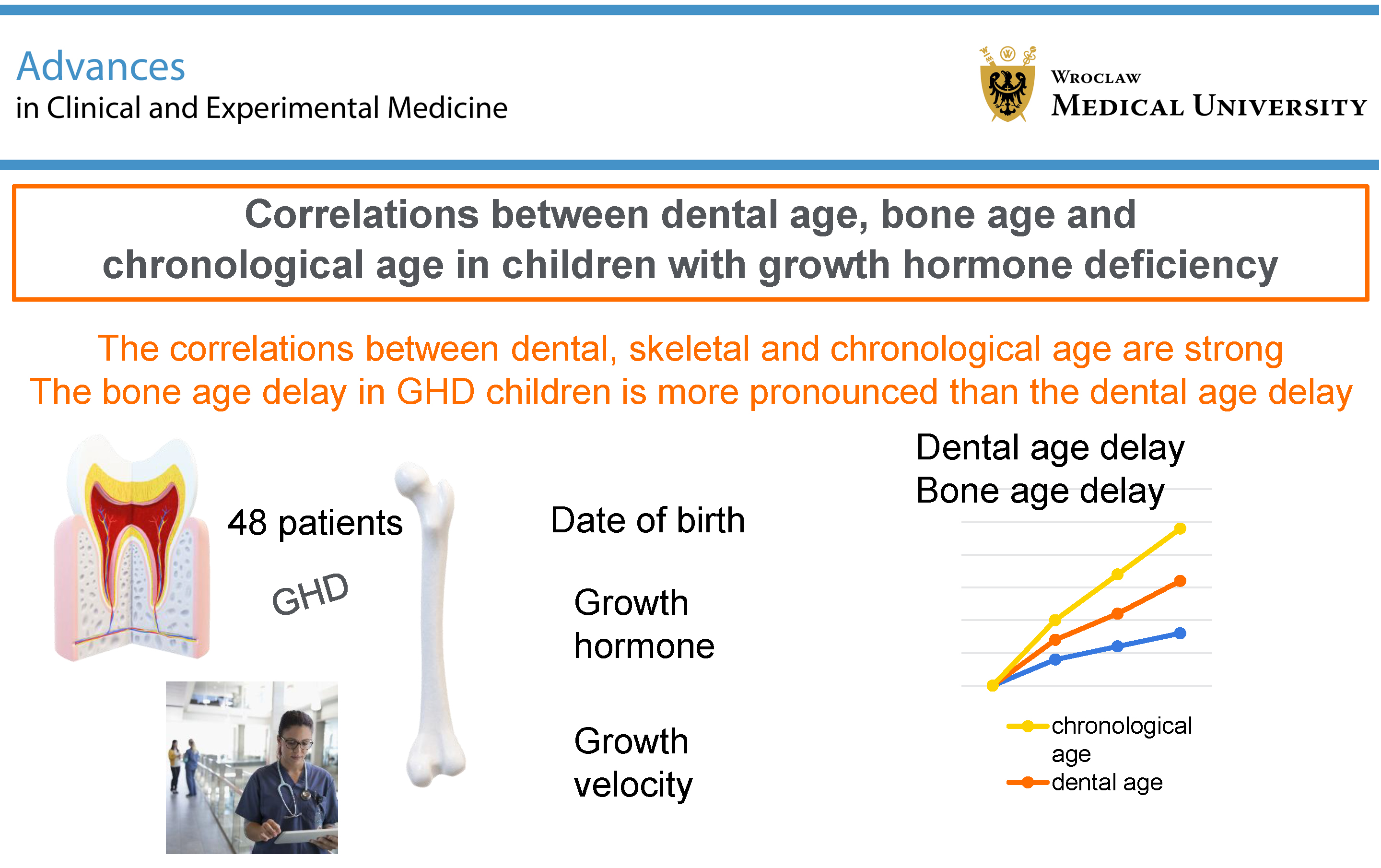
Results. The correlations between dental, skeletal, and chronological age were strong and statistically significant (p < 0.05). No significant correlation was found between the extent of dental age delay and bone age delay. Dental age delay was significantly greater in patients with GHD before treatment than in healthy children (−0.31 vs 0.41, p = 0.024). Bone age delay in children with GHD was more pronounced than dental age delay; it was 1.99 years before treatment and 1.32 years during treatment.
Conclusions. Our results revealed significant differences between bone age and chronological age, as well as between bone age and dental age, in children with GHD, and these differences were reduced during treatment. Significant correlations between bone, dental, and chronological ages indicate that as chronological age increases, dental age and bone age also increase in a predictable manner. Dental and skeletal age delays in patients with GHD are not synchronized, and clinicians should therefore be aware of these discrepancies, particularly when planning dental interventions.
Key words: growth hormone deficiency, bone age, dental age, chronological age, child development
Background
The growth of children is multifactorial and influenced by a combination of disease, nutrition, and environmental conditions.1 Reliable growth indicators that can be easily compared are essential for guiding therapeutic interventions. Bone age and dental age serve as key metrics used to assess the biological maturity and developmental stage of a child. Factors such as malnutrition,2 chronic diseases, and genetic syndromes can adversely affect biological age, particularly dental and skeletal maturation, thereby altering growth velocity and the development of the stomatognathic system.3, 4
Dental development, from tooth bud formation to eruption, can be influenced by endocrine disorders and may vary across different regions of the world.5 Interestingly, although systemic diseases may delay physiological growth, dental maturation is generally less delayed than skeletal maturation.3, 6 The relationships among chronological age, bone age, and dental age are crucial for understanding the determinants of growth and development in children.
Studies have shown inconsistent correlations among growth indicators in both healthy children and children with chronic diseases. Some research indicates a positive correlation among skeletal, dental, and chronological age in children.7, 8, 9, 10, 11 Conversely, some authors have reported little correlation among biological age markers in children.12, 13, 14 Certain congenital diseases, such as cleidocranial dysplasia, Silver–Russell syndrome, and Williams syndrome, can disrupt typical patterns of growth and development, and the correlation among biological age indicators may decrease due to variability in how these diseases affect different individuals.15, 16, 17
Among young patients diagnosed with isolated growth hormone deficiency (GHD), evaluating discrepancies among dental, skeletal, and chronological ages, as well as correlations among these growth indicators, can provide valuable insights into the extent of developmental delays across various organs and the effectiveness of therapeutic interventions. Understanding these relationships helps in tailoring treatments to address specific developmental issues associated with GHD.3
Clinical studies involving children with GHD rarely include, in addition to general medical aspects, an evaluation of potential abnormalities in the stomatognathic system.3, 18, 19, 20, 21 Although the clinical manifestations of certain conditions associated with growth disorders have been described in detail in recent years, there is still insufficient information in the medical literature regarding patients with isolated GHD.11, 22 The results of such studies could contribute to providing more comprehensive care for these patients.
Objectives
This study aimed to assess how dental and skeletal age correspond to chronological age in children with GHD. We hypothesized that, in children with isolated GHD, delayed dental age would be positively correlated with delayed bone age, suggesting that both developmental processes are similarly influenced by GHD.
Materials and methods
Research design and studied population
This cross-sectional observational study was conducted in children with GHD aged 6–14 years. Patients were recruited and diagnosed at a tertiary referral endocrine center (Department of Pediatric Diabetes, Auxology and Obesity, Poznan University of Medical Sciences, Poland) between October 2015 and May 2024. The correlation among dental age, skeletal age, and chronological age was the primary objective of the clinical investigation. Based on previous literature and biological rationale, a strong positive correlation (r = 0.60) was anticipated. Therefore, a one-tailed test was considered appropriate. Using an alpha level of 0.05 and a power of 0.80, the minimum required sample size to detect a statistically significant correlation was calculated to be 15 participants. To enhance the generalizability of the findings, we recruited a larger sample, including all eligible participants available during the study period.
Inclusion and exclusion criteria
The inclusion criteria comprised: age between 6 and 14 years; short stature (height ≤ −2 standard deviation scores (SDS) for age and sex); diagnosis of GHD; and the presence of more than 1 erupted permanent tooth or up to 3 erupted permanent second molars. To limit the influence of factors other than pituitary disorders on tooth development, strict exclusion criteria were applied. Patients with any systemic, metabolic, or syndromic diseases, as well as those with malnutrition, were excluded.
For the control group, male and female children were selected from among patients treated at the pediatric dental clinic. Every 3rd generally healthy patient, with a height between the 10th and 90th percentiles according to local Polish growth charts appropriate for age and sex and with no history of any systemic or metabolic disease affecting normal growth, was invited to participate in the study. Patients were matched for sex and age relative to the study group.
Socioeconomic status was not analyzed in detail in our study. However, all included children came from the same geographical region, attended public schools, and had equal access to public healthcare. Therefore, their socioeconomic status can be considered similar.
Medical and dental outcomes
Growth hormone (GH) deficiency diagnosis was based on 2 stimulation tests: glucagon (0.03 mg/kg body weight), insulin (0.1 units/kg body weight), or clonidine (100 mcg/m2). According to the currently applicable Polish national therapeutic program, the GH cutoff was set at 10 ng/mL; therefore, GHD was diagnosed in children whose peak serum GH level did not exceed 10 ng/mL.
Thyroid status was assessed based on thyroid-stimulating hormone (TSH) and free thyroid hormone (free thyroxine – fT4) levels. To assess bone age, hand-wrist radiographs were evaluated using the Greulich and Pyle method. These examinations were performed only in children with GHD during hospital-based diagnostic evaluation.
In both the study and control groups, height measurements (in cm) were performed using a stadiometer (SECA, Hamburg, Germany), with an accuracy of 1 mm. For all children included in the study, the hSDS was calculated in accordance with the guidelines.23
The oral cavity was examined by a pediatric dentist (N.T.-W.) who had previously been calibrated using children not included in the study. A high level of agreement was observed for repeated measurements (kappa value >0.80).
To evaluate dental age, the Matiegka and Lukasova method was selected. Clinical age estimation was necessary due to the lack of panoramic radiographs for all patients.24 For ethical reasons, performing orthopantomograms solely for research purposes was not possible. In the present study, calculation of dental age was based on tooth eruption status – a tooth was considered present if any part of its crown was visible in the oral cavity.25, 26 The eruption status was then converted into dental age using sex-specific conversion tables.
Dental age delay and bone age delay were defined as the differences between dental or bone age and chronological age. For data analysis, the GHD group was divided into 2 subgroups (before and during rhGH (recombinant human growth hormone) therapy) due to the suspected influence of GH treatment on dental and bone age.
The legal guardians of the patients provided written informed consent for participation in the study. The clinical investigation was conducted in accordance with the Declaration of Helsinki, and the protocol was approved by the Bioethical Committee of Poznan University of Medical Sciences, Poland (approvals No. 785/15 and No. 777/23).
Statistical analyses
The groups were matched using propensity score matching, comparing the GHD group with the control group. Propensity scores were calculated as the probability of group membership conditional on age and sex. Using a nearest-neighbor matching algorithm without replacement and with a caliper of 0.01, the most appropriately matched pairs were selected.
The normality of the distribution was tested using the Shapiro–Wilk test, and equality of variances was assessed using Levene’s test (see shared data). Descriptive statistics are presented as percentages for categorical variables, means with standard deviations (SDs) for normally distributed continuous variables, and medians with minimum and maximum values for non-normally distributed continuous variables. Differences between 2 groups were assessed using the paired-samples Student’s t-test or the Wilcoxon signed-rank test. For comparisons involving more than 2 groups, we applied the Friedman’s test with Dunn’s post hoc test or a two-way analysis of variance (two-way ANOVA) with a post hoc least significant difference (LSD) test. This analysis was used to assess the main and interaction effects of group (study vs control) and time point (before vs after hormone administration). Correlations among chronological age, dental age, and bone age in short-statured children and the control group were evaluated using Spearman’s rank correlation coefficient (rho) or Pearson’s correlation coefficient (r). All results were considered statistically significant at p < 0.05. Statistical analyses were conducted using Statistica v. 13.0 (StatSoft Inc., Round Rock, USA), PQStat Software (2024), v. 1.8.6.120 (Poznań, Poland), or MedCalc® Statistical Software v. 22.014 (MedCalc Software Ltd., Ostend, Belgium).
Results
Out of a total of 93 examined patients with short stature, 48 (18 females and 30 males) fulfilled the inclusion criteria. Thus, the present study included 48 patients with GHD, either before or during treatment, and 48 healthy controls. Table 1 presents the clinical data, including means and SDs (or medians and ranges, where appropriate), for all groups. There were more male than female patients. Most patients presented with mixed dentition.
Table 2 presents the results of the two-way ANOVA, LSD post hoc test, and Student’s t-test among the examined groups. Chronological age and dental age did not differ significantly between the control and GHD groups (p = 0.715 and p = 0.784, respectively).
The difference in dental age delay between the study and control groups was statistically significant (p = 0.045). The post hoc LSD test revealed significant differences between the GHD during-treatment group and the control group (−0.48 vs 0.41 years, p = 0.014) and between the GHD before-treatment group and the control group (−0.31 vs 0.41 years, p = 0.024).
For children with GHD before treatment, the dental age delay was −0.31 ±1.25 years, whereas the bone age delay was significantly greater at −1.99 ±0.97 years (p < 0.001). During treatment, the dental age delay was −0.48 ± 0.98 years, and the bone age delay was −1.32 ±1.28 years, with a significant difference between them (p = 0.042).
Table 3 presents a comparison of dental age, chronological age, and bone age in children with GHD and matched controls. For children with GHD before treatment, the median dental age was 10 years (range: 6.50–14.00 years), the chronological age was 10.45 ±2.21 years, and the bone age was 8.47 ±2.52 years. There was a highly significant difference between chronological age and bone age (p < 0.001) and between bone age and dental age (p < 0.001). For children with GHD during treatment, the dental age was 10.27 ±2.31 years, the chronological age was 10.75 ±2.32 years, and the bone age was 9.43 ±2.93 years. There was a significant difference between chronological age and bone age (p = 0.022). For the matched control children, the median dental age was 10.29 years (range: 6.08–14.00 years), and the chronological age was 10.44 ±2.33 years. There was no significant difference between chronological age and dental age (p = 0.167) (Table 3).
Table 4 shows the correlation coefficients between skeletal age, dental age, and chronological age according to sex and treatment status (before treatment vs during therapy). Skeletal age, chronological age, and dental age were significantly correlated in all patients and subgroups. Spearman’s and Pearson’s correlation coefficients were greater than 0.69 in both groups and in both sexes, indicating a strong correlation. Dental age delay and bone age delay were not correlated (Table 4).
Discussion
This manuscript emphasizes the strong correlation among dental, bone, and chronological age in patients with isolated GHD. Interestingly, our results did not confirm the initial hypothesis. Although dental age was significantly delayed in children with GHD compared to healthy peers, we found no significant correlation between the extent of dental age delay and bone age delay. This indicates that, despite both being affected by GHD, the degree of delay in dental and skeletal development may not progress in a synchronized manner in individual patients. This finding suggests that dental and skeletal maturation may be regulated by partially independent biological mechanisms and that dental development is less sensitive to GH levels than bone development. From a clinical perspective, these results emphasize the need to independently assess both dental and skeletal maturity when managing patients with GHD, rather than assuming that one parameter reliably reflects the other.
Interestingly, the clinical implications of discrepancies between dental and bone age remain relatively underexplored. However, recent studies27, 28 have reported delayed tooth eruption in children with GHD, which may be associated with a mismatch between tooth and bone size. Furthermore, the consequences of such discrepancies appear to be supported by a previous study by Torlińska et al., in which greater dental crowding was observed in children with delayed bone age in both idiopathic short stature (ISS) and GHD cohorts.3 It is also worth noting that a relationship exists among skeletal maturation, dental development, and the formation of the permanent dentition.21, 29 Nevertheless, further research is needed to fully elucidate the mechanisms underlying these interrelated processes.
Data on the extent of dental age delay in short-stature patients and the impact of GH replacement therapy on dental development are scarce and frequently conflicting. The incomplete and inconsistent information on this topic prompted us to explore the relationships among maturation indicators in children with GHD.
In the study by Torlińska et al., dental age delay, bone age delay, and occlusal traits were assessed in a cohort of short-stature children with GHD and ISS. The findings indicated significant dental age delay in children with GHD before treatment compared to healthy peers, as well as a more pronounced bone age delay.3 To expand on these findings, we decided to recruit a larger sample of participants with isolated GHD and analyze correlations within specific subgroups to gain deeper insights into the developmental delays associated with GHD. Dental age assessment is essential in the daily practice of dentists and orthodontists. Planning conservative treatment and potential tooth extractions as part of an orthodontic treatment plan must be based not only on a dental examination but also on comprehensive knowledge of the patient’s general health and growth patterns associated with the underlying condition.
Skeletal maturation occurs in consistent and defined stages across different ethnic groups, although the timing may vary due to genetic, environmental, regional, and climatic factors.30 For this reason, all participants in our study were from the same geographical region and ethnic group. Skeletal maturation is commonly assessed based on the stages of skeletal development observed in hand-wrist radiographs.30 Compared with skeletal maturation, tooth mineralization and eruption are less affected by endocrine, environmental, and nutritional factors. This makes them very useful for age estimation,31 which can be based on the stage of development of dental germs and the progressive sequence of their eruption in the oral cavity.2, 32 Nevertheless, it should be noted that dental development and eruption can vary due to factors such as climate, nutrition, race, and genetic inheritance.5, 32 The advantage of methods based on tooth mineralization is that they can be applied at any stage of dental development. In contrast, clinical dental age cannot be determined once all permanent teeth have erupted. In this study, because panoramic radiographs were not available, we based our analysis on clinical examination, which allows for a quick and non-invasive assessment.
Only a few researchers have evaluated the biological age of individuals with hypopituitarism or impaired growth velocity. Valejo-Bolaños et al. investigated the relationships among dental, bone, and chronological age in children with isolated GHD. They reported a significant difference of 1.52 years between chronological age and bone age and 0.92 years between chronological age and dental age. The study concluded that both dental and bone age were significantly delayed compared to chronological age after 2.5 years of GH therapy.33
Our current results confirm the presence of significant differences between skeletal age and chronological age in young patients with GHD. The delay in bone age in the study group was as much as 1.99 years, but this delay was reduced during treatment. In contrast, we did not observe similarly large differences between the dental and chronological ages of the patients studied. A statistically significant difference indicating a slight delay in dental age in patients with GHD was observed when they were compared with an age- and sex-matched control group.
In the previous study by Torlińska et al., dental, skeletal, and chronological age in children with GHD and ISS was examined. The clinical investigation was carried out in 46 children (aged 5–14 years): 15 with ISS, 17 with GHD before GH replacement therapy, and 14 with GHD during therapy. The study confirmed dental age delay in children with GHD before GH therapy. Both children with ISS and untreated GHD showed marked bone age delay.3 In the present study, which focused solely on patients with GHD, these findings were confirmed in a larger cohort.
More extensive analyses have been conducted in syndromic patients with short stature. Pinchi et al. carried out research to understand the influence of genetic syndromes (Down syndrome and Williams syndrome) on dental maturation. The study sample included 159 children with chromosomal abnormalities and a control group of 157 healthy children aged 4.49–19.8 years. The results revealed no statistically significant differences in dental age estimates between syndromic and healthy individuals or between sexes and age cohorts.1
Diz et al. examined 155 patients with cerebral palsy, intellectual disability, and Down syndrome. The control group comprised 688 children. No significant differences between dental age and chronological age were found in boys with the studied disorders. However, dental age was significantly delayed compared to chronological age in girls with cerebral palsy or Down syndrome.6
This finding was confirmed by de Moraes et al., who observed that the mean dental age was slightly lower than the chronological age in children with Down syndrome, with a significant difference observed only in females.32
Another study by Pozsonyi et al. found delayed bone development up to the age of 8 years in children with Down syndrome compared with healthy controls.34 De Moraes et al. reported that the skeletal age of subjects with Down syndrome aged 6–16 years was significantly delayed compared to their chronological age, particularly at the age of 7 years.35
The delay in dental age among patients with Williams syndrome is not uniform, reflecting the syndrome’s broad range of dental manifestations. Some individuals exhibit eruption delays of approx. 1–2 years,36 whereas others may have more typical eruption timelines.16, 36 Regarding cleidocranial dysplasia, patients present with a range of dental abnormalities, such as delayed exfoliation of primary teeth, delayed eruption of permanent teeth by up to several years, and often the presence of supernumerary teeth. Not all patients experience the same degree of dental age delay. This variability can be attributed to genetic differences, phenotypic expression, the presence and number of supernumerary teeth, and differences in skeletal development.15
Children with Silver–Russell syndrome may exhibit delayed dental age. However, some may have relatively minor delays, whereas others exhibit more significant delays. This variability can be influenced by the extent of growth restriction and the presence of other craniofacial abnormalities. Bergman et al. reported that the dental maturity of children with Silver–Russell syndrome was within normal limits, whereas the timing of tooth eruption was delayed by approx. 1 year.16 Ioannidou-Marathiotou et al. reported an atypical pattern of tooth eruption in a girl with Silver–Russell syndrome who, at the age of 10 years, was in the early mixed dentition stage, with normal eruption of permanent upper and lower incisors only.37 These findings highlight the considerable research attention given to syndromic forms of short stature. In contrast, studies involving children with isolated GHD remain limited. The present study aims to address this gap in the literature.
Notably, even if dental development occurs later than usual, there can still be a high correlation between dental age and chronological age if the delay is consistent across the population. This means that although dental age or skeletal age might be lower than expected for a given chronological age, the relationship between the 2 ages can remain strong and predictable. For orthodontists and pediatric dentists, understanding the mismatch among dental, skeletal, and chronological age is crucial for treatment planning. Treatments may need to be adjusted based on dental and skeletal maturity rather than the patient’s chronological age.
Some research has been conducted on the correlation among indicators of biological age in children from different geographical regions. In a study by Bala et al.,2 160 healthy North Indian children aged 8–14 years, equally divided by sex, were examined. The study revealed that skeletal age and dental age were not strongly correlated with chronological age across all age groups. However, there was a significant correlation between skeletal age and dental age in children aged 12–14 years of both sexes. Conversely, another Indian study reported a very strong correlation among chronological, skeletal, and dental age.7
Poulsen et al.5 reported significant correlations between dental and skeletal maturation in healthy children with a mean age of 11.48 years. In the Swedish population, a study revealed strong correlations between wisdom tooth development and chronological age, skeletal age, and dental age.10 Similarly, significant correlations were found between mandibular third molar mineralization and chronological age in Croatian subjects.9
A study of Yugoslavian children with cerebral palsy revealed varying degrees of delay in dental and skeletal age compared with chronological age, highlighting poor correlation in some cases due to the diverse impact of the condition on development.38
In the study by de Moraes et al.,32 Pearson’s correlation coefficient for dental age compared to skeletal age was greater than 0.9 in both the Down syndrome group and the healthy control group, regardless of sex. In the present study, strong correlations of 0.69 or greater were found among dental age, chronological age, and bone age in Caucasian children with GHD. This means that as chronological age increases, dental age and bone age also increase in a predictable manner.
Limitations of the study
Our study has several limitations. First, the method used for dental age estimation is clinically acceptable but not widely adopted, making it unclear to what extent the results are applicable to other populations, as tooth eruption patterns may vary among ethnic groups. Future studies should include dental age assessment based on radiographic images in this patient population. Second, the sample size, particularly for patients with GHD during treatment, was relatively small. Third, we did not account for variables such as socioeconomic background or physical activity, which could influence both dental and skeletal development. Despite these limitations, this study contributes to understanding the developmental patterns of children with GHD. By highlighting discrepancies among different indicators of biological age, it provides valuable insights into how GHD affects physical development. These findings may help clinicians better monitor and manage the growth and development of children with GHD, particularly with regard to treatment planning.
Conclusions
This study underscores the importance of considering multiple indicators of biological age in the management of children with GHD. Our results confirm the presence of significant differences between bone age and chronological age, as well as between bone age and dental age, in children with GHD. These differences were reduced during treatment. Significant correlations among bone age, dental age, and chronological age indicate that, as chronological age increases, dental age and bone age also increase in a predictable manner. Although delays in both dental age and bone age are observed in children with isolated GHD, the expected correlation between these delays was not confirmed. This suggests that dental and skeletal development may not always progress in parallel in patients with GHD. Therefore, when assessing growth and planning orthodontic or medical interventions, clinicians should consider that dental age and bone age may provide complementary but independent information.
Data Availability Statement
The datasets supporting the current study are openly available in Zenodo at https://doi.org/10.5281/zenodo.1
5065743.
Consent for publication of personal information
Not applicable
Use of AI and AI-assisted technology
Not applicable