Abstract
Background. Cardiac hypertrophy refers to the compensatory response of the heart to various physiological or pathological stimuli in order to maintain its function. Let-7e-5p has been reported to be upregulated in patients with cardiac hypertrophy.
Objectives. This study aimed to investigate the role of let-7e-5p in the progression of cardiac hypertrophy using animal and cell models.
Materials and methods. Peripheral blood was collected from patients with cardiac hypertrophy and healthy controls to detect let-7e-5p expression via reverse transcription quantitative polymerase chain reaction (RT-qPCR). An aortic banding (AB)-induced cardiac hypertrophy rat model was established to explore let-7e-5p expression in vivo. Antagomir let-7e-5p was used to investigate the effects of let-7e-5p silencing on cardiac hypertrophy progression in an AB-induced rat model. Angiotensin II (Ang II) was used to induce hypertrophy in H9c2 rat cardiomyocytes in vitro. The downstream regulatory mechanism of let-7e-5p was investigated in H9c2 cells.
Results. We first examined and verified that let-7e-5p was upregulated in blood samples from patients with hypertrophic cardiomyopathy (HCM) compared to healthy controls and showed good diagnostic performance according to receiver operating characteristic (ROC) analysis. In vivo, let-7e-5p was overexpressed in the heart tissues of AB-induced cardiac hypertrophy rats. Let-7e-5p deficiency alleviated AB surgery-induced cardiac hypertrophy in rat models. In vitro, let-7e-5p expression was higher in Ang II-induced H9c2 rat cardiomyocytes. Let-7e-5p inhibition reversed the Ang II-induced increase in cardiomyocyte size and the upregulation of ANP, BNP, and β-MHC expression, whereas let-7e-5p overexpression showed the opposite effects. Mechanistically, limb-bud and heart (LBH) was identified as a target of let-7e-5p, and LBH overexpression reversed the promotive effects of let-7e-5p on hypertrophy in Ang II-treated H9c2 cells. IGF–PI3K–AKT signaling was activated in Ang II-treated H9c2 cells, and let-7e-5p silencing suppressed its activation by targeting LBH. The IGF1R inhibitor PQ401 reversed the enhancement of H9c2 hypertrophy induced by let-7e-5p upregulation or LBH silencing.
Conclusions. Let-7e-5p promotes cardiac hypertrophy by targeting LBH and regulating the IGF–PI3K–AKT signaling pathway, which may provide novel insights into targeted therapy.
Key words: Let-7e-5p, cardiac hypertrophy, LBH protein, IGF–PI3K–AKT signaling pathway, microRNA regulation
Background
Cardiac hypertrophy refers to the compensatory response of the heart to various physiological or pathological stimuli in order to maintain its function. This process is accompanied by an increase in cardiomyocyte size, changes in cardiomyocyte morphology, modifications in gene expression, and cytoskeletal remodeling.1 Pathological cardiac hypertrophy can be induced by various factors, such as hypertension, pressure or volume overload, drug toxicity, and others, leading to the development of heart failure and affecting the clinical outcomes and quality of life of patients.2, 3 Currently, classical drugs, including β-adrenergic receptor blockers and renin–angiotensin–aldosterone system inhibitors, are commonly used for the treatment of hypertrophic cardiomyopathy (HCM). However, their therapeutic effects remain limited due to the high incidence of heart failure.4, 5 Thus, the exploration of novel therapeutic targets is needed to develop effective treatment strategies.
MicroRNAs (miRNAs) are endogenous small non-coding RNAs (~22 nucleotides) that regulate the expression of protein-coding genes at the post-transcriptional level.6 MiRNAs also serve as important regulators of biological processes such as cell proliferation, differentiation, and survival. Substantial evidence has revealed that dysregulated miRNAs may affect the development of diverse cardiovascular diseases, including pathological cardiac hypertrophy.7 For example, miR-30d has been reported to be downregulated in cardiac hypertrophy both in vivo and in vitro, and its overexpression attenuates pathological cardiomyocyte hypertrophy in vitro and in vivo.8 MiR-27b-3p is highly expressed during the process of cardiac hypertrophy, and its depletion significantly ameliorates cardiac fibrosis, inflammation, and hypertrophy in TAC-induced mice by targeting FGF1.9 MiR-214 is highly expressed in the myocardial tissue of Ang II-stimulated mice. Its overexpression has been shown to downregulate SIRT3, thereby inducing mitochondrial dysfunction and contributing to cardiac hypertrophy.10 Let-7e-5p has previously been reported to be highly expressed in the blood of patients with congestive heart failure, suggesting its role in heart disease.11 Bioinformatics analyses have also revealed that let-7e-5p is a biologically meaningful miRNA involved in the regulation of molecular mechanisms underlying cardiac hypertrophy.12 Additionally, a previous study demonstrated that let-7e-5p is upregulated in isoproterenol (ISO)-induced cardiac hypertrophy in rats. However, the function and precise mechanisms of let-7e-5p in cardiac hypertrophy remain unclear.
Limb-bud and heart (LBH) is a protein-coding gene involved in embryonic growth and heart development.13 Aberrant expression of LBH is closely linked to the progression of a variety of cardiovascular diseases. For example, LBH is upregulated in response to transforming growth factor (TGF)-β1 stimulation in cardiac fibroblasts, and ectopic expression of LBH promotes the activation of cardiac fibroblasts under hypoxic conditions, suggesting its potential as a target for cardiac repair and antifibrotic therapy.14, 15 Additionally, LBH has also been suggested to protect against myocardial ischemia/reperfusion (I/R) injury by reducing cardiomyocyte apoptosis and ferroptosis. Bioinformatics analysis suggests that LBH is a potential target of let-7e-5p, and LBH has been shown to be downregulated in the heart tissue of patients with HCM based on the GSE89714 dataset from the Gene Expression Omnibus (GEO) database (https://www.ncbi.nlm.nih.gov/geo/query/acc.cgi?acc=GSE89714). The regulatory mechanism between let-7e-5p and LBH still requires further investigation.
Objectives
The current study aimed to validate the expression of let-7e-5p in blood samples from human patients and to explore the potential function and downstream mechanisms of let-7e-5p in cardiac hypertrophy using aortic banding (AB)-induced rat models in vivo and angiotensin II (Ang II)-induced rat cardiomyocytes (H9c2) in vitro. We hypothesized that let-7e-5p exerts detrimental effects on cardiac hypertrophy by targeting LBH.
Material and methods
Animals
Sprague Dawley rats (200 g; Vital River, Beijing, China) were housed at 22 ±3°C under a 12 h/12 h light/dark cycle. Aortic banding surgery was performed to induce cardiac hypertrophy in rats as previously described.16 Briefly, rats were anesthetized with pentobarbital sodium (50 mg/kg; MilliporeSigma, St. Louis, USA) via intraperitoneal injection. Next, the aorta was exposed by opening the left thoracic intercostal space between the 2nd and 3rd ribs and then ligated with a 7-0 silk suture and a 26- or 27-gauge needle. After gently withdrawing the needle, the aorta was constricted. Rats in the sham group underwent the same surgical procedure, but no aortic ligation was performed. To explore the effects of let-7e-5p on cardiac hypertrophy in vivo, chemically modified antisense oligonucleotides (antagomirs) targeting let-7e-5p and the negative control (NC) were synthesized by GenePharma (Shanghai, China). Rats in the model group were injected daily with antagomir let-7e-5p or antagomir NC via the tail vein for 3 days beginning 24 h after surgery. The body weight of the rats was measured throughout the experiment. At the end of the experiment, rats were euthanized by inhalation of 5% isoflurane, and their hearts and tibias were collected and measured. The ratios of heart weight to body weight (HW/BW) and heart weight to tibia length (HW/TL) were calculated. Ventricular tissue was harvested and either frozen at −80°C or embedded in paraffin.
All procedures were approved by the Ethics Committee of Shanghai Tenth People’s Hospital, Tongji University (Shanghai, China; approval No. 2023-0721L issued on July 21, 2023) and were conducted in strict accordance with the recommendations of the National Institutes of Health (NIH) Guide for the Care and Use of Laboratory Animals. The study is reported in accordance with the ARRIVE (Animal Research: Reporting of In Vivo Experiments) guidelines.17
Echocardiography and histological analyses
Echocardiography was performed in rats as previously described.18 After light general anesthesia, left ventricular (LV) functional parameters were monitored and recorded. Fractional shortening (FS%) and ejection fraction (EF%) were subsequently calculated.
Paraffin-embedded heart tissues from each group of rats were subjected to hematoxylin and eosin (H&E) staining using a Hematoxylin and Eosin Staining Kit (Beyotime, Shanghai, China) according to the manufacturer’s instructions. Image-Pro Plus software (Media Cybernetics, Bethesda, USA) was used to analyze cardiomyocyte size.
Immunohistochemistry
Paraffin-embedded rat heart tissues were sectioned, dewaxed in xylene, and rehydrated through graded alcohol solutions. After antigen retrieval, the sections were rinsed with phosphate-buffered saline (PBS), treated with 3% H2O2 solution, and then incubated with primary antibodies against atrial natriuretic peptide (ANP; PA5-29559, 1:200, Thermo Fisher Scientific, Waltham, USA), B-type natriuretic peptide (BNP; PA5-96084, 1:200; Thermo Fisher Scientific), and beta-myosin heavy chain (β-MHC; PA5-100023, 1:200, Thermo Fisher Scientific) at 37°C for 2 h. The sections were then rinsed 3 times with PBS and incubated with secondary antibodies for 60 min at room temperature. After staining with diaminobenzidine (DAB) and hematoxylin, the sections were observed and imaged under a BX51 light microscope (Olympus Corp., Tokyo, Japan).
Clinical samples collection
A total of 30 patients with HCM and 30 healthy controls matched for age and sex were enrolled in this study. The inclusion criteria were as follows: 1) age between 18 and 75 years; 2) diagnosis confirmed by echocardiography, electrocardiography, and magnetic resonance imaging (MRI) by 2 cardiologists, with left ventricular maximal wall thickness (LVMWT) >15 mm; and 3) provision of written informed consent and willingness to participate in the study. The exclusion criteria were as follows: 1) diabetes mellitus, hyperlipidemia, infectious diseases, cancer, or other severe medical conditions; 2) pregnancy; and 3) a history of transplantation. Venous blood was collected at enrollment and centrifuged at 2,000 × g for 10 min to obtain serum.
All participants signed written informed consent before enrollment. The study was conducted in full accordance with the principles outlined in the Declaration of Helsinki (2013 revision).
Cell culture and treatment
Rat cardiomyocytes (H9c2) were obtained from Procell (Wuhan, China). Cells were cultured in Dulbecco’s modified Eagle’s medium (DMEM) containing 10% fetal bovine serum (FBS; Gibco, Waltham, USA) and 1% penicillin/streptomycin (P/S; MilliporeSigma) at 37°C in a humidified atmosphere containing 5% CO2. To induce cardiomyocyte hypertrophy in vitro, H9c2 cells were exposed to 1 μM Ang II (MilliporeSigma) for 48 h.
Cell transfection
The let-7e-5p inhibitor and mimics, NC inhibitor, and NC mimics were synthesized by RiboBio Co. (Guangzhou, China). For LBH overexpression or knockdown, pcDNA3.1/LBH (oe-LBH), sh-LBH, and the corresponding empty control vectors (oe-NC and sh-NC) were provided by GenePharma. H9c2 cells were transfected with these vectors using Lipofectamine 3000 reagent (Invitrogen, Waltham, USA) for 48 h.
Reverse transcription quantitative PCR
Total RNA was isolated from human blood samples and rat heart tissues using TRIzol Reagent (Thermo Fisher Scientific). RNA was then reverse-transcribed using the TaqMan miRNA Reverse Transcription Kit for miRNAs and an appropriate cDNA synthesis kit for mRNAs. Reverse transcription quantitative polymerase chain reaction (RT-qPCR) was performed using a SYBR Green I Master Mix kit (Invitrogen) on a 7500 Real-Time PCR System (Applied Biosystems, Waltham, USA). Relative gene expression levels were calculated using the 2−ΔΔCt method, with U6 serving as the internal control for miRNAs and GAPDH serving as the internal control for mRNAs.
Western blot
Total protein was extracted from H9c2 cardiomyocytes and rat cardiac tissues using radioimmunoprecipitation assay (RIPA) buffer (Epizyme, Shanghai, China), followed by determination of protein concentration using a bicinchoninic acid (BCA) assay kit. Sodium dodecyl sulfate–polyacrylamide gel electrophoresis (SDS-PAGE) was then used for protein separation, and the proteins were subsequently transferred onto polyvinylidene difluoride (PVDF) membranes (MilliporeSigma). After incubation with primary antibodies (Thermo Fisher Scientific) overnight at 4°C, the membranes were incubated with a secondary antibody (31460; 1:10,000; Thermo Fisher Scientific) at room temperature for 60 min. Enhanced chemiluminescence (ECL) was then used for protein band visualization, and ImageJ software v. 6.1.0 (National Institutes of Health (NIH), Bethesda, USA) was used for data analysis.
Immunofluorescence
Immunofluorescence staining was used to evaluate the size of H9c2 cardiomyocytes. Briefly, H9c2 cells were collected 48 h after the indicated transfection and then stimulated with Ang II. After fixation with formaldehyde and permeabilization with Triton X-100 (0.1%; Beyotime), the cells were incubated with anti-α-actinin antibody (ab137346; 1:1,000; Abcam, Cambridge, UK) at 4°C overnight and then incubated with the secondary antibody (ab150077; 1:1,000; Abcam) the following day. Cell nuclei were stained with 4′,6-diamidino-2-phenylindole (DAPI). Images were captured using a fluorescence microscope (Olympus IX73; Olympus Corp.). The cell surface area was quantified using ImageJ software (NIH).
Luciferase reporter assays
The wild-type (WT) or mutant (MUT) LBH 3′UTR sequences containing the let-7e-5p binding site were cloned into the pmirGLO reporter vector, generating pmirGLO-LBH-WT and pmirGLO-LBH-MUT constructs. H9c2 cardiomyocytes were co-transfected with the NC inhibitor or let-7e-5p inhibitor together with pmirGLO-LBH-WT or pmirGLO-LBH-MUT using Lipofectamine 3000 (Invitrogen). After 48 h, luciferase activity was measured using a dual-luciferase reporter assay system (Promega, Madison, USA) according to the manufacturer’s protocol.
RNA pulldown assay
Biotinylated let-7e-5p provided by RiboBio was incubated with cardiomyocytes for 48 h. Afterward, H9c2 cardiomyocytes were harvested and lysed, followed by incubation with beads (MilliporeSigma). After washing, the bound RNA was purified, and RT-qPCR was performed to analyze the enrichment of candidate mRNAs.
Statistical analyses
Results were analyzed using SPSS v. 17.0 (SPSS Inc., Chicago, USA) and are presented as mean ± standard deviation (SD). Each experiment was independently performed at least 3 times, with at least 3 biological samples included in each assay. Normality was evaluated using the Shapiro–Wilk test, with p > 0.05 indicating a normal distribution. Homogeneity of variance was assessed using the F test for comparisons between 2 groups and the Brown–Forsythe test or Levene’s test for comparisons among 3 or more groups.
If the data conformed to a normal distribution and exhibited homogeneous variance, comparisons between 2 groups or among multiple groups were performed using a two-tailed unpaired Student’s t-test or one-way analysis of variance (ANOVA) followed by Tukey’s post hoc test. Two-way ANOVA followed by Bonferroni’s post hoc test was used for comparisons involving 2 independent variables. If the data did not conform to a normal distribution or exhibited heterogeneous variance, the Mann–Whitney U test or Welch’s t-test was applied for comparisons between 2 groups, and the Kruskal–Wallis test followed by Dunn’s post hoc test was applied for comparisons among 3 or more groups. Receiver operating characteristic (ROC) curves were used to assess the diagnostic performance of let-7e-5p in cardiac hypertrophy. A p < 0.05 was considered statistically significant.
Results
Let-7e-5p aggravates aortic banding-induced cardiac hypertrophy in rats
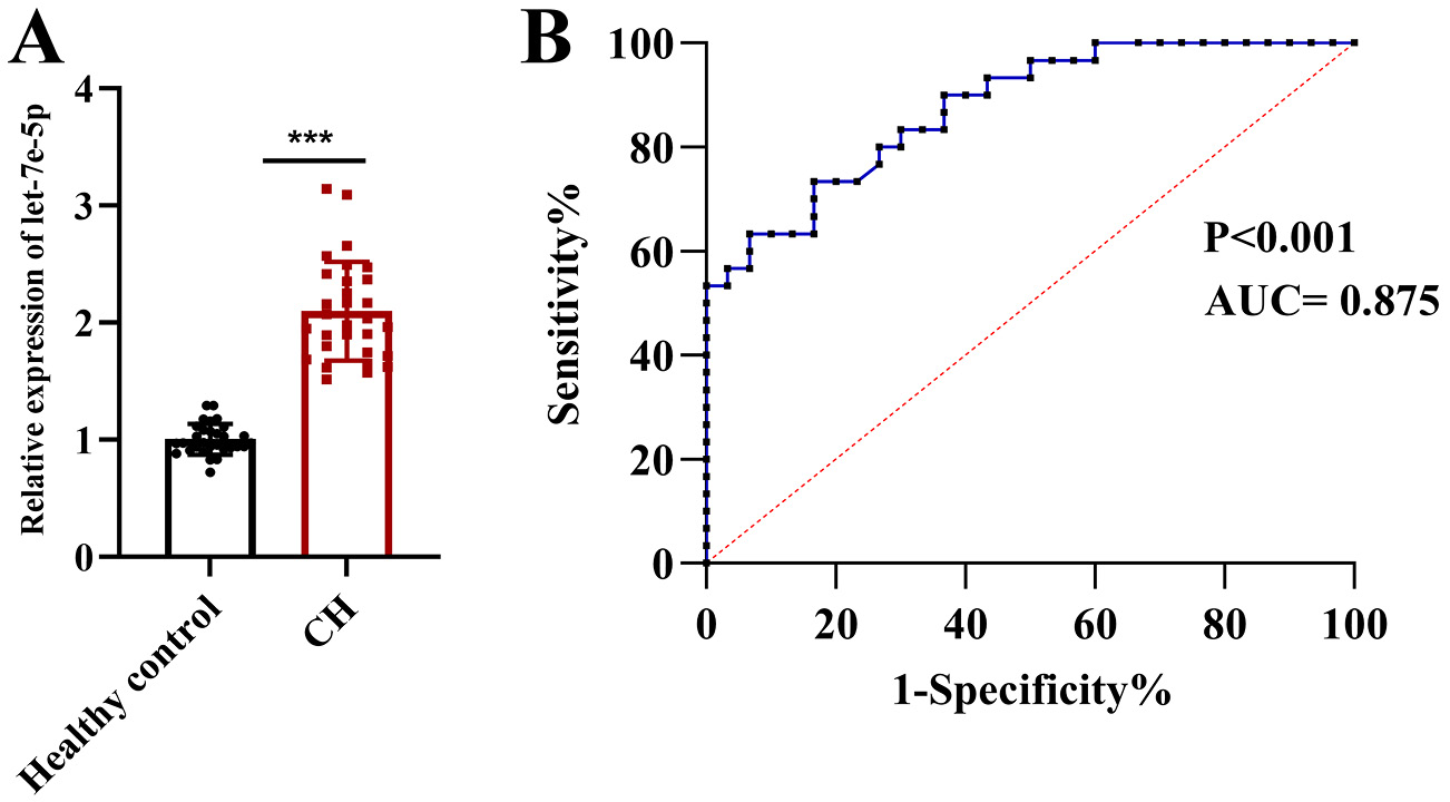
The let-7e-5p level in patients with HCM (n = 30) and healthy control individuals (n = 30) was detected using RT-qPCR. It was found to be dramatically upregulated in cardiac hypertrophy (Figure 1A). As shown by the ROC curve, let-7e-5p may serve as a robust biomarker for the diagnosis of cardiac hypertrophy, with an area under the curve (AUC) of 0.875 (p < 0.001; 95% confidence interval (95% CI): 0.791–0.959) (Figure 1B). Overall, let-7e-5p is aberrantly overexpressed in cardiac hypertrophy and may have high predictive value for the diagnosis of cardiac hypertrophy.
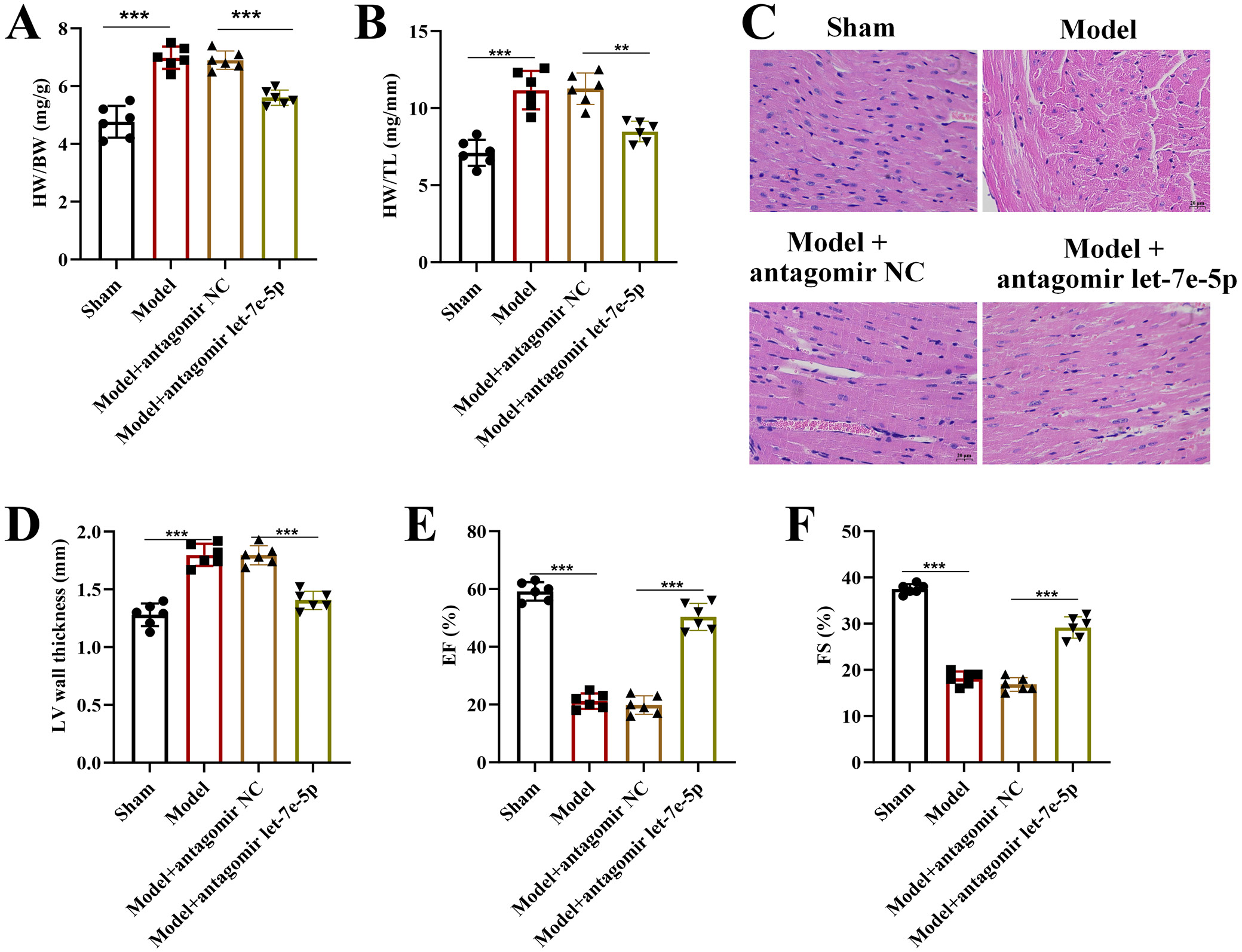
Aortic banding surgery was performed in rats to induce cardiac hypertrophy. We found that let-7e-5p was upregulated in the model group and was successfully silenced by the administration of antagomir let-7e-5p (Supplementary Fig. 1A). Next, we measured heart weight (HW), body weight (BW), and tibial length (TL) in each group of rats. We found that the hypertrophy indices HW/BW and HW/TL were both elevated in the model group and were reversed by antagomir let-7e-5p treatment, indicating that its deficiency significantly attenuated pathological cardiac enlargement in rats (Figure 2A,B).
Hematoxylin and eosin staining showed a significant increase in heart size in the model group, accompanied by histological changes such as tissue damage and fibrosis, whereas antagomir let-7e-5p treatment reduced heart size, LV wall thickness, and the pathological changes induced by AB surgery (Figure 2C,D). Echocardiographic analysis showed that the EF% and FS% of rats in the model group were both decreased relative to the sham group, whereas administration of antagomir let-7e-5p significantly restored EF% and FS% levels (Figure 2E,F).
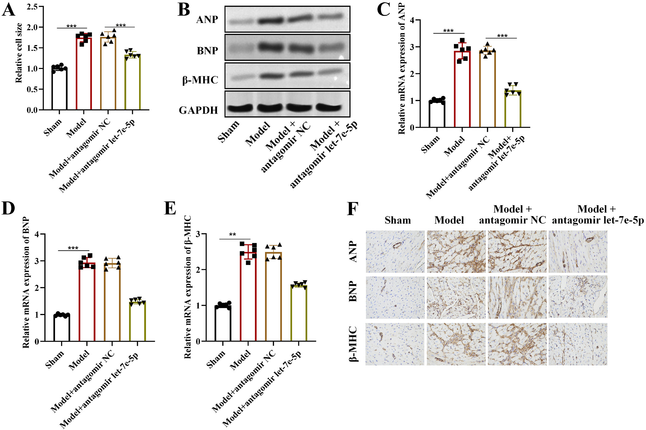
Similarly, we observed an increase in cardiomyocyte size in response to AB surgery, and antagomir let-7e-5p significantly inhibited cardiomyocyte enlargement in the model group (Figure 3A). Furthermore, we detected the expression levels of cardiac hypertrophy markers in rat hearts. According to western blot analysis, the elevated expression of ANP, BNP, and β-MHC proteins observed in the model group was markedly reduced by antagomir let-7e-5p treatment (Figure 3B). RT-qPCR results also indicated that the increased mRNA levels of ANP, BNP, and β-MHC induced by AB surgery were restored by administration of antagomir let-7e-5p in rat hearts (Figure 3C–E). Consistently, immunohistochemical analysis showed enhanced staining intensity of ANP, BNP, and β-MHC in rat hearts from the model group, which was reduced by antagomir let-7e-5p treatment (Figure 3F).
Let-7e-5p promotes Ang II-stimulated cardiomyocyte hypertrophy
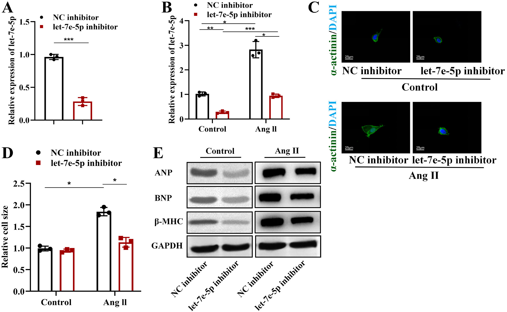
Rat cardiomyocytes (H9c2) were treated with Ang II to induce hypertrophy in vitro. The transfection efficiency of the let-7e-5p inhibitor or mimics was confirmed using RT-qPCR (Figure 4A). The let-7e-5p level was increased in Ang II-treated H9c2 cells and was restored by transfection with the let-7e-5p inhibitor (Figure 4B). According to the immunofluorescence staining results, the increase in cardiomyocyte size induced by Ang II treatment was significantly reversed by let-7e-5p inhibition (Figure 4C,D). Meanwhile, we demonstrated that the protein levels of ANP, BNP, and β-MHC, which were upregulated by Ang II treatment, were reversed by let-7e-5p silencing in H9c2 cells (Figure 4E).
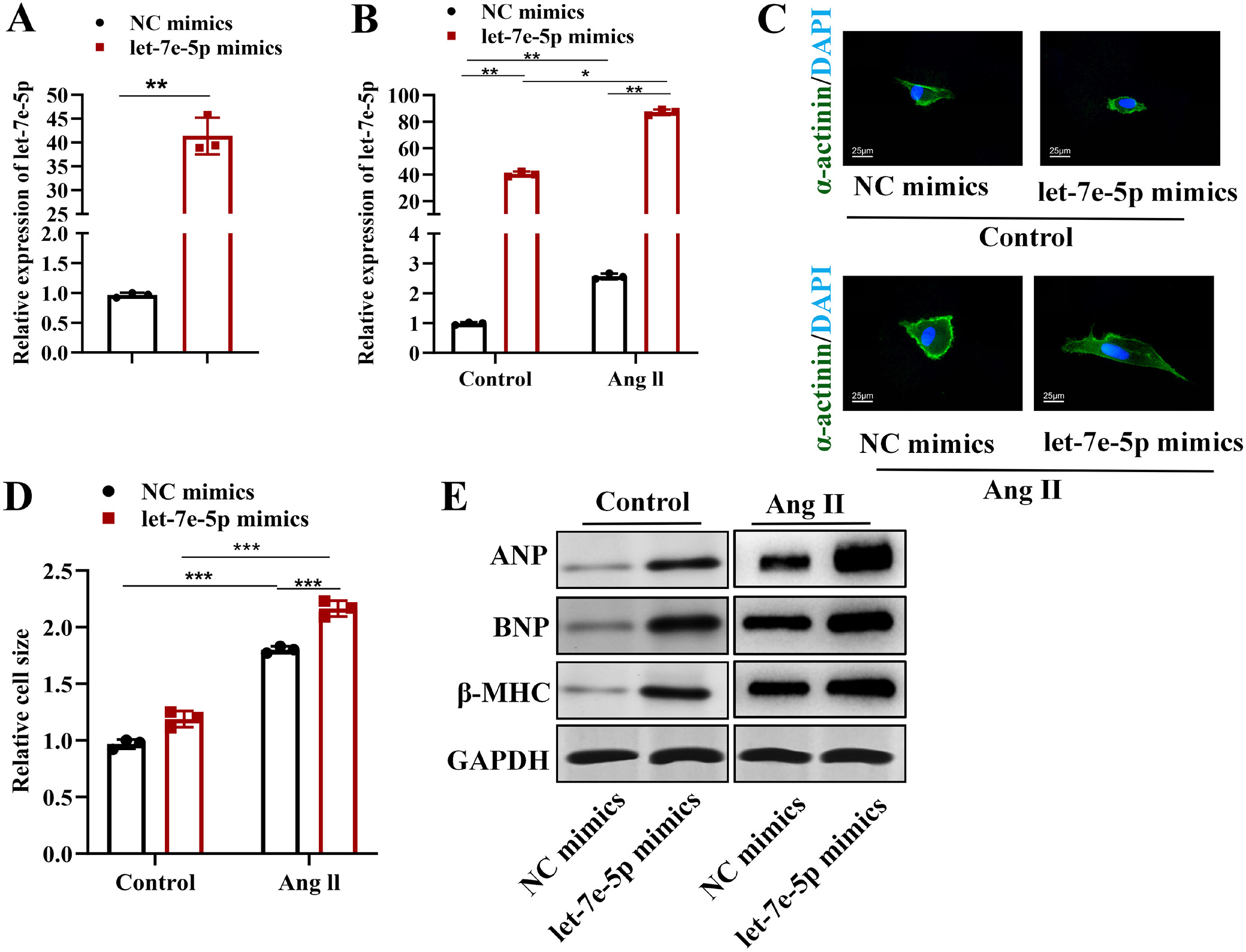
Moreover, we explored the effects of let-7e-5p overexpression on Ang II-stimulated cardiomyocytes to further confirm the role of let-7e-5p. The transfection efficiency of let-7e-5p mimics was verified with RT-qPCR (Figure 5A). We found that transfection with let-7e-5p mimics significantly increased let-7e-5p levels in H9c2 cells in the presence or absence of Ang II exposure (Figure 5B). Immunofluorescence staining revealed that the increase in cardiomyocyte size induced by Ang II treatment was further enhanced by let-7e-5p upregulation (Figure 5C,D). Consistently, the protein levels of ANP, BNP, and β-MHC, which were upregulated by Ang II, were further enhanced by let-7e-5p overexpression (Figure 5E). Overall, let-7e-5p enhances Ang II-stimulated cardiomyocyte hypertrophy.
LBH is targeted by Let-7e-5p in H9c2 cardiomyocytes
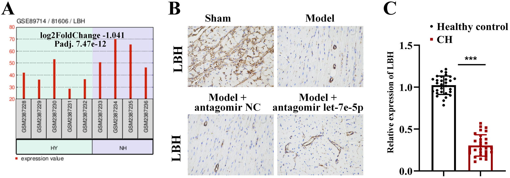
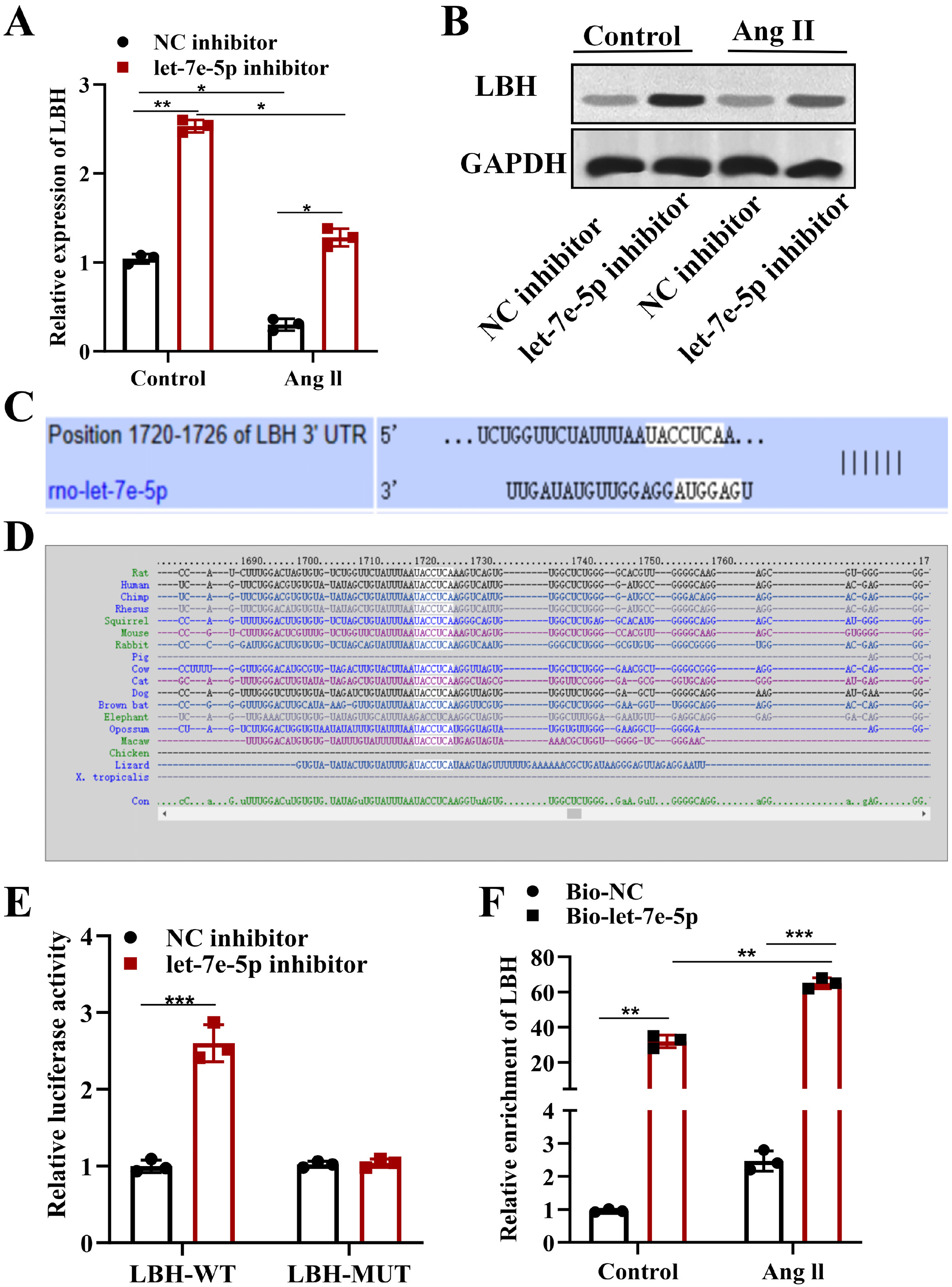
The downstream targets of rno-let-7e-5p were explored using the TargetScan database (https://www.targetscan.org/vert_80). A total of 2,022 genes were predicted to contain potential binding sites for let-7e-5p. We then explored dysregulated genes in cardiac hypertrophy based on analysis of the GSE89714 dataset from the GEO database, and 311 downregulated genes were identified. Next, we intersected the downregulated genes identified in cardiac hypertrophy with the predicted target genes of let-7e-5p, yielding 21 genes for further analysis (Supplementary Fig. 2A), including RBFOX1, LBH, ATP2A2, FRK, CCDC141, ADRB1, NCEH1, MEIS1, VSNL1, SCUBE3, TMEM178B, GRIN3A, NFXL1, HOOK1, SCD, SYT2, HAS2, B3GNT7, CADM2, GRIK2, and VASH2. We then examined the expression of the candidate mRNAs in H9c2 cells following let-7e-5p inhibition, and LBH, ADRB1, and HOOK1 were found to be upregulated in H9c2 cells after let-7e-5p silencing (Supplementary Fig. 2B). Next, we evaluated the expression of these 3 candidate genes in cardiac tissues from rats in the sham and model groups. LBH was found to be downregulated in AB-induced rats, whereas ADRB1 and HOOK1 were both upregulated, and the expression of all 3 genes was significantly increased by antagomir let-7e-5p treatment (Supplementary Fig. 2C–E). The results for ADRB1 and HOOK1 were not consistent with the database analysis. Additionally, previous studies have identified ADRB1 and HOOK1 as potential biomarkers of cardiac hypertension.19, 20 Therefore, LBH was selected as a potential target of let-7e-5p in the regulation of cardiac hypertrophy. The expression pattern of LBH in the GSE89714 dataset is shown in Figure 6A, demonstrating that LBH was expressed at low levels in the heart tissue of patients with HCM. We then detected LBH expression in rat cardiac tissues from each group using immunohistochemistry (IHC) and found that antagomir let-7e-5p treatment significantly restored LBH expression in the model group of rats (Figure 6B). We also measured serum LBH expression in patients with HCM and healthy controls, and LBH was likewise found to be downregulated in HCM (Figure 6C). Furthermore, we examined LBH expression in Ang II-exposed H9c2 cells. LBH expression was shown to be decreased following Ang II stimulation and increased by transfection with the let-7e-5p inhibitor (Figure 7A,B).
The binding site between let-7e-5p and LBH was predicted using the TargetScan database (Figure 7C,D). Luciferase reporter assays showed that the luciferase activity of WT LBH was elevated after let-7e-5p silencing, whereas that of mutant LBH was not significantly altered, confirming the interaction between LBH and let-7e-5p (Figure 7E). We then explored the effects of Ang II treatment on the interaction between LBH and let-7e-5p in H9c2 cells using RNA pull-down assays. The results showed that the enrichment of LBH in the RNA complex pulled down by let-7e-5p probes was significantly increased in the Ang II group relative to the control group, indicating that Ang II treatment enhanced the interaction between let-7e-5p and LBH (Figure 7F). Overall, these results indicate that let-7e-5p targets LBH and negatively regulates its expression, which may be related to the progression of cardiac hypertrophy.
Let-7e-5p enhances Ang II-stimulated cardiomyocyte hypertrophy by targeting LBH
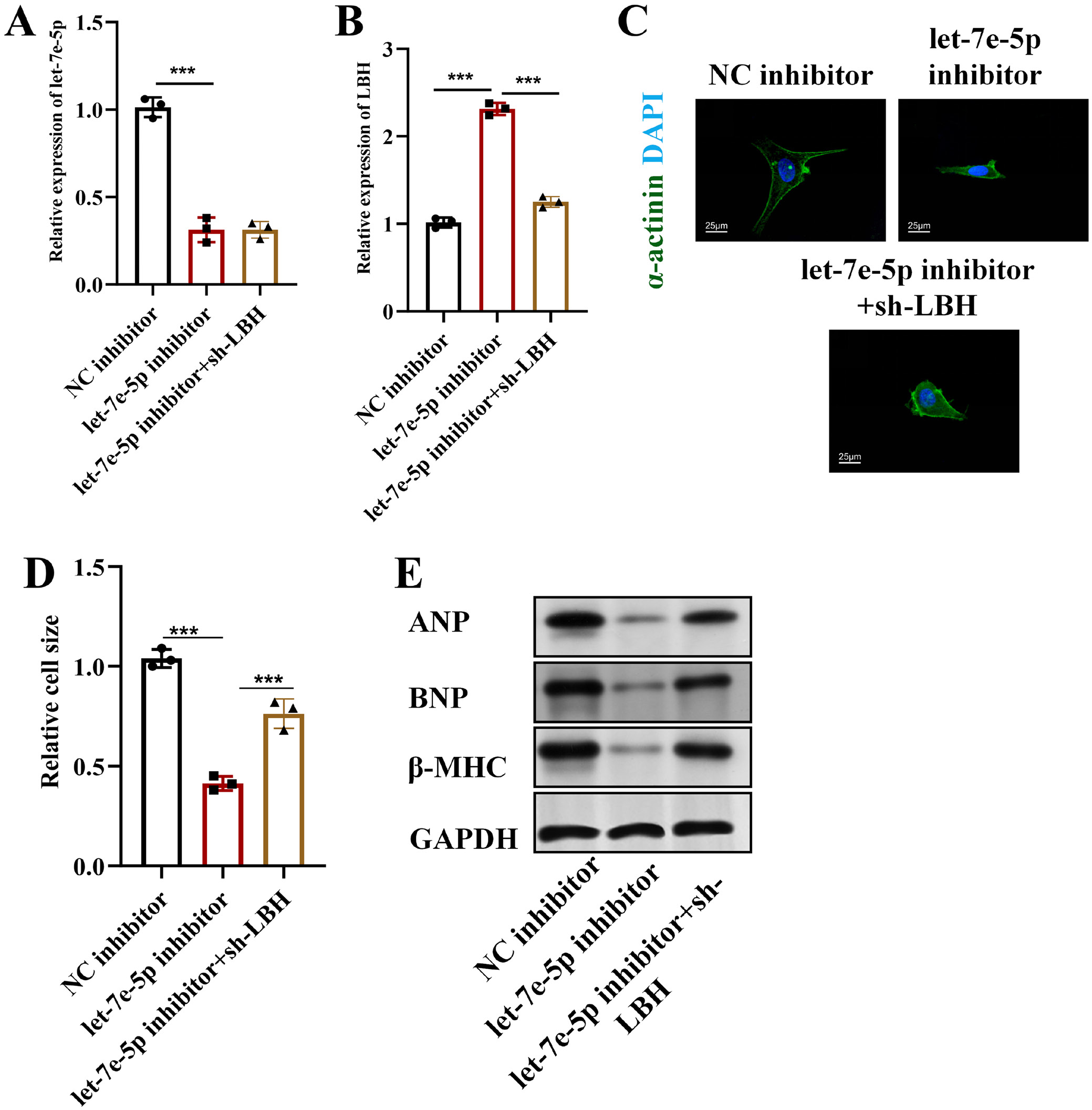
Rescue assays were performed to explore whether let-7e-5p regulates cardiac hypertrophy by modulating LBH in Ang II-exposed H9c2 cardiomyocytes. As shown in Figure 8A,B, let-7e-5p was downregulated after transfection with the let-7e-5p inhibitor, whereas no significant alteration was observed after LBH knockdown. In contrast, LBH was found to be upregulated in response to let-7e-5p inhibition in H9c2 cells and was significantly reduced after transfection with sh-LBH. According to the immunofluorescence results, the size of Ang II-stimulated H9c2 cardiomyocytes was reduced by let-7e-5p silencing and was significantly increased after co-transfection with sh-LBH relative to the let-7e-5p inhibitor group (Figure 8C,D). Similarly, we found that the reduced protein expression levels of ANP, BNP, and β-MHC induced by let-7e-5p inhibition were significantly restored after LBH silencing (Figure 8E).
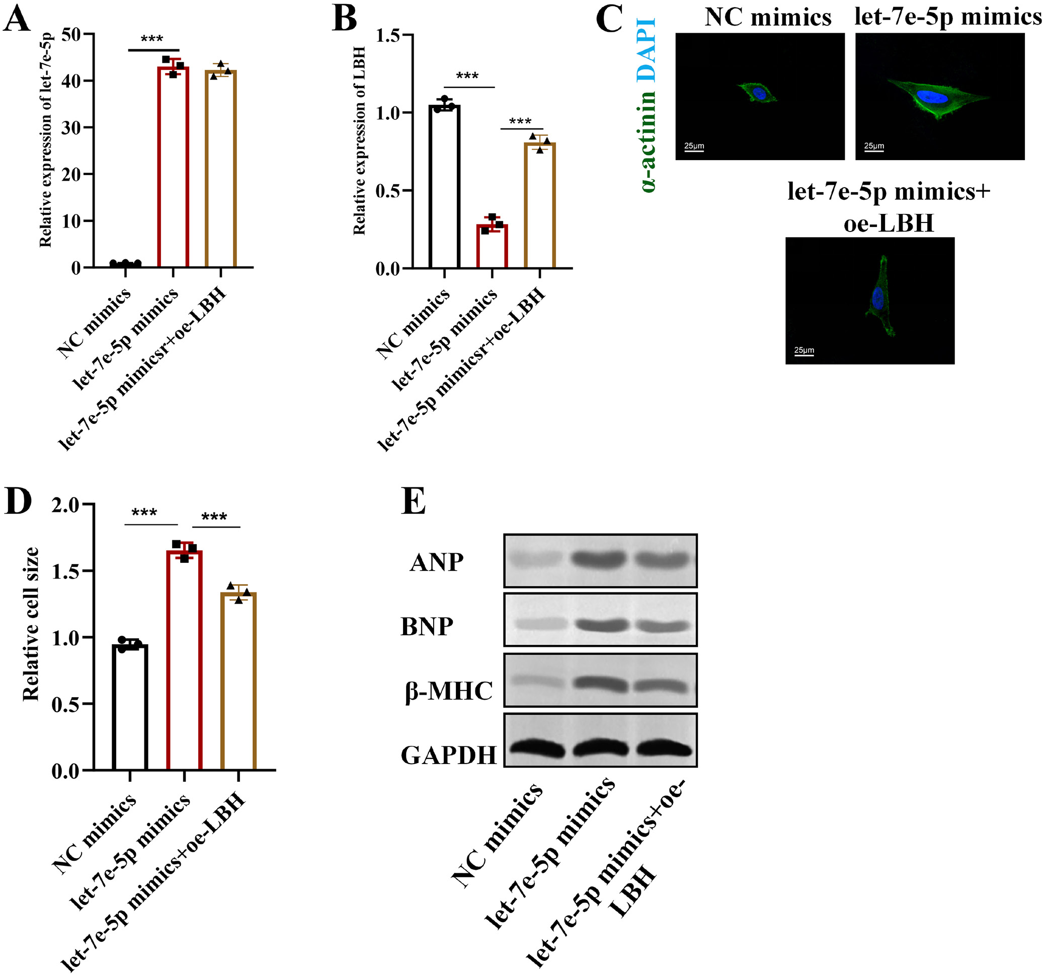
Furthermore, let-7e-5p was overexpressed using let-7e-5p mimics, and LBH was overexpressed using the pcDNA/LBH vector (oe-LBH) to verify the role of the let-7e-5p/LBH axis in cardiac hypertrophy. RT-qPCR results confirmed that let-7e-5p was upregulated by let-7e-5p mimics and was not significantly altered by oe-LBH, whereas LBH was downregulated by let-7e-5p overexpression, which was significantly reversed by oe-LBH transfection in H9c2 cells (Figure 9A,B). Moreover, we demonstrated that the increase in the size of Ang II-exposed H9c2 cells induced by let-7e-5p overexpression was partially reversed by LBH upregulation (Figure 9C,D). Similarly, the upregulation of ANP, BNP, and β-MHC proteins induced by transfection with let-7e-5p mimics was reversed by LBH overexpression (Figure 9E). Overall, let-7e-5p targets LBH to promote the development of cardiac hypertrophy.
Let-7e-5p facilitates the activation of the IGF–PI3K–Akt pathway via LBH
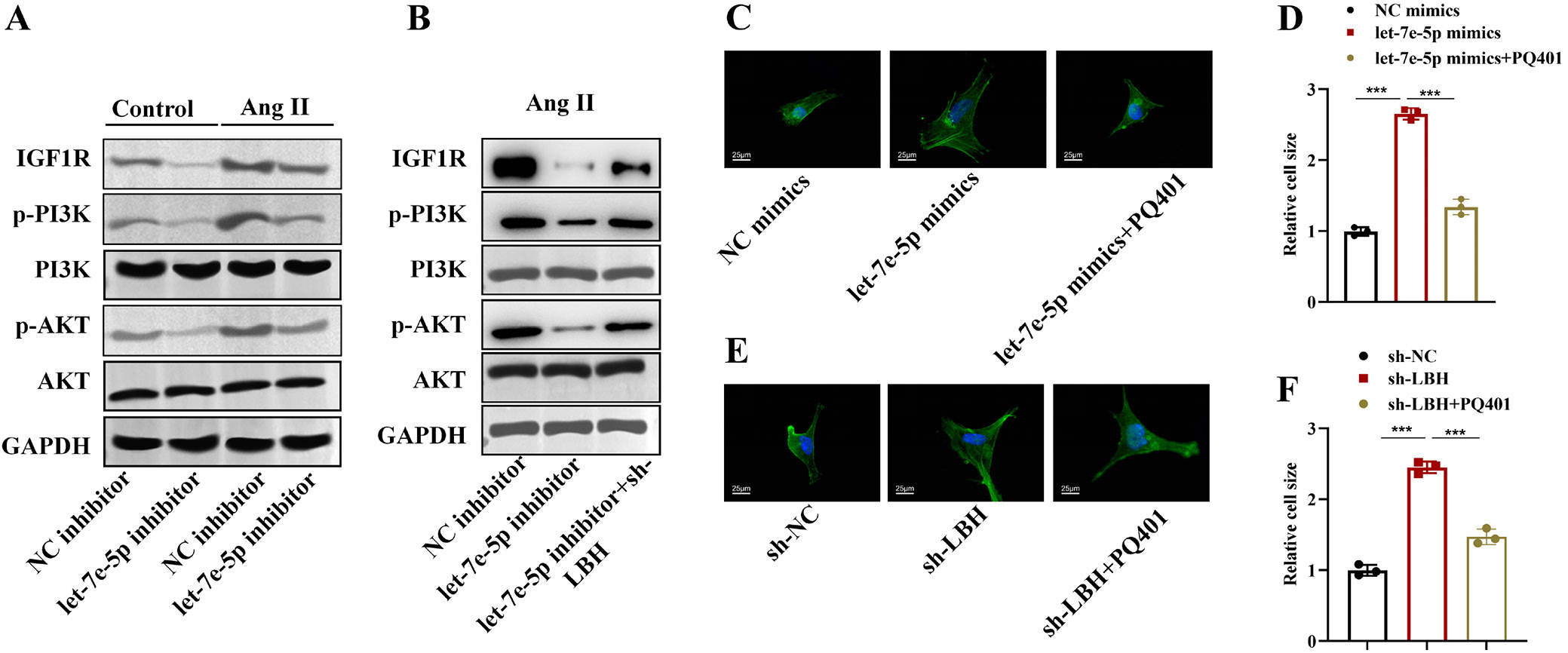
Previous studies have reported that the IGF–PI3K–Akt signaling pathway plays a crucial role in the development of cardiac hypertrophy and that let-7e-5p may activate the PI3K–Akt pathway in the heart.21, 22 Therefore, we investigated the impact of the let-7e-5p/LBH axis on IGF–PI3K–Akt signaling in Ang II-induced H9c2 cells. As shown in Figure 10A, Ang II treatment was found to upregulate the levels of IGF1R, p-PI3K/PI3K, and p-Akt/Akt in H9c2 cells, whereas let-7e-5p silencing exerted the opposite effects. Moreover, the reduced expression of IGF1R, p-PI3K/PI3K, and p-Akt/Akt induced by let-7e-5p silencing was significantly reversed after LBH knockdown in Ang II-treated H9c2 cells, suggesting that let-7e-5p targets LBH to activate the IGF–PI3K–Akt pathway (Figure 10B).
Additionally, the IGF1R inhibitor PQ401 was used in rescue assays. Immunofluorescence results showed that H9c2 cell size was increased after let-7e-5p overexpression, whereas a marked reduction was observed in the let-7e-5p mimics + PQ401 group, indicating that inhibition of the IGF–PI3K–Akt pathway partially reversed the promotive effect of let-7e-5p on cardiomyocyte hypertrophy (Figure 10C,D). Meanwhile, we also found that the increase in H9c2 cardiomyocyte size induced by LBH knockdown was significantly reversed by PQ401 treatment (Figure 10E,F). These results indicate that let-7e-5p promotes cardiomyocyte hypertrophy by targeting LBH and activating IGF–PI3K–Akt signaling.
Discussion
Currently, effective treatment options for cardiac hypertrophy remain limited, and it is imperative to develop therapeutic strategies to improve cardiac function and prevent progression to heart failure. Understanding the potential mechanisms involved in cardiac hypertrophy is essential for discovering diagnostic biomarkers and therapeutic targets. In this study, we found that let-7e-5p was aberrantly upregulated in patients with cardiac hypertrophy. Further analyses demonstrated that let-7e-5p plays a detrimental role and facilitates cardiac hypertrophy both in vitro and in vivo, which may provide novel insights into the diagnosis and treatment of this condition in clinical practice.
MicroRNAs are critical regulatory RNAs involved in the progression of a variety of heart diseases. Growing evidence has indicated that circulating miRNAs are promising noninvasive and accessible biomarkers for HCM diagnosis.23 In our study, ROC curve analysis revealed that let-7e-5p expression showed good diagnostic performance for cardiac hypertrophy, suggesting its potential utility in HCM diagnosis. The function of miRNAs can be modulated through systemic or local delivery of miRNA mimics or inhibitors to block the interaction between miRNAs and their target genes.24 Preclinical studies have been conducted to explore their efficacy against cardiac pathology. For example, miR-218 has been reported to be downregulated in cardiac hypertrophy, and its upregulation significantly suppresses isoprenaline-induced cardiomyocyte hypertrophy by targeting REST.25 MiR-27a-3p levels have been shown to be increased in Ang II-treated cardiomyocytes, and transfection with a miR-27a-3p inhibitor significantly reduced myocardial hypertrophy and electrical remodeling through regulation of Hoxa10.26
Let-7e-5p dysregulation has been identified in many diseases, including multiple cancers.27, 28 Let-7e-5p is also aberrantly expressed after myocardial infarction and may be involved in the pathophysiology of cardiovascular diseases.29, 30 In our study, we demonstrated the upregulation of let-7e-5p in the heart tissues of AB-induced rats. Administration of antagomir let-7e-5p effectively alleviated cardiac hypertrophy in rats subjected to AB surgery. Additionally, let-7e-5p was shown to be upregulated by Ang II in H9c2 cardiomyocytes. Let-7e-5p inhibition exerted suppressive effects on Ang II-induced cardiomyocyte hypertrophy, whereas let-7e-5p overexpression significantly enhanced the cardiomyocyte hypertrophy induced by Ang II treatment. These results indicate a critical role of let-7e-5p in cardiac hypertrophy.
LBH was identified as a target gene of let-7e-5p. LBH is recognized as being crucial for embryonic and cardiac development.13 Accumulating evidence has revealed that LBH is aberrantly expressed and implicated in the progression of diverse diseases, including cancers, liver diseases, rheumatoid arthritis, and others.15, 31, 32 Additionally, LBH has been shown to mediate the activation of cardiac fibroblasts and has been suggested as a valuable therapeutic target for myocardial I/R injury.14, 15, 33
In this study, we demonstrated that LBH was downregulated in the serum of patients with HCM, and bioinformatics analysis of the GSE89714 dataset similarly showed that LBH was expressed at low levels in heart tissues from these patients. Moreover, we found downregulation of LBH in the heart tissues of rats subjected to AB surgery and in H9c2 cells treated with Ang II. The interaction between let-7e-5p and LBH was confirmed, and let-7e-5p was shown to negatively regulate LBH expression in both rat heart tissues and H9c2 cells.
Furthermore, we demonstrated that LBH is essential for let-7e-5p-mediated cardiac hypertrophy, as evidenced by rescue assays. LBH silencing reversed the suppressive effects of let-7e-5p silencing on Ang II-treated H9c2 cells, whereas LBH overexpression reversed the promotive effects of let-7e-5p overexpression on H9c2 hypertrophy.
The IGF–PI3K–Akt pathway has previously been reported to be associated with endothelial dysfunction in the heart. Insulin-like growth factor (IGF) is a peptide hormone with a structure similar to that of insulin and is associated with cardiac function. It binds to its receptor, IGF1R, followed by the phosphorylation of PI3K and Akt.34 Multiple studies have revealed that activation of the IGF–PI3K–Akt pathway can exert protective effects against cardiac injury by suppressing cardiomyocyte apoptosis.35, 36 Furthermore, a study has demonstrated the promotive effect of activation of this pathway on cardiomyocyte hypertrophy.37 Consistently, our study demonstrated that let-7e-5p promotes activation of this pathway by targeting LBH, and inhibition of the pathway using the IGF1R inhibitor PQ401 reversed the effects of let-7e-5p mimics or the sh-LBH vector on H9c2 hypertrophy.
Limitations of the study
First, although AB mimics pressure overload-induced hypertrophy, this model cannot fully replicate the complex pathogenesis of cardiac hypertrophy in humans, and the role and mechanisms of let-7e-5p should be further validated in other models of cardiac hypertrophy. Second, the upstream regulatory mechanisms responsible for let-7e-5p upregulation in cardiac hypertrophy remain unclear and warrant further investigation in future studies.
Conclusions
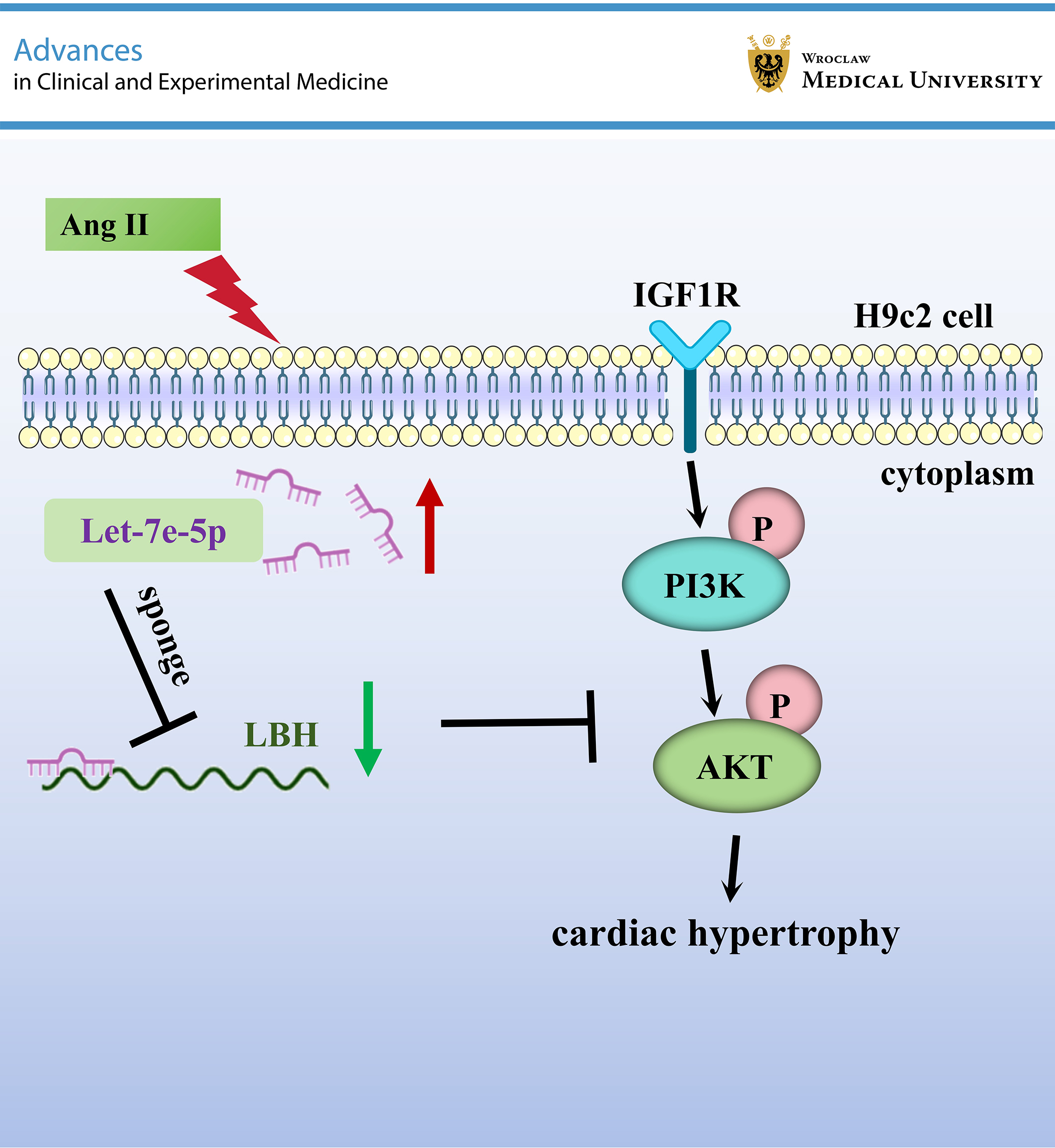
Let-7e-5p is highly expressed in cardiac hypertrophy and aggravates the development of cardiac hypertrophy by targeting LBH and activating the IGF–PI3K–Akt pathway. The results of our study may provide new insights into targeted therapy for cardiac hypertrophy.
Supplementary data
The supplementary materials are available at https://doi.org/10.5281/zenodo.17733961. The package contains the following files:
Supplementary Table 1. Results of the Shapiro–Wilk normality test.
Supplementary Table 2. Results of the homogeneity of variance test.
Supplementary Table 3. Results of the statistical analyses.
Supplementary Fig. 1. Expression of let-7e-5p in rat heart tissues in vivo. RT-qPCR was used to detect let-7e-5p expression in the heart tissues of rats in the sham, model, model + antagomir NC, and model + antagomir let-7e-5p groups (***p < 0.001).
Supplementary Fig. 2. Identification of candidate mRNA targets of let-7e-5p. A. Venn diagram showing the overlap between downregulated genes in the heart tissue of patients with HCM from the GSE89714 dataset and predicted targets of let-7e-5p in the TargetScan database. A total of 21 candidate mRNAs were identified. B. RT-qPCR analysis of the expression of 21 candidate mRNAs in H9c2 cells transfected with NC inhibitor or let-7e-5p inhibitor. C. RT-qPCR analysis of the expression levels of 3 candidate mRNAs in rat heart tissues from each group (**p < 0.01, ***p < 0.001).
Data Availability Statement
The datasets supporting the findings of the current study are openly available in the Figshare repository at https://doi.org/10.6084/m9.figshare.30453719.
Consent for publication of personal information
Not applicable.
Use of AI and AI-assisted technology
Not applicable.