Abstract
Background. Diabetic retinopathy (DR) is one of the most common complications of diabetic microvascular disease and its pathogenesis is complicated. The PI3k/Akt signaling pathway plays an important role in the angiogenesis of DR.
Objectives. To explore the molecular mechanisms of the ACO3 protein and related proteins in DR, in order to provide a scientific theoretical basis for the clinical treatment of this disease.
Materials and methods. A DR rat model was used in this study. One group (anti-ACO3 group, n = 10) was injected with protein ACO3 antagonist; the 2nd study group (the DR group, n = 10) was injected with the same amount of normal saline; the control group (n = 10) did not undergo any procedures. We used hematoxylin and eosin (H&E) staining to observe the pathological features of the eye tissues. Immunohistochemistry was used to analyze the expression of ACO3 and AKT. Western blot was used to analyze the expression of ANGPT1, ANGPT4 and KDR; reverse transcription polymerase chain reaction (RT-PCR) was used to assess the mRNA expression of AKT, ACO3 and SMOX in the eye tissues of the rats.
Results. In the anti-ACO3 group, the results of H&E staining showed that there was a decrease in retinal edema and no obvious abnormality in the blood vessels. The immunohistochemistry analysis of proteins proved that ACO3 and AKT were strongly expressed in the DR group. The western blot analysis of ANGPT1, ANGPT4 and KDR expression showed that in the DR group, the expression of all 3 proteins was higher than in the anti-ACO3 group, and much higher than in the control group.
Conclusions. The mRNA expression of AKT, ACO3 and SMOX was strong in the DR group, but decreased in the anti-ACO3 group.
Key words: diabetic retinopathy, mechanism, ACO3, PI3k/Akt signaling pathway
Background
Along with social and economic development, improved living standards and changes in eating habits, diabetes has become a common disease affecting people’s health and life. Diabetic retinopathy (DR) is one of the most universal complications of diabetic microvascular disease, often causing serious visual impairment.1 According to the research conducted by the World Health Organization (WHO), the number of diabetic patients in the world reached 366 million cases in 2011, and by 2025 this number will exceed 500 million.2 It was recently reported that the global morbidity of DR was 34.6%, while in developed countries it was nearly 40.3%.3 The number of patients with DR in China has been reported to be as high as 13.16 million.4 The most common features of DR patients include decreased vision, microhemangioma, bleeding spots, hard exudate, retinal neovascularization, venous beading, and macular edema.5, 6 In some severe cases, vitreous hemorrhage, fibrogenesis and retinal angiogenesis have been noted, which might cause neovascular glaucoma or retinal detachment.7 If effective treatment measures are not taken in time, retinal cell death and fibrosis are likely to occur, resulting in a permanent loss of vision. Therefore, an early and accurate diagnosis and treatment of DR is particularly important.8
The pathogenesis of DR is complicated. It is related to glucose metabolism, growth hormone and angiogenic growth factors. In terms of glucose metabolism, when blood glucose level is elevated, the osmotic pressure inside cells is higher than that of the outside cells due to the failure of glycolysis, leading to an imbalance of water electrolytes in eye cells.9, 10 Retinal microvascularization would interfere with the metabolism of inositol and accumulate advanced glycation end products (AGEs) under the stimulus of long-term high-sugar environment. At the same time, this process affects the activity of Na+/K+-ATPase in cells; increases the permeability of capillaries; thickens the basement membrane; narrows capillary lumens; increases retinal ischemia, hypoxia and the release of vascular proliferation factors; and promotes the formation of new blood vessels.11
As one of the important pathways involved in endothelial cell migration, proliferation and vascular dysfunction, the PI3k/Akt signaling pathway also plays an important role in the process of DR neovascularization.12 Among proteins related to PI3k/Akt signaling pathway, the ACO3 protein is involved in regulating cell proliferation and inducing neovascularization.
Objectives
The aim of the study was to explore the molecular mechanisms of the ACO3 protein and related proteins in DR, in order to provide a scientific theoretical basis for the clinical treatment of this disease.
Materials and methods
The study used 30 Wistar rats (purchased from Harbin Medical University, China); streptozotocin (STZ; Sigma-Aldrich, St. Louis, USA); ACO3 antagonist (Thermo Fisher Scientific, Waltham, USA); hematoxylin (Invitrogen, Carlsbad, USA); phosphate-buffered saline (PBS) (0.1 m, pH 7.0); TRIzol RNA agent; 6-, 12- and 48-plate cell culture dishes (Thermo Fisher Scientific); 5 mL and 10 mL sterile pipettes (Corning, New York, USA); 10 μL, 20 μL, 200 μL, and 1000 μL transfer pipettes (Eppendorf, Enfield, USA); 10 mL and 50 mL centrifuge tubes (Thermo Fisher Scientific); a conventional polymerase chain reaction (PCR) instrument (Agilent Technologies Inc., Santa Clara, USA); western blotting instruments (Eppendorf AG, Hamburg, Germany); a confocal microscope (Olympus Corp., Tokyo, Japan); a LKB-V ultra-thin slicer (JEOL Ltd., Tokyo, Japan); and a Jem-2000EX fundus fluorescein angiograph (JEOL Ltd.).
Rat model of diabetic retinopathy
We divided 30 Wistar rats (8–9-week old) into 3 groups of 10 rats each. One was the control group, while the other 2 were used to establish the DR model. Twenty rats were randomly assigned to the 2 model diabetic groups. These rats were administered STZ (60 mg/kg) by intraperitoneal injection to induce diabetes. Their fasting glucose was monitored and glucose level ≥16.65 mmol/L was considered the standard model. After 1 month, the diabetic rats were injected with 0.05 μg of vascular endothelial growth factor (VEGF), 2 mm behind the temporal limbic cornea using a microinjector. It passed through the ciliary body and entered the vitreous cavity. The injection was done slowly to keep the needles in for 10 s, in order to create a model of ophthalmic lesions in proliferative diabetes mellitus. The fundus changes in the rats were examined to judge whether the DR model had been successfully constructed. One DR group (called anti-ACO3 group) was injected with 5 μL of ACO3 antagonist (10 μg/mL); the other DR group (called DR group) was injected with the same amount of saline in vitreous body.
Fundus fluorescein angiography
Fundus fluorescein angiography (FFA)13 was carried out as follows: before the examination, 1 mL of 1% fluorescein sodium was injected intravenously for an allergy test, and was observed for 15 min to ensure that there was no abnormal reaction. Then, 5 mL of 10% fluorescein sodium was immediately injected into each rat and 1 eye was selected as the main eye. After 10–15 s of injection, the early image was shot continuously. After 1–10 min of injection, the middle image was shot. After 10 min, the late image was shot and stored for analysis.
H&E staining to examine the pathological features of the eye tissues
Eye tissues were taken from the 2 DR groups and the control group, and hematoxylin and eosin (H&E) staining was used to examine the pathological features of the tissues.14 Four weeks after the administration of the ACO3 antagonist or saline to the diabetic rats, pentobarbital sodium was injected intraperitoneally for anesthesia and the retinal tissues of the rats were taken. The tissues were fixed with 4% paraformaldehyde and decalcified with 15% ethylenediamine tetraacetic acid (EDTA) at room temperature. After dehydration, the retinal tissues were embedded in paraffin and 4-μm tissue sections were prepared for pathological observation. The section samples of the 3 groups of rats were stained and dewaxed with xylene twice. Then, the samples were hydrated with gradient ethanol for 3 min and stained with hematoxylin for 5 min. The tissues were then rinsed with 1% hydrochloric acid-ethanol for 30 s, followed by reflux treatment in 0.2% ammonia for 2 min, stained with 0.5% eosin for 10 min, and washed with water once. Gradient ethanol was used for dehydration treatment, and finally, neutral gum was used for sealing. The image was analyzed using an optical microscope.
Immunohistochemistry analysis of ACO3 and AKT expression
Immunohistochemistry staining was used to detect the expression of ACO3 and AKT in the eye tissues.15 The paraffin-embedded islet tissue of the rats was sectioned at a thickness of 4 μm. The two-step immunohistochemistry method was used for staining. The tissue sections were roasted at 65°C for 12 h, dewaxed and hydrated. Endogenous peroxidase was inactivated by 3% hydrogen peroxide, and the specimens were washed twice with PBS. Mouse anti-human primary antibody ACO3 and AKT were added and washed twice with PBS. Next, the streptomycin working solution (labeled with horseradish peroxidase (HRP)) was added and incubated at 37°C for 25 min. Then, it was washed with PBS, stained with hematoxylin and sealed with neutral chicle.
Western blot analysis of ANGPT1, ANGPT4 and KDR expression
Total proteins were extracted from the eye tissue and blood vessels of each group of rats, and 20-μg protein samples were prepared. We used 5% concentrated gel and 12% isolated gel to isolate proteins using sodium dodecyl sulphate–polyacrylamide gel electrophoresis (SDS-PAGE). Objective and internal proteins were transferred to nitrocellulose (NC) membranes, then sealed with 5% skimmed milk powder sealing fluid for 2 h at room temperature. Rat anti-human primary antibody ANGPT1 (1:500), rat anti-human primary antibody ANGPT4 (1:500), rat anti-human primary antibody KDR (1:500), and rat anti-human primary antibody β-actin (1:1000) were added and incubated overnight at 4°C. The specimens were washed 4 times in Tris-buffered saline with Tween (TBST), and HRP-labeled sheep anti-rat secondary antibody (1:5000) was added and incubated at 37°C for 1 h. The specimens were again washed 4 times in TBST. Color was developed with enhanced chemiluminescence (ECL) solution; protein bands were exposed using ECL blotting detection reagent (Beyotime, Shanghai, China); and the images were photographed and analyzed quantitatively. The experiment was repeated 3 times.
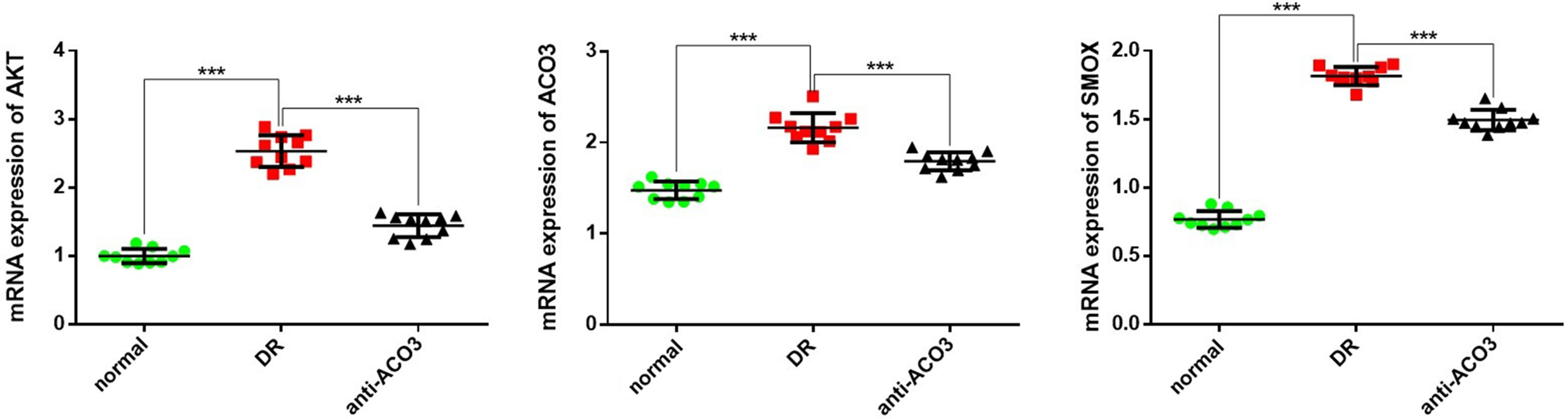
RT-PCR analysis of AKT, ACO3 and SMOX mRNA expression
The analysis of AKT, ACO3 and SMOX mRNA expression was conducted as described by Rouhani et al.16: total RNA was extracted with TRIzol reagent (Shanghai Pufei Biotech, Shanghai, China), according to the manufacturer’s instructions. Nucleic acid-protein complexes were extracted with chloroform and precipitated in isopropanol. The complexes were cleaned using 75% ethanol and purified with RNase-free water. The expression level of GAPDH was selected as the internal reference. Primers for PCR detection were designed and synthesized according to the information of the target gene sequences, as shown in Table 1. The amplifications were performed in a 96-well plate at 95°C for 10 min, followed by 40 cycles of 95°C for 15 s and 60°C for 1 min. Each sample was run in triplicate. The relative miRNA-126 and mRNA expression was expressed using the 2–ΔΔCt method.
Statistical analyses
One-way analysis of variance (ANOVA) followed by Tukey’s post hoc test were used to analyze the data for comparison among 3 groups. The variance homogeneity was tested using Levene’s variance homogeneity test. The distribution of the data was analyzed with Kolmogorov–Smirnov and Shapiro–Wilk method, and normally distributed data were expressed as mean ± standard deviation (SD). Statistical significance was defined as p < 0.05. All calculation was conducted using SPSS v. 18.0 software (SPSS Inc., Chicago, USA).
Results
Rat model of diabetic retinopathy
Rats with the blood glucose value ≥16.7 mmol/L were defined as diabetic rats. Then, the diabetic rats injected with vitreous cavity were subjected to fundus fluorescein examination to observe the obvious retinal hemorrhage or exudation, telangiectasia, arteriovenous abnormalities, and other conditions in the fundus of rats, indicating that the DR model of the rats was successfully established.
Fundus fluorescein angiography
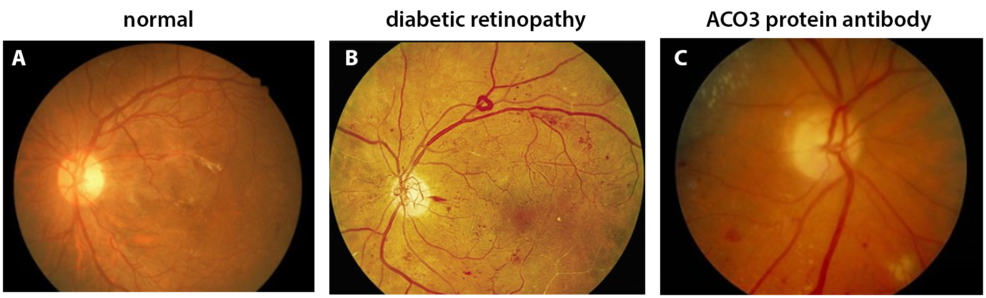
The FFA results showed that there was no significant DR and no abnormal phenomena in the control group (Figure 1A). However, background fluorescence enhancement, vascular tortuosity and dilatation, and fluorescence leakage from neovascularization, intraretinal hemorrhage and microhemangioma were observed in the DR group (Figure 1B). The characteristics of the eye tissue of the DR rats in the anti-ACO3 group were significantly better than in the DR group, and the number of new microvessels was lower in the anti-ACO3 group (Figure 1C). This indicates that the ACO3 protein is involved in regulating cell proliferation and inducing neovascularization.
H&E staining to examine pathological features of the eye tissues
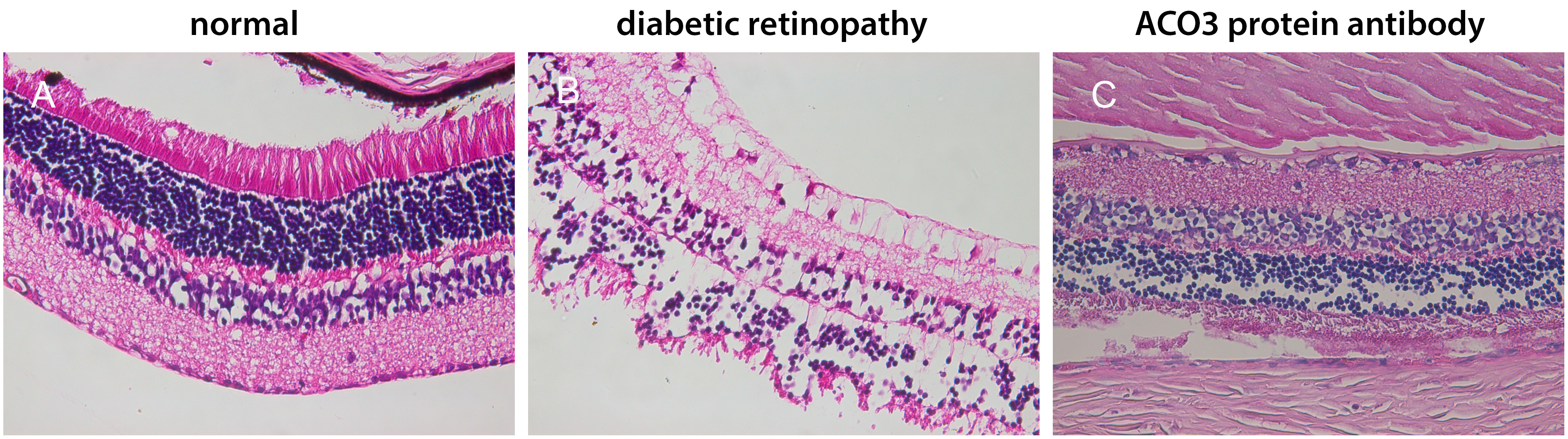
The pathological histology of the retinas in all 3 groups was examined. It could be seen that the surface of the retina in the control group was smooth, and the cells in the inner and outer nuclear layer were arranged closely and orderly (Figure 2A). In the DR group, there was obvious edema in the inner retina boundary; the cells in the inner and outer nuclear layers were not arranged neatly, and more synaptic membranes of vascular endothelial cells could be seen (Figure 2B). However, in the anti-ACO3 group, there was a decrease in retinal edema, and loose arrangements of cells were observed only in the inner core layer (Figure 2C).
Immunohistochemistry analysis of ACO3 and AKT expression
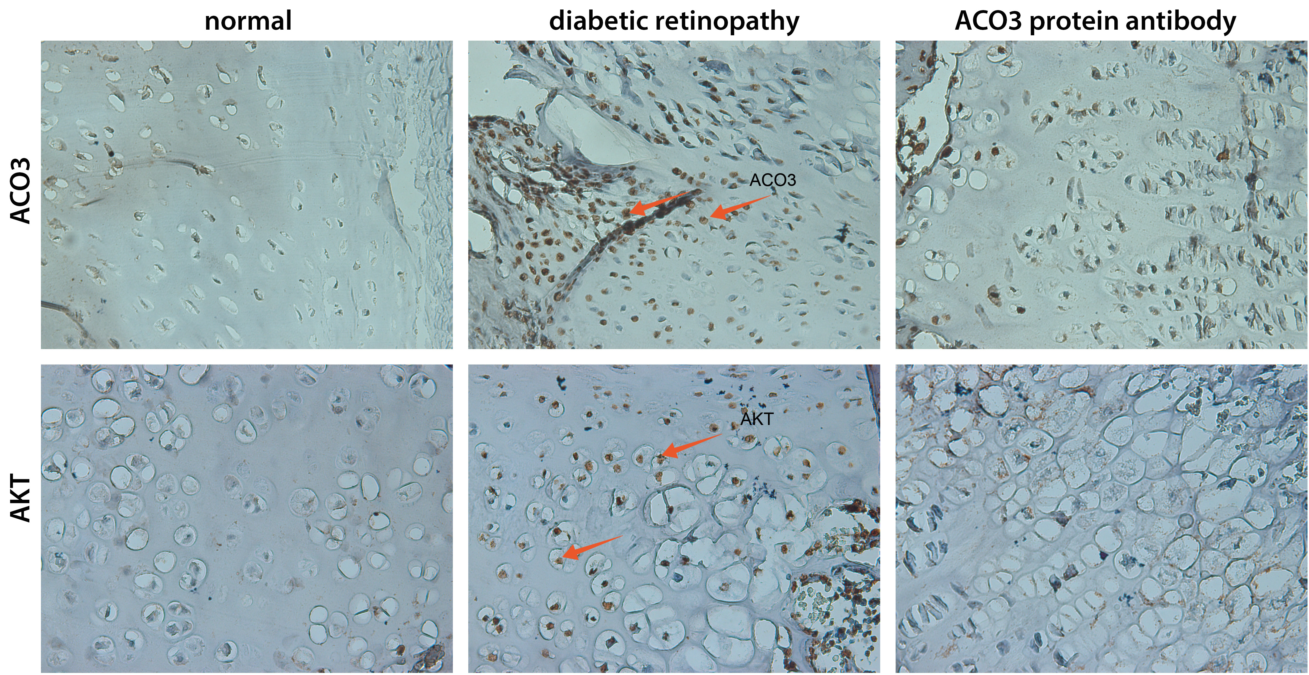
The expression levels of proteins ACO3 and AKT were the lowest in the control group, and were highly expressed in the DR group, as indicated by the red arrow in Figure 3. In the anti-ACO3 group, the expression levels of ACO3 and AKT were decreased.
Western blot analysis of ANGPT1,
ANGPT4 and KDR expression
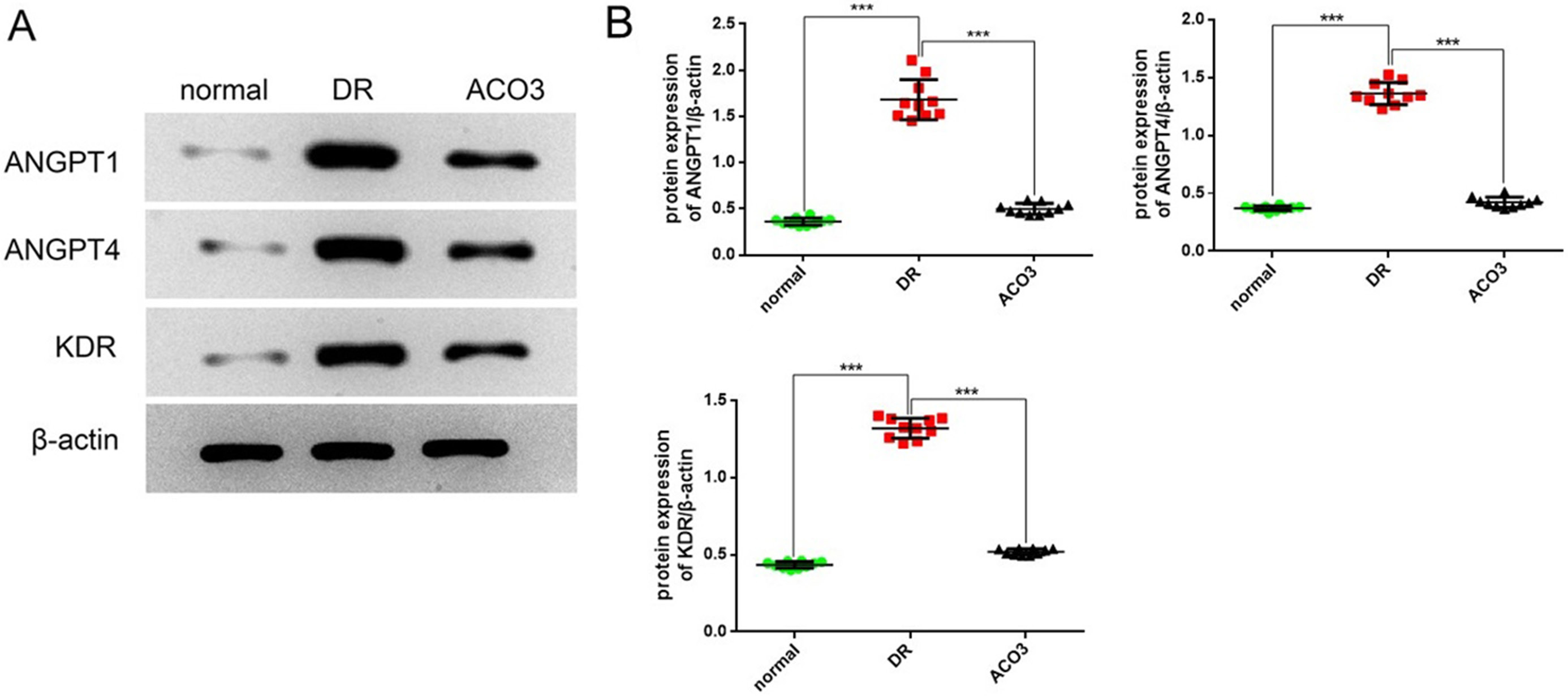
The expression levels of ANGPT1, ANGPT4 and KDR in the rat eye tissues are shown in Figure 4. The expression of these proteins was higher in DR group than in the anti-ACO3 group (ANGPT1 p < 0.001, ANGPT4 p < 0.001, KDR p < 0.001, ANOVA followed by Tukey’s post hoc test), and much higher than in the control group (ANGPT1 p < 0.001, ANGPT4 p < 0.001, KDR p < 0.001, ANOVA followed by Tukey’s post hoc test). However, the expression levels of these proteins were high in anti-ACO3 group compared with the control group (p < 0.05). The original statistical data of western blot are shown in Table 2, Table 3, Table 4, Table 5.
RT-PCR analysis of AKT, ACO3
and SMOX mRNA expression
The mRNA expression levels of AKT, ACO3 and SMOX in the DR rats with eye lesions were all increased, but were lower in the anti-ACO3 group compared with the DR group (AKT p < 0.001, ACO3 p < 0.001, SMOX p < 0.001, ANOVA followed by Tukey’s post hoc test) (Figure 5). Cell proliferation in the DR group indicates that ACO3 is involved in regulating cell proliferation and inducing neovascularization. The ACO3 and SMOX have characteristics of mutual regulation, and synergistically participate in the regulation of amino acid metabolism and promotion of cell proliferation. The high expression levels of AKT indicate the the activation of the PI3K/Akt signaling pathway, which regulates the increase of NOS3, an enzyme related to vascular endothelial cells, and produces NO, which affects the permeability of blood vessels and increases damage to retinal cells. The original statistical data of PCR are shown in Table 2, Table 3, Table 4, Table 5.
Discussion
The occurrence and development of DR is a complex pathological process, and a series of pathological changes of DR are caused by abnormal changes of cytokines, signal transduction metabolic enzymes, inflammation, ion channels, and related genes.17, 18 Neovascularization is a characteristic pathological marker of the entry of DR into the proliferative phase. Therefore, preventing and inhibiting the formation of DR neovascularization is a key target to delay the progression of DR. The PI3K/Akt signaling pathway is one of the signaling pathways that participate in the regulation of cell growth, proliferation and differentiation.19, 20 When the PI3K/Akt signaling pathway is activated, it can not only accelerate the survival cycle of endothelial cells, but also cooperate with VEGF in cell survival and migration, and finally induce the formation of new blood vessels.21 The AKT regulates serine antiapoptotic signaling protein in endothelial cells from cell cycle G1 phase to S phase. Therefore, AKT regulates endothelial cell proliferation, migration and has a key role in inducing angiogenesis.22, 23 In this study, the ischemia and hypoxia induced by high glucose level in the rat model activated and accelerated the expression of AKT in the PI3k/Akt signaling pathway, and increased its phosphorylation, thus accelerating the formation of neovascularization. The results of this experiment showed that the expression of AKT and related proteins in the DR group and anti-ACO3 group was significantly higher than in the control group, indicating that the expression of AKT was enhanced and the PI3k/Akt signaling pathway was activated during the formation of DR lesions.
Limitations of the study
This study is mainly based on the basic experiment of animal model. It explored the involvement of ACO3 protein in DR through the PI3k/Akt signaling pathway of rat model of DR. However, there are still great structural and pathological differences between animals and humans. Therefore, this study should also examine the differences and effects of various animal models, and further clinical studies should be performed.
Conclusions
As an important pathway involved in endothelial cell migration, proliferation and vascular dysfunction, the PI3K/Akt signaling pathway also plays an important role in the process of DR neovascularization. The ACO3 protein, also known as VAP1, is involved in regulating cell proliferation and inducing neovascularization.24 Proteins ACO3 and SMOX have the characteristics of mutual regulation, and are synergistically involved in the regulation of amino acid metabolism.25 The expression levels of ACO3, SMOX and AKT proteins were decreased in the anti-ACO3 group, which indicated that the PI3K/Akt signaling pathway was inhibited, and the formation of retinal neovascularization was inhibited to a certain extent. This delayed the progression of DR and played a certain therapeutic role in the proliferation phase.