



Abstract
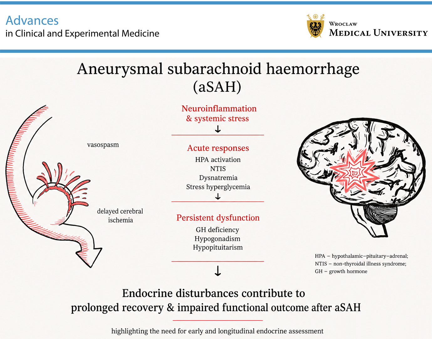
Aneurysmal subarachnoid hemorrhage (aSAH) is a life-threatening cerebrovascular event associated with profound neurological and systemic disturbances. Among these, alterations in hypothalamic–pituitary function, water–electrolyte balance, and glucose metabolism are frequently observed but remain difficult to interpret, particularly in the setting of critical illness. Many endocrine abnormalities detected during the acute phase reflect adaptive neuroendocrine stress responses rather than true hormonal failure, whereas persistent deficiencies may emerge during recovery and contribute to long-term morbidity. This narrative review summarizes current evidence on endocrine disturbances after aSAH, emphasizing the distinction between acute neurocritical care physiology and chronic endocrine sequelae. We discuss the hypothalamic–pituitary–adrenal (HPA), thyroid, somatotropic, and gonadal axes, as well as dysnatremia and stress hyperglycemia, addressing their prevalence, pathophysiological background, diagnostic challenges, and therapeutic considerations in both intensive care unit (ICU) and post-acute settings. Available data remain heterogeneous and largely observational. Current evidence does not support routine hormonal intervention during the acute phase except in clearly defined endocrine emergencies. Careful interpretation of biochemical findings and structured endocrine follow-up after neurological stabilization appear essential for identifying clinically meaningful long-term dysfunction.
Key words: subarachnoid hemorrhage, hypopituitarism, critical care, neuroendocrinology, endocrine system diseases
Introduction
Aneurysmal subarachnoid hemorrhage (aSAH) represents a severe subtype of stroke, accounting for approx. 5% of all cerebrovascular events, with an annual incidence of 6–9 cases per 100,000 individuals in developed countries.1, 2 Epidemiological analyses continue to demonstrate a global burden with regional variation.3 Despite advances in neurosurgical treatment and neurocritical care, aSAH remains associated with substantial mortality and morbidity,1, 4 and nearly 1/3 of survivors experience long-term functional impairment.5 Importantly, the condition frequently affects relatively young individuals during their most productive years, resulting in considerable personal, social, and economic consequences.1, 5 Although aneurysm rupture accounts for the vast majority of spontaneous SAH cases, other vascular entities such as perimesencephalic hemorrhage,6 moyamoya disease,7 or dural arteriovenous fistulas8 may present with different clinical and systemic profiles, which should be distinguished from true aSAH when interpreting endocrine sequelae.
Clinical management has traditionally focused on the prevention of rebleeding, treatment of vasospasm and delayed cerebral ischemia (DCI), and stabilization of intracranial dynamics.4 However, this primarily neurological perspective may underrecognize the systemic consequences of the hemorrhagic event, including disturbances in neuroendocrine regulation.
Endocrine dysfunction, particularly involving the hypothalamic–pituitary axis, has been described as a potential consequence of aSAH in both the acute and chronic phases of the disease.9, 10 These abnormalities may remain clinically unapparent because their manifestations – such as fatigue, cognitive slowing, or reduced stress tolerance – overlap with neurological sequelae and post-critical illness recovery.9, 11 As a result, endocrine disturbances may be underdiagnosed or recognized only later in the course of rehabilitation.
Reports published over the past 2 decades describe highly variable prevalence rates of hormonal abnormalities after aSAH, ranging widely across prospective cohorts12, 13 and systematic reviews.2 Many detected alterations appear transient or subclinical, further complicating their interpretation and clinical relevance.3, 12 Consequently, the true burden and significance of endocrine sequelae remain incompletely understood. Longitudinal observations suggest that some abnormalities resolve spontaneously, whereas others become apparent only during the later phases of recovery, reflecting the dynamic nature of neuroendocrine adaptation after brain injury.9, 11
Renewed interest in this field, reflected in recent systematic reviews and observational studies, highlights the need to better characterize alterations in cortisol regulation, thyroid function, growth hormone (GH) secretion, gonadal axis activity, and water–electrolyte balance after aSAH while acknowledging the lack of standardized screening strategies or consensus regarding optimal follow-up.10, 12, 14
Objectives
This article was designed as a clinically oriented narrative review aimed at synthesizing current knowledge on endocrine alterations following aSAH and providing a clinically relevant framework for interpreting these disturbances across different phases of the disease, from acute neurocritical illness to long-term recovery.
Materials and methods
The literature was selected pragmatically based on its clinical relevance to endocrine disturbances observed after aSAH, with particular attention to studies addressing the timing of dysfunction, interpretation in critically ill patients, and long-term outcomes. Given the heterogeneity of the available evidence and the absence of standardized endocrine assessment protocols, findings were synthesized descriptively rather than through a formal systematic methodology.
Results
Endocrine disturbances observed after aSAH represent a spectrum of dynamic neuroendocrine alterations driven by primary brain injury, the systemic stress response, and the effects of intensive care management. Their interpretation requires careful distinction between transient adaptive changes occurring during critical illness and less frequent chronic hypothalamic–pituitary dysfunction emerging during recovery. Importantly, many biochemical abnormalities reported in the literature are derived from studies conducted outside the immediate neurocritical care setting and cannot be directly extrapolated to unstable intensive care unit (ICU) patients. Consequently, endocrine evaluation strategies derived from outpatient endocrinology cannot be directly translated to the neurocritical care environment, where most patients with high-grade aSAH are managed.
Reported prevalence varies considerably due to heterogeneity in diagnostic criteria, the timing of endocrine evaluation, and patient selection. Overall, pituitary dysfunction has been described in approx. 30–55% of patients when assessed early after hemorrhage,3, 13 decreasing to roughly 15–25% at medium- and long-term follow-up,2, 10 supporting the concept that many abnormalities are functional and reversible rather than structural. Selective hormonal deficits are more common than complete hypopituitarism.10, 12
From a clinical perspective, this temporal evolution is essential for decision-making: misinterpreting early adaptive responses may lead to unnecessary endocrine interventions, whereas failing to recognize persistent dysfunction may adversely affect long-term recovery.
Effects of hemorrhage on endocrine regulation
Rupture of a cerebral aneurysm releases blood into the subarachnoid space, producing an abrupt rise in intracranial pressure, transient global ischemia, and microvascular dysfunction, which initiate a cascade of secondary brain injury mechanisms characteristic of aSAH.15 Secondary injury mechanisms – including excitotoxicity, oxidative stress, blood–brain barrier (BBB) disruption, and neuroinflammation – have been proposed as contributors to hypothalamic dysregulation after aSAH,12, 14, 15 with subsequent effects on pituitary perfusion and hormonal signaling being discussed in neuroendocrine-focused analyses.12
The pituitary gland is anatomically vulnerable because of its proximity to the circle of Willis and its dependence on the low-pressure hypophyseal portal circulation, which is particularly sensitive to changes in perfusion, vasospasm, and intracranial hypertension.12, 14 Additional contributors may include local edema, hydrocephalus, and the effects of neurosurgical or endovascular treatment.2, 16
Together, these processes provide a biological substrate for both transient neuroendocrine dysregulation during acute illness and delayed endocrine sequelae observed in selected survivors, forming the pathophysiological basis for the axis-specific disturbances discussed in the following sections.
Hypothalamic–pituitary–adrenal axis
Pathophysiology, prevalence and temporal pattern
The hypothalamic–pituitary–adrenal (HPA) axis is regulated through hypothalamic secretion of corticotropin-releasing hormone (CRH), stimulation of pituitary adrenocorticotropic hormone (ACTH) secretion, and subsequent adrenal cortisol production.17 Structural or functional disturbances at the hypothalamic or pituitary level after aSAH may therefore lead to secondary adrenal insufficiency, although such permanent injury appears uncommon.2, 12, 14 In the acute phase, however, this axis is predominantly characterized by stress-induced activation rather than failure.12, 14, 17
Activation of the HPA axis is an expected component of the systemic stress response following aSAH. Acute brain injury induces hypothalamic stimulation, sympathetic activation, and increased cortisol secretion, which support hemodynamic stability and metabolic adaptation during critical illness.12, 14, 17 Consequently, biochemical alterations in cortisol dynamics are frequently observed during the acute phase and should be interpreted predominantly as adaptive physiology rather than as evidence of structural pituitary injury.
This neuroendocrine activation is mediated not only by direct hypothalamic stimulation but also by inflammatory cytokines such as interleukin-6 (IL-6) and tumor necrosis factor alpha (TNF-α), which enhance CRH secretion and modify adrenal steroidogenesis during critical illness. In parallel, reduced cortisol-binding globulin concentrations and altered peripheral cortisol metabolism increase free cortisol availability, further complicating biochemical interpretation in ICU patients. These changes reflect a coordinated systemic stress response rather than true hypothalamic–pituitary failure.13, 18, 19
Persistent secondary adrenal insufficiency appears uncommon in longitudinal follow-up and is reported in fewer than 10–15% of patients in prospective cohorts13 and confirmed by meta-analytic data.2, 10
Clinical meaning in acute aSAH (ICU context)
Elevated or fluctuating cortisol levels should generally be interpreted as part of critical illness physiology rather than endocrine failure. This response supports cardiovascular function and inflammatory modulation during the neurocritical phase.12, 14, 17
Diagnostic considerations and limitations
Assessment of adrenal function in critically ill patients is challenging because cortisol-binding globulin levels, metabolism, and assay reliability are altered during systemic illness.17 Standard endocrine diagnostic thresholds derived from outpatient endocrinology are therefore not applicable to unstable ICU patients. True secondary adrenal insufficiency should be suspected only when clinical features are disproportionate to the expected stress response, particularly persistent or unexplained hypotension, hyponatremia, hypoglycemia, or failure to wean from vasopressor support after resolution of the acute neurological insult.2, 12, 14, 17 In such cases, delayed endocrine evaluation after stabilization is appropriate, as early biochemical testing during critical illness lacks diagnostic reliability.
Treatment considerations
Biochemical abnormalities alone do not justify corticosteroid therapy. Treatment should follow general critical care indications, such as refractory shock, rather than endocrine testing alone.12, 20 This reflects the recognized limitations of cortisol interpretation in critical illness, where altered binding proteins, tissue resistance, and inflammatory signaling modify measured hormone levels without indicating true adrenal failure.12, 14, 17
When chronic secondary adrenal insufficiency is confirmed after recovery, treatment follows standard endocrine practice with physiologic glucocorticoid replacement (e.g., hydrocortisone administered in divided daily doses), with dose individualization based on clinical status rather than biochemical normalization alone. Management principles are consistent with established endocrine guidelines for hypopituitarism.19
Such management should be supervised by endocrinology specialists and is not initiated during the acute neurocritical phase. Evidence from neurocritical care and post-aSAH cohorts indicates that most early abnormalities represent transient stress physiology rather than permanent ACTH deficiency.12, 14
Relevance in the chronic phase
Formal assessment of ACTH reserve should be deferred until neurological stabilization. When present, persistent deficiency – although uncommon – may contribute to nonspecific symptoms such as fatigue, orthostatic intolerance, hyponatremia, or reduced stress tolerance, which frequently overlap with post-SAH recovery.10, 13
Thyroid axis alterations
Pathophysiology, prevalence, and temporal pattern
Alterations in thyroid hormone homeostasis following aSAH are predominantly attributable to non-thyroidal illness syndrome (NTIS), an adaptive neuroendocrine response widely observed in critical illness rather than a manifestation of intrinsic thyroid or pituitary disease. This phenomenon has been described across multiple intensive care populations and is also reported in cohorts of patients with aSAH.12, 14, 18
This adaptive response is characterized by an early decrease in circulating triiodothyronine (T3), normal or suppressed thyroid-stimulating hormone (TSH), and initially preserved thyroxine (T4) concentrations that may fall in more severe or prolonged critical illness.18, 19 These changes result from a coordinated set of central and peripheral mechanisms, including decreased hypothalamic thyrotropin-releasing hormone (TRH) secretion, altered pituitary responsiveness, reduced activity of type 1 deiodinase, increased conversion of T4 to reverse T3 via type 3 deiodinase, and cytokine-mediated modulation of thyroid hormone transport and receptor signaling.21, 22, 23
Rather than representing endocrine failure, these changes are now understood as a form of metabolic reprogramming aimed at reducing energy expenditure and optimizing substrate utilization during systemic stress.19, 22
In the context of aSAH, acute brain injury, sympathetic activation, inflammatory signaling, and critical care interventions converge to trigger this adaptive response.12, 14, 18 Thyroid test abnormalities are therefore frequently observed early after aSAH, whereas true central hypothyroidism remains uncommon and is usually identified only during long-term follow-up.12, 13, 14
Importantly, NTIS evolves dynamically over time. During the acute phase, T3 reduction predominates, followed by gradual normalization of thyroid hormone metabolism as systemic inflammation resolves and patients recover clinically.21, 22 This temporal reversibility supports the concept that early thyroid abnormalities after aSAH are functional and potentially protective rather than structural.
Clinical meaning in acute aSAH (ICU context)
In neurocritical care settings, reduced T3 concentrations should be interpreted primarily as a marker of illness severity and metabolic adaptation rather than as evidence of hypothalamic–pituitary–thyroid axis failure.23, 24 Similar hormonal patterns are observed in sepsis, trauma, and other forms of critical illness, underscoring their limited disease specificity.18, 23
Accordingly, thyroid hormone changes in acute aSAH should be viewed within the broader framework of systemic stress physiology rather than as an isolated endocrine disorder.
Diagnostic considerations, and limitations
Interpretation of thyroid function tests in critically ill patients is inherently challenging. Alterations in binding proteins, changes in hormone distribution, impaired peripheral deiodination, and assay-related limitations reduce the specificity of TSH, FT3, and FT4 measurements in the ICU environment.18, 24 Although reverse T3 may be elevated as part of this adaptive response, its routine measurement is not recommended because of its limited standardization and unclear impact on clinical decision-making.18, 22 Consequently, laboratory abnormalities alone cannot reliably distinguish adaptive non-thyroidal illness syndrome from true central hypothyroidism during the acute phase, and endocrine evaluation is generally deferred until clinical stabilization.12, 14
Evaluation for central hypothyroidism should be reserved for patients with persistent biochemical abnormalities beyond the acute phase, especially when accompanied by unexplained fatigue, bradycardia, hyponatremia, or impaired rehabilitation despite neurological recovery.12, 14 Transient NTIS-pattern changes during ICU stay do not constitute an indication for treatment and typically resolve spontaneously.18, 22
When interpreted in a clinical context, these alterations may nevertheless serve as markers of systemic stress burden and reduced physiological reserve, helping to identify patients who require closer hemodynamic and metabolic monitoring rather than endocrine-specific interventions.18, 21, 23
Treatment considerations
Current evidence does not support routine thyroid hormone replacement in patients with NTIS-pattern abnormalities. Interventional studies in critically ill populations have failed to demonstrate clinical benefit, and exogenous hormone administration may disrupt adaptive metabolic responses.23, 25
Levothyroxine therapy should therefore be reserved for patients with confirmed central hypothyroidism diagnosed after neurological stabilization, following standard endocrinology guidelines and with careful weight-based dose titration based on free T4 levels rather than TSH levels.26 Prior exclusion or treatment of adrenal insufficiency is essential to avoid precipitating an adrenal crisis, as thyroid hormone replacement increases metabolic demand and accelerates cortisol clearance, potentially unmasking an inadequate adrenal reserve. In patients with unrecognized secondary adrenal insufficiency, initiation of levothyroxine may therefore lead to hypotension, electrolyte disturbances, and hemodynamic deterioration. Accordingly, evaluation of adrenal function – or empirical glucocorticoid coverage when clinically indicated – should precede thyroid hormone therapy in the post-acute setting.19, 26
Relevance in the chronic phase
After recovery from the acute neurological insult, reassessment of thyroid function may identify the rare cases of persistent central hypothyroidism requiring long-term management.13 In most patients, however, NTIS resolves spontaneously with clinical improvement, reflecting restoration of hypothalamic–pituitary–thyroid axis regulation and peripheral hormone metabolism.21, 22
This distinction between transient adaptive alterations and true chronic endocrine sequelae is critical to avoid both overtreatment during critical illness and underrecognition of late dysfunction.
Dysnatremias with emphasis on cerebral salt wasting
Pathophysiology, prevalence, and temporal pattern
Hyponatremia after aSAH is most commonly attributed to cerebral salt wasting (CSW), a syndrome characterized by renal sodium loss and secondary hypovolemia occurring in the setting of acute brain injury.27, 28 Proposed mechanisms include increased release of natriuretic peptides, altered sympathetic regulation of renal sodium handling, and neuroinflammatory signaling triggered by subarachnoid blood exposure, leading to inappropriate natriuresis despite volume depletion.4, 14, 27 These processes appear to reflect brain–kidney crosstalk specific to acute neurological injury rather than primary endocrine dysfunction.
Typically, CSW develops within the first 1–2 weeks after hemorrhage and has been reported in approx. 30–50% of patients with aSAH, depending on diagnostic criteria and monitoring intensity.4, 20, 27 This disturbance is closely linked to the acute phase of the disease and rarely persists beyond early recovery.
Hyponatremia in patients with aSAH is most commonly explained by CSW but may also result from alternative, non-volume-depleting mechanisms, making differentiation from other causes clinically essential. Among these, the most relevant differential diagnosis is the syndrome of inappropriate antidiuretic hormone secretion (SIADH), which may develop after aSAH as a consequence of hypothalamic irritation and non-osmotic vasopressin release triggered by subarachnoid blood and intracranial stress.4, 14
Clinical meaning in acute aSAH (ICU context)
In contrast to adaptive hormonal responses observed in other neuroendocrine axes, CSW represents a clinically significant cause of hypovolemia that may compromise cerebral perfusion if unrecognized. Because the maintenance of adequate intravascular volume is a central principle of aSAH management, untreated natriuresis may exacerbate the risk of DCI, a major determinant of poor functional outcome.4, 5, 27, 28
Diagnostic considerations and limitations
Differentiation between CSW and SIADH is essential, as the 2 entities require opposite management strategies. While SIADH is characterized by euvolemia or mild hypervolemia, CSW involves true extracellular volume depletion with ongoing natriuresis. Misclassification may lead to inappropriate fluid restriction, potentially worsening cerebral hypoperfusion in patients with aSAH.4, 27 In practice, the diagnosis remains clinical and is based on the combination of hyponatremia, elevated urinary sodium excretion, and signs of volume contraction in the appropriate neurological setting.
Treatment considerations
Unlike other neuroendocrine alterations, CSW represents a condition that requires prompt recognition and treatment because ongoing natriuresis and hypovolemia may directly compromise cerebral perfusion. Therapy is therefore guided by volume status rather than sodium concentration alone, with treatment indicated in patients with clinical hypovolemia or a negative fluid balance.4, 27, 28 Management focuses on restoration of intravascular volume and sodium balance through isotonic or hypertonic fluid administration and sodium supplementation rather than fluid restriction. The therapeutic goal is the maintenance of euvolemia and adequate cerebral perfusion, not rapid biochemical normalization of serum sodium.4, 27, 28
Relevance in the chronic phase
Importantly, CSW is a transient neurocritical care phenomenon and does not represent chronic hypothalamic–pituitary dysfunction.4, 27, 28 Electrolyte disturbances typically resolve as the acute brain injury stabilizes and therefore should not be interpreted as evidence of persistent endocrinopathy requiring long-term endocrine follow-up.
Stress hyperglycemia
Pathophysiology, prevalence, and temporal pattern
Stress hyperglycemia is a common metabolic consequence of acute brain injury and reflects activation of the systemic stress response rather than primary endocrine pathology. Following aSAH, a catecholamine surge, HPA axis activation, inflammatory cytokine release, and increased hepatic gluconeogenesis contribute to transient insulin resistance and elevated plasma glucose levels.29, 30, 31, 32 Direct hypothalamic injury and autonomic dysregulation after subarachnoid hemorrhage further amplify sympathetic output, contributing to the characteristic “neurogenic stress hypermetabolism” described in acute brain injury.4, 21, 29, 30 These mechanisms resemble metabolic responses observed in other critically ill populations and represent an adaptive attempt to ensure substrate availability during acute physiological stress.
Clinical studies indicate that up to 70–90% of patients with aSAH develop hyperglycemia during the first days to weeks after hemorrhage, with glucose levels often exceeding 126–144 mg/dL and correlating with initial neurological severity and systemic stress burden.31, 32, 33
Clinical meaning in acute aSAH (ICU context)
In the neurocritical care setting, hyperglycemia should be interpreted primarily as a marker of illness severity rather than as a manifestation of intrinsic hypothalamic–pituitary dysfunction. Similar metabolic patterns occur in sepsis, trauma, and other forms of critical illness, limiting its specificity as a neuroendocrine indicator.29, 30 Observational studies have shown associations between elevated glucose levels and poorer outcomes; however, these relationships likely reflect the magnitude of physiological stress rather than a causal endocrine mechanism.31, 32
Diagnostic considerations and limitations
Because stress hyperglycemia is driven by systemic metabolic adaptation, routine endocrine evaluation is not indicated during the acute phase. Measurement of insulin, cortisol dynamics, or other hormonal parameters does not aid clinical decision-making and may lead to overinterpretation of adaptive responses.29, 30
Further evaluation is warranted when hyperglycemia persists after clinical stabilization, when insulin requirements remain unexpectedly high, or when glycated hemoglobin (HbA1c) levels suggest pre-existing dysglycemia. In such cases, assessment should follow standard diabetology criteria rather than neuroendocrine investigation.29, 30, 33
Treatment considerations
Randomized trials in critically ill populations have not demonstrated benefit from strict glycemic normalization, and overly aggressive glucose control may increase the risk of hypoglycemia and adverse outcomes. Therefore, current management favors moderate glycemic targets aimed at avoiding both severe hyperglycemia and treatment-related hypoglycemia rather than achieving tight metabolic correction.30, 31 This strategy aligns with general critical care practice rather than endocrine-specific intervention.
Relevance in the chronic phase
Glucose metabolism typically normalizes with neurological recovery, and persistent dysglycemia should prompt evaluation for preexisting diabetes or standard metabolic disorders rather than being attributed to hypothalamic–pituitary injury.29, 30, 33 Stress hyperglycemia itself does not constitute evidence of chronic endocrine dysfunction after aSAH.29, 30 Persistent hyperglycemia beyond the acute recovery phase should not be attributed to neuroendocrine stress alone. When elevated glucose levels continue after clinical stabilization, standard diagnostic criteria for diabetes mellitus should be applied, including fasting plasma glucose, HbA1c measurement, or oral glucose tolerance testing, once the influence of acute illness has resolved.29, 30 Measurement of HbA1c during or shortly after hospitalization may help distinguish previously unrecognized diabetes from transient stress hyperglycemia, as it reflects pre-morbid glycemic status rather than acute metabolic fluctuations.29 Patients with ongoing dysglycemia therefore require reassessment according to general diabetology guidelines rather than neuroendocrine evaluation.33
Chronic anterior pituitary dysfunction (somatotropic and gonadal axes)
In contrast to the previously described acute neuroendocrine responses, somatotropic dysfunction is a delayed phenomenon emerging during recovery rather than during the neurocritical phase.12, 13 Suppression of the hypothalamic–pituitary–gonadal axis in severe illness, by contrast, is typically an adaptive response aimed at conserving energy for vital organ function.18, 24 Proinflammatory cytokines, hypercortisolemia, and altered hypothalamic signaling inhibit gonadotropin-releasing hormone pulsatility, leading to transient reductions in sex hormone secretion commonly observed during the acute phase of neurological injury.18, 19, 24
Pathophysiology, prevalence and temporal pattern
Delayed hypothalamic–pituitary dysfunction may develop as a consequence of ischemia, microvascular compromise, or inflammatory injury affecting the adenohypophysis.12, 14 Unlike acute endocrine alterations, these abnormalities typically become clinically relevant only after stabilization of the primary neurological insult.11, 12
Growth hormone deficiency has been reported in the chronic phase of recovery and may manifest as reduced muscle mass, increased adiposity, dyslipidemia, fatigue, and apathy.12, 13 In adults, these symptoms are often subtle and may remain underrecognized in the context of neurological recovery,12 and have been associated with adverse metabolic profiles and increased cardiovascular risk in hypopituitary populations.13
Luteinizing hormone (LH) and follicle-stimulating hormone (FSH) deficiencies lead to secondary hypogonadism. In men, clinical features include reduced libido, erectile dysfunction, infertility, and loss of muscle and body hair, whereas in women, symptoms may include menstrual irregularities, infertility, vasomotor symptoms, and mood disturbances.10, 34 In both sexes, prolonged sex steroid deficiency may contribute to reduced bone mineral density and impaired quality of life.10, 34 Long-term pituitary dysfunction has been reported in approx. 15–25% of survivors, with GH deficiency and hypogonadotropic hypogonadism among the most frequently described findings.2, 10, 13 These disorders emerge during recovery rather than during acute hospitalization and may contribute to fatigue, reduced exercise tolerance, and impaired quality of life.
Diagnostic considerations and limitations
Endocrine testing is unreliable during critical illness and should not be used to diagnose somatotropic deficiency in the ICU setting. Dynamic stimulation tests should be performed only after recovery, as random GH or insulin-like growth factor-1 (IGF-1) measurements are not diagnostically valid in critically ill patients due to stress-related suppression of pulsatile GH secretion and altered binding proteins.12, 13 During critical illness, GH secretion loses its normal pulsatile pattern and becomes dysregulated, while hepatic resistance to GH signaling leads to reduced IGF-1 production despite preserved or even elevated circulating GH levels. This dissociation renders static measurements physiologically uninterpretable and explains why somatotropic testing is deferred until recovery.19, 21, 23
The hypothalamic–pituitary–gonadal axis is evaluated using LH, FSH, estradiol in women, and total testosterone in men, with secondary hypogonadism suggested by low sex steroid levels accompanied by low or inappropriately normal gonadotropin levels.26
Treatment considerations
Importantly, GH therapy has no role during acute critical illness, as randomized trials in ICU populations have demonstrated increased mortality with pharmacologic GH administration.35 Sex steroid replacement, when indicated, should be considered only in the chronic recovery phase after confirmation of persistent hypogonadism and in accordance with general endocrinology practice rather than during the acute neurocritical stage.12, 26
Relevance in long-term follow-up
Targeted endocrine follow-up should be considered in patients with persistent fatigue, reduced exercise tolerance, sarcopenia, sexual dysfunction, menstrual disturbance, or otherwise unexplained decline in functional recovery after neurological stabilization. Screening of all survivors is not supported by current evidence; however, symptom-driven evaluation allows identification of the minority of patients with clinically meaningful hypopituitarism.2, 10, 13, 14, 15 These chronic deficits may influence quality of life and functional recovery, although causal relationships and the benefits of replacement therapy remain insufficiently established.14, 15
Discussion
Hormonal abnormalities may remain clinically unrecognized because their manifestations – fatigue, apathy, reduced exercise tolerance, or cognitive slowing – overlap with neurological sequelae and post-intensive care syndrome, making attribution difficult without structured reassessment.2, 10, 14
Reported prevalence and temporal patterns of the major endocrine disturbances after aSAH are summarized in Table 1.2, 10, 11, 12, 14, 17, 19, 20, 21, 25, 27, 28, 29, 30, 31, 32, 33 Collectively, these alterations resemble a coordinated endocrine reprogramming of critical illness, in which multiple hormonal axes are transiently downregulated or redistributed to prioritize cardiovascular stability, substrate delivery, and cellular survival rather than long-term anabolic or reproductive functions.18, 19, 21
The available literature does not provide high-quality evidence that routine endocrine intervention improves neurological outcomes after aSAH, and persistent uncertainty remains regarding the true prevalence, mechanisms, and clinical relevance of post-hemorrhagic pituitary dysfunction.10, 13, 14, 20 Interpretation is further complicated by the complex metabolic and hemodynamic environment of critical illness itself. These interactions highlight the difficulty of attributing early biochemical abnormalities solely to structural endocrine damage.
In addition, fluctuations in volume status, nutritional support, and metabolic expenditure characteristic of the acute phase further modify endocrine laboratory profiles, reinforcing the concept that early abnormalities must be interpreted within the broader physiology of critical illness rather than as isolated glandular failure.
The principal clinical challenge is therefore to avoid both overdiagnosis during the acute phase and underrecognition of persistent dysfunction during recovery. Structured endocrine follow-up after neurological stabilization appears more clinically meaningful than routine testing during critical illness.11, 15
Limitations of the study
This review and the underlying evidence base are subject to several limitations. Most available studies are single-center, involve small patient groups, and use heterogeneous diagnostic criteria and testing methodologies, which substantially limit comparability across studies and contribute to ongoing debate regarding optimal screening strategies.10, 13, 20 As this review follows a narrative format, there is also a potential element of subjectivity in the selection of the literature. Differences in hormonal assays and diagnostic thresholds across studies further contribute to variability in the reported outcomes. Long-term endocrine consequences are insufficiently studied, which limits conclusions about chronic outcomes in aSAH survivors. These limitations highlight the need for updated, large-scale, and standardized studies in this field.
Conclusions
Endocrine disturbances after aSAH represent a continuum ranging from adaptive neuroendocrine activation during acute critical illness to delayed hypopituitarism in selected survivors. Time-sensitive interpretation and targeted follow-up, rather than routine intervention during the acute phase, appear essential for appropriate management.
Use of AI and AI-assisted technologies
During the preparation of this work, the authors used ChatGPT-5 (OpenAI, 2026) in order to check for language and stylistic errors. ChatGPT was solely used to improve the language and readability of the text. After using this tool, the authors carefully reviewed and edited the content and take full responsibility for the publication.