


Abstract
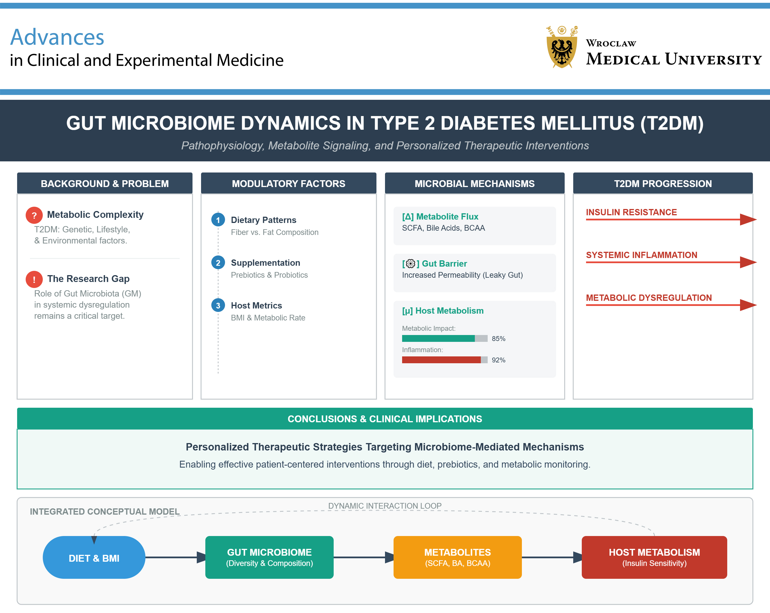
Type 2 diabetes mellitus (T2DM) is a complex metabolic disorder influenced by genetic, environmental, and lifestyle factors. Emerging evidence highlights the crucial role of the gut microbiota (GM) in the pathophysiology of T2DM, with alterations in microbial diversity and composition contributing to insulin resistance, systemic inflammation, and metabolic dysregulation. This review explores the current literature on the role of the GM, its metabolites, including short-chain fatty acids (SCFAs), bile acids (BAs), and branched-chain amino acids (BCAAs), as well as the impact of diet and body mass index (BMI) on the progression of T2DM. We examine how dietary patterns, including fiber intake, fat composition, and the use of prebiotics and probiotics, modulate the GM. Furthermore, we analyze evidence regarding the influence of the microbiome on host metabolism, gut permeability, and the pathophysiology of T2DM. Understanding the dynamic interactions among the GM, microbial metabolites, diet, and BMI may provide promising avenues for the development of personalized therapeutic strategies targeting microbiome-mediated mechanisms in the prevention and management of T2DM. Such approaches may facilitate more effective patient-centered interventions.
Key words: probiotics, diet, type 2 diabetes mellitus, body mass index, gut microbiota
Introduction
Type 2 diabetes mellitus (T2DM) is a prevalent chronic metabolic disorder characterized by hyperglycemia resulting from insulin resistance (IR) and/or inadequate insulin secretion.1 The global prevalence of T2DM has increased rapidly, with approx. 537 million adults affected in 2021. Projections suggest that this number will rise to approx. 783 million by 2045.2 Type 2 diabetes mellitus is closely associated with obesity and a sedentary lifestyle and is frequently accompanied by chronic low-grade inflammation and metabolic dysregulation.3 In recent years, accumulating evidence has implicated the gut microbiota (GM) – a complex community of trillions of microorganisms residing in the gastrointestinal tract – in both the pathogenesis and progression of T2DM.4 The human GM plays a critical role in nutrient digestion, energy harvest, immune modulation, and the production of bioactive metabolites, including short-chain fatty acids (SCFAs), bile acids (BAs), branched-chain amino acids (BCAAs), and trimethylamine N-oxide (TMAO). Disruption of its composition, referred to as dysbiosis, may adversely affect host metabolism.2 Studies have shown that individuals with T2DM often exhibit a distinct gut microbial profile compared with non-diabetic individuals.5
These alterations include changes in microbial diversity and shifts in the relative abundance of specific bacterial taxa, some of which have been associated with IR, impaired glucose metabolism, and increased gut permeability.6 These observations raise the question of whether gut dysbiosis contributes to the development or progression of T2DM. Conversely, T2DM itself, together with its common comorbidities, such as obesity, may also drive alterations in GM composition, suggesting a bidirectional relationship. Understanding this reciprocal interaction is crucial, as it may pave the way for novel therapeutic strategies targeting the GM. Dietary interventions, including high-fiber diets, the Mediterranean diet, ketogenic diets, probiotics, prebiotics, and even fecal microbiota transplantation, are currently being explored as approaches to modulate the GM and potentially improve metabolic outcomes in T2DM.6
This paper presents a literature review of recent evidence on GM alterations associated with T2DM, focusing on meta-analyses and systematic reviews published over the past 2 years. We systematically mapped current findings across microbial diversity and taxonomic composition, GM-derived metabolites, dietary influences, gut permeability, and microbiota-targeted therapeutic strategies.
Objectives
The objective of this review was to evaluate recent scientific evidence regarding alterations in GM composition and diversity associated with obesity and T2DM, as well as to analyze the role of microbial metabolites, including SCFAs, BAs, and BCAAs, in the pathophysiology of T2DM. The review further examines the impact of dietary patterns (such as fiber intake, fat composition, and the use of prebiotics and probiotics) on GM in individuals with T2DM, as well as the association between GM, BMI, and metabolic outcomes. In addition, it assesses the relationships among gut permeability, systemic inflammation, and IR as influenced by the GM in T2DM, and identifies potential microbiota-targeted therapeutic strategies, including dietary modulation, probiotics, and fecal microbiota transplantation, for the prevention and management of T2DM.
Methodology
The literature search was conducted using 2 electronic databases, PubMed and Embase, covering the period from January 1, 2022, to February 25, 2025. The search strategy included the following keywords: “type 2 diabetes and gut microbiota” and “changes in gut microbiota and type 2 diabetes.” The initial database search identified 2,087 records. Duplicate publications were removed prior to screening. Four authors (K.W., A.P., J.K., and R.S.) independently screened titles and abstracts to identify the most relevant studies. A total of 114 publications were selected based on the predefined inclusion criteria.
Diversity of gut microbiome
Microbial diversity can be assessed using alpha and beta diversity metrics. Alpha diversity reflects the richness and abundance of microbial species within a specific microbial community, whereas beta diversity measures the degree of variation between GM communities.5
Alpha diversity
A recent systematic review of cross-sectional studies involving 65,754 individuals, including 1,800 with T2DM and 2,770 with prediabetes, reported that alpha diversity was lower in T2DM and prediabetic cohorts in 86% of the included studies compared with healthy controls.6 These findings suggest a reduction in microbial richness and/or evenness associated with the diabetic state. However, a 2025 meta-analysis of 13 case-control studies found no consistent differences in alpha diversity between patients with T2DM and healthy control groups, highlighting substantial inter-study heterogeneity (p > 0.05; I2 > 75%).7 Similarly, a meta-analysis by Pinart et al. reported lower alpha diversity in obese individuals in a substantial proportion of the included studies; however, analysis of the Shannon index revealed no significant differences (mean difference (MD)= 0.06, p > 0.05, I2 = 81%).8 These inconsistencies likely reflect differences in BMI, medication use, dietary patterns, geographic location, and sequencing methodologies across studies.5, 9
Beta diversity
Nevertheless, numerous studies consistently indicate that beta diversity differs between individuals with and without diabetes. Thus, even when changes in overall microbial diversity are modest or inconsistent, shifts in the relative abundance of specific microbial taxa are frequently observed in diabetes, suggesting that the GM of individuals with diabetes differs from that of healthy individuals.5 Nevertheless, numerous studies consistently indicate that beta diversity differs between individuals with and without diabetes. Thus, even when changes in overall microbial diversity are modest or inconsistent, shifts in the relative abundance of specific microbial taxa are frequently observed in diabetes, suggesting that the GM of individuals with diabetes differs from that of healthy individuals.5 Slouha et al.1 analyzed studies involving populations of diverse ethnic backgrounds and observed similar outcomes. These findings are further supported by other authors.10, 11, 12, 13 Furthermore, Xu et al. demonstrated that dietary patterns, including the Mediterranean diet and ketogenic diets, significantly influence the beta diversity of the GM.14
Firmicutes:Bacteroidetes ratio
Firmicutes and Bacteroidetes are the 2 predominant bacterial phyla in the human gut, collectively accounting for approx. 90% of the GM.5 An early hypothesis suggested that obesity and metabolic disorders were associated with an increased Firmicutes:Bacteroidetes ratio. In a systematic review by Slouha et al., a trend toward a higher Firmicutes:Bacteroidetes ratio in individuals with elevated BMI was reported; however, this association was not statistically significant (Spearman’s ρ = 0.042, p = 0.579).1
Given that dietary fiber is predominantly metabolized by Bacteroidetes species, which produce beneficial SCFAs, this hypothesis appears biologically plausible.4 Moreover, although lipopolysaccharides (LPS) derived from Gram-negative bacteria were previously thought to promote inflammation, recent evidence suggests that Bacteroidetes may produce less pro-inflammatory or even anti-inflammatory LPS subtypes.15 Some studies have suggested that certain Firmicutes taxa may exhibit greater efficiency in extracting energy from dietary substrates, potentially contributing to obesity. Additionally, reductions in the abundance of Firmicutes or Bacteroidetes may promote the overgrowth of pro-inflammatory Proteobacteria, thereby exacerbating inflammatory processes.16 However, findings have been inconsistent, and the Firmicutes:Bacteroidetes ratio is now widely regarded as an oversimplified marker of gut dysbiosis.5
Taxonomic changes
As phylum-level analyses are increasingly regarded as oversimplified, research has shifted toward the investigation of specific taxonomic alterations. A systematic review encompassing 20 cohorts confirmed statistically significant changes in GM composition among individuals with T2DM, characterized by reduced abundances of beneficial taxa, including Akkermansia, Faecalibacterium, Bifidobacterium, Bacteroides, and certain Clostridiales. Conversely, T2DM was associated with significant enrichment of pro-inflammatory and opportunistic bacteria, particularly Escherichia–Shigella (often analyzed as a combined group due to 16S rRNA gene sequence similarity), Fusobacterium, Enterococcus, Ruminococcus, and Dorea. Interestingly, some studies also reported increased abundance of Lactobacillus species. Increased abundance of Gram-negative bacteria may contribute to enhanced endotoxin release, whereas reduced levels of Faecalibacterium may decrease butyrate production, a key anti-inflammatory metabolite and primary energy source for colonocytes.5, 9, 17
Moreover, studies indicate that, despite geographic and ethnic variability, common microbial patterns consistently emerge in T2DM, including reduced abundance of Faecalibacterium and increased abundance of Escherichia.9 A metagenomic meta-analysis further identified microbial gene signatures associated with butyrate synthesis and LPS production, which distinguished individuals with T2DM from healthy controls.2, 9
Bacteroides
The human GM can be broadly categorized into Bacteroides- and Prevotella-dominant enterotypes, with Bacteroides-dominant enterotypes being associated with high-fat, high-protein dietary patterns and reduced microbial diversity.18 Although Bacteroides-dominant enterotypes broadly correlate with dietary patterns and metabolic health, the effects at the genus and species levels vary considerably. The Bacteroides enterotype has been reported to be negatively correlated with levels of the tight junction scaffolding protein zonula occludens-1 (ZO-1) (r = −0.22 to −0.53, p < 0.05) and SCFAs (p < 0.05). Elevated serum ZO-1 levels in individuals with T2DM may indicate impaired gut barrier integrity.19
Five cross-sectional studies20, 21, 22, 23, 24 reported negative associations. For example, Munnuka et al.20 identified a significant inverse correlation between Bacteroides abundance and BMI (r = −0.28, p = 0.011). In contrast, the 3 interventional studies25, 26, 27 reported positive associations, potentially reflecting the antibiotic-like effects of metformin on the GM (p < 0.05). Additionally, Bacteroides abundance has been shown to be inversely correlated with BCAA levels, while therapeutic interventions may improve BCAA metabolism and reduce obesity. Animal studies have demonstrated that administration of Bacteroides significantly enhances valine oxidation in brown adipose tissue (p < 0.05), thereby promoting more efficient BCAA catabolism.4, 28 Notably, He et al. reported that 21 of 23 Bacteroides operational taxonomic units were negatively associated with T2DM.29
At the species level, Bacteroides intestinalis and Bacteroides vulgatus have been reported to be reduced in T2DM, whereas Bacteroides stercoris increases following surgery in patients with diabetes remission.24, 25, 30, 31 Animal studies have shown that Bacteroides acidifaciens and Bacteroides uniformis improve glucose intolerance (p < 0.05),32 whereas Bacteroides fragilis induces interleukin-10 (IL-10), thereby protecting against IR (p < 0.05). This effect appears to be mediated through the toll-like receptor 2 (TLR2) signaling pathway, as no significant increase in IL-10 was observed in mice lacking this receptor.33 Bacteroides fragilis has also been proposed as a key mediator of metformin’s metabolic effects.27 Furthermore, Bacteroides thetaiotaomicron enhances regulatory T-cell (Treg) gene expression, thereby reducing inflammation,33 while B. vulgatus and Bacteroides dorei improve gut barrier integrity, lowering LPS production and endotoxemia (p < 0.05).34 Additionally, B. acidifaciens enhances fatty acid oxidation through the TGR5–PPAR-α signaling pathway, thereby alleviating obesity and T2DM.35 These findings underscore the importance of distinguishing the roles of Bacteroides across different taxonomic levels to better understand their metabolic and immunological effects.
Faecalibacterium
Faecalibacterium, which constitutes more than 5% of the total intestinal bacterial population,36 is a SCFA-producing genus that appears to exert protective anti-inflammatory effects and is frequently reported to be reduced in individuals with T2DM.5, 37 Faecalibacterium prausnitzii has also shown potential in reducing IR by inducing glucagon-like peptide-1 (GLP-1) secretion through the free fatty acid receptor 2 (FFAR2).4 Additionally, F. prausnitzii has been shown to reduce intestinal inflammation by inhibiting nuclear factor kappa B (NF-κB) activation, suppressing the production of pro-inflammatory cytokines, and enhancing secretion of the anti-inflammatory cytokine IL-10. A recent study further demonstrated that butyrate produced by F. prausnitzii exerts anti-inflammatory effects by increasing the expression of Dact3, a gene associated with the Wnt/c-Jun N-terminal kinase (JNK) signaling pathway.38
Akkermansia muciniphila
Akkermansia muciniphila also produces SCFAs and influences metabolic pathways such as AMP-activated protein kinase (AMPK) and peroxisome proliferator-activated receptor alpha (PPARα), thereby regulating metabolism, lipid storage, and inflammation. It promotes gut barrier integrity through interactions with dendritic cells, activation of the ALPK1/TIFA signaling pathway, and modulation of mucus layer properties. Consequently, A. muciniphila is considered a promising next-generation probiotic.2, 5, 39
Liu et al. conducted a meta-analysis of 15 animal studies investigating the effects of A. muciniphila administration as a probiotic. Supplementation was associated with an average reduction in body weight of 10.4%; however, no clear dose–response relationship was observed, and the effects were more pronounced during the early stages of obesity (6–12 weeks). Administration of A. muciniphila also reduced fasting blood glucose levels by 21.2% and improved glucose tolerance by 22.1% (p < 0.001), with a pooled effect size of 0.43 (95% CI: 0.18–0.63). Moreover, circulating insulin levels increased by 26.9% (p < 0.001).40 Zhang et al. identified a significant co-occurrence relationship between Akkermansia and F. prausnitzii (ρ = 0.36, p < 0.05). This finding suggests potential synergistic benefits of combining these microorganisms as next-generation probiotics.41
Bifidobacterium
Bifidobacterium is a genus of fiber-fermenting bacteria.9, 42, 43 The relationship between Bifidobacterium abundance and T2DM appears to be complex. Several studies have reported reduced abundance of Bifidobacterium species in individuals with metabolic disorders, along with significant associations with greater bacterial richness and reduced intestinal permeability.2 However, some studies have linked higher Bifidobacterium abundance to hyperlipidemia. Moreover, in individuals with T2DM, correlations have been observed between Bifidobacterium abundance, melatonin-related metabolites, and inflammatory markers. Specifically, Bifidobacterium abundance was negatively correlated with serum melatonin levels (p < 0.01) and positively correlated with inflammatory markers, including LPS and IL-10 (false discovery rate (FDR) < 0.05).1
Escherichia–Shigella
Bednarska and Håberg reported a positive association between Escherichia–Shigella abundance and T2DM across multiple cohort studies, likely mediated by LPS and endotoxin-induced release of pro-inflammatory cytokines through nucleotide-binding oligomerization domain (NOD) and TLR signaling pathways.9
A meta-analysis of 32 studies also reported increased abundance of Escherichia–Shigella in individuals with obesity (p < 0.05).8 Additionally, Escherichia abundance has been associated with animal-based dietary patterns, whereas fiber-rich, plant-based dietary interventions may reduce its abundance.44
Lactobacillus
Lactobacillus and Bifidobacterium are common probiotic genera known to contribute to SCFA production, support intestinal barrier integrity, and reduce oxidative stress and inflammation in T2DM animal models, likely through modulation of inflammatory pathways.45 However, available evidence remains mixed, with some studies linking Lactobacillus abundance to higher fasting blood glucose and glycated hemoglobin (HbA1c) levels in individuals with T2DM. Other authors5, 46, 47 have suggested that Lactobacillus may contribute to T2DM progression by promoting GM-related inflammation (p < 0.05). Consistent with these findings, a Mendelian randomization study by Liu et al.48 also reported a potential association between Lactobacillus and increased risk of T2DM. This association may reflect species-specific effects, as certain Lactobacillus species may preferentially thrive in high-glucose environments. Alternatively, metformin treatment may promote their growth, suggesting that increased Lactobacillus abundance could represent a consequence of treatment rather than a contributing factor in T2DM pathogenesis.9
Other species associated with T2DM
Dorea, a genus within the Lachnospiraceae family, is frequently reported to be elevated in individuals with T2DM and has been associated with a pro-inflammatory gut environment, potentially expanding as beneficial microbial populations decline. In addition, increased abundances of Fusobacterium and Enterococcus have been reported in T2DM, along with Ruminococcus gnavus, a mucus-degrading species associated with inflammatory processes.5, 9
Gut microbiota-derived metabolites in T2DM
Short-chain fatty acids
Short-chain fatty acids, primarily acetate, propionate, and butyrate, are produced mainly through bacterial fermentation of dietary fiber and resistant starch in the colon.2 They serve as an energy source for colonic epithelial cells and function as signaling molecules by interacting with G protein-coupled receptors, including FFAR2, FFAR3, and G protein-coupled receptor 109A (GPR109A).2, 49
Moreover, SCFAs exert beneficial metabolic effects by enhancing gut barrier integrity, stimulating the secretion of GLP-1 and peptide YY (PYY), regulating glucose and lipid metabolism in peripheral tissues, and modulating the host metabolic state, including appetite regulation.1, 2, 17 For example, butyrate improves insulin sensitivity and exerts anti-inflammatory effects by inhibiting NF-κB activation. Evidence suggests that butyrate-producing bacteria are depleted in individuals with prediabetes and T2DM.2, 9, 50 Candela et al. reported that a fiber-rich diet significantly increased the abundance of SCFA-producing bacteria, including Faecalibacterium and Akkermansia (p = 0.01).23 Furthermore, SCFAs activate the SIRT1/AMPK/PGC-1α signaling pathway, which may represent a key mechanism underlying their anti-aging effects.51 Aging-associated alterations in the GM are characterized by reduced SCFA production and amylolytic activity, alongside increased proteolytic activity, thereby contributing to intestinal inflammaging in older individuals.52, 53 Overall, current evidence suggests that SCFAs may exert beneficial effects in T2DM by enhancing insulin secretion and satiety while reducing inflammation.2
One study further suggests that disturbances in SCFA production or absorption may contribute to the development of T2DM by inducing epigenetic modifications, particularly through alterations in DNA methylation.54
Lipopolysaccharide
Lipopolysaccharide, a structural component of the outer membrane of most Gram-negative bacteria, serves as a protective barrier and can translocate into the bloodstream, typically through impaired intestinal barrier integrity, thereby triggering an immune response. Numerous studies have implicated this mechanism in the development of systemic inflammation.55
Metabolic endotoxemia is characterized by persistently elevated circulating LPS levels that promote chronic low-grade inflammation and has been implicated in obesity-associated IR and T2DM. In the setting of gut dysbiosis, e.g., induced by high-fat dietary patterns or frequent antibiotic exposure, LPS-producing bacteria may proliferate, while intestinal barrier function becomes compromised, resulting in increased systemic LPS concentrations.56, 57 This, in turn, may trigger chronic inflammation within pancreatic islets, ultimately contributing to pancreatic β-cell dysfunction and reduced insulin sensitivity.2, 58, 59
Lipopolysaccharide activates toll-like receptor 4 (TLR4) expressed on immune and other cell types, thereby triggering the NF-κB and mitogen-activated protein kinase (MAPK) signaling pathways. In conjunction with TNF receptor-associated factor (TRAF) family E3 ligases, the TLR signaling cascade activates multiple transcriptional regulators, including NF-κB, activator protein 1 (AP-1), and interferon regulatory factors.60 Collectively, these factors drive the expression of genes involved in host immune responses. Activation of these pathways promotes the production of pro-inflammatory cytokines, including tumor necrosis factor alpha (TNF-α), interleukin-1 beta (IL-1β), and interleukin-6 (IL-6). These cytokines disrupt insulin signaling in insulin-responsive tissues and may consequently contribute to pancreatic β-cell dysfunction.2, 60, 61 In individuals with T2DM, elevated levels of inflammatory cytokines and markers of endotoxemia are frequently observed, supporting a link between gut-derived LPS and the chronic inflammatory state associated with the disease.2, 17, 61 Lipopolysaccharide may contribute to the pathogenesis of T2DM by perpetuating systemic inflammation. Mouse models have demonstrated that low-dose LPS infusion significantly promotes weight gain and IR (p < 0.05), whereas TLR4-knockout mice or administration of TLR4 antagonists significantly reduce LPS-induced production of pro-inflammatory cytokines, such as IL-6 and TNF-α (p < 0.01). Additionally, the GM composition observed in T2DM, characterized by enrichment of Gram-negative Enterobacteriaceae, including Escherichia coli, may favor increased LPS release.2, 9
Furthermore, individuals with T2DM frequently exhibit reduced expression of tight junction proteins in the intestinal epithelium, which may reflect dysbiosis-associated alterations in gut barrier integrity and facilitate LPS translocation.1 Taken together, these changes contribute to metabolic endotoxemia in T2DM, thereby exacerbating IR. Although therapeutic strategies aimed at reducing LPS burden have demonstrated improvements in metabolic outcomes, a recent animal study targeting macrophages showed that suppression of the inflammatory response alone was insufficient to improve insulin sensitivity (p > 0.05).62
Bile acids
Bile acids, the principal components of bile, are essential for the digestion and absorption of lipids, including the absorption of fat-soluble vitamins.63 In addition to their digestive functions, BAs act as signaling molecules involved in a variety of metabolic processes, particularly those regulating lipid and glucose metabolism.64
Bile acids also activate multiple BA receptors, including the farnesoid X receptor (FXR), G protein-coupled BA receptor 1 (GPBAR1), pregnane X receptor (PXR), and vitamin D receptor (VDR). Through these receptors, BAs regulate signaling pathways within a complex metabolic network controlling lipid, glucose, steroid, xenobiotic, and energy metabolism.65
In recent years, increasing attention has been directed toward the bidirectional relationship between BAs and the GM. Collins et al. emphasized the importance of BA–GM interactions not only in metabolic disorders, including T2DM, but also in neoplastic diseases and infectious conditions. Probiotic supplementation and antibiotic exposure may also substantially influence this interaction.66
Bile acids are frequently highlighted in the literature as key regulators in metabolic disorders, including T2DM, obesity, and prediabetic states. Bishay et al. emphasized the role of BA accumulation in the development of IR.67
Recent research from the University Medical Center Schleswig-Holstein (Kiel, Germany) further underscored the interaction between BA metabolism and the GM in metabolic disorders, including T2DM and prediabetes. In a study involving 492 individuals, impaired glycemic control was associated with elevated circulating BA levels, a higher proportion of secondary BAs, and an increased glycine-to-taurine conjugation ratio, particularly for cholic acid, chenodeoxycholic acid, and deoxycholic acid (p < 0.001, Kruskal–Wallis test).68
Alterations in GM composition, particularly in beta diversity, were significantly associated with changes in the BA pool (p = 0.0004), especially with secondary BAs such as deoxycholic acid (p = 0.001), independently of T2DM or obesity. Metabolic pathway differences associated with T2DM were primarily observed in bacterial taxa involved in cell membrane synthesis and polyamine production. Collectively, these findings suggest that targeting BA metabolism may represent a promising therapeutic strategy for prediabetes and T2DM.68
Dysbiosis in T2DM, particularly in cases characterized by depletion of Clostridia and Bifidobacterium species possessing bile salt hydrolase activity, may result in reduced BA deconjugation, thereby affecting farnesoid X receptor (FXR) signaling. Interestingly, interventions aimed at modulating BA pools have demonstrated metabolic effects. For example, a recent study involving a non-absorbed BA sequestrant in prediabetic animal models delayed diabetes onset by altering gut BA composition and improving glycemic control.27
Although not all studies have demonstrated a significant association between GM-mediated BA alterations and T2DM development, the available evidence consistently supports an interaction between these mechanisms in metabolic regulation. Nonetheless, targeting BA receptors, such as FXR and Takeda G protein-coupled receptor 5 (TGR5), through microbiota-based or pharmacological interventions remains an active area of research in metabolic disease.
Trimethylamine N-oxide
Trimethylamine N-oxide is predominantly generated from dietary substrates, including phosphatidylcholine, choline, carnitine, betaine, dimethylglycine, and ergothioneine, through GM-mediated metabolism in the colon. Its circulating levels are influenced by multiple factors, including age, sex, dietary patterns, GM composition, renal function, and hepatic flavin-containing monooxygenase activity.69
Trimethylamine (TMA), produced by gut bacterial metabolism of choline and L-carnitine, is absorbed into the bloodstream and subsequently rapidly oxidized to TMAO by hepatic flavin-containing monooxygenase 3 (FMO3).70
In recent years, increasing attention has been directed toward the association between the GM, its metabolites, including TMAO, and a broad range of diseases, particularly metabolic and cardiovascular disorders.71 Elevated TMAO levels may result from dietary factors, alterations in GM composition, dysbiosis, or impaired gut barrier function. Advances in sequencing technologies have substantially expanded our understanding of the GM and its role in health and disease. Diet plays a crucial role in shaping GM composition, thereby influencing the production of metabolites such as SCFAs and TMAO. While TMAO has been associated with an increased risk of cardiovascular disease (CVD) and mortality (p < 0.001), SCFAs are generally considered protective in metabolic health, and these metabolites should therefore not be grouped together in terms of risk interpretation. Bacterial taxa implicated in TMA production include members of Clostridia, Proteus, Shigella, and Aerobacter.70
In the gastrointestinal tract, certain bacterial taxa, such as Acinetobacter, can metabolize lecithin and carnitine to produce TMA, which is subsequently oxidized to TMAO in the liver. Analyses of fecal samples have shown that individuals with higher abundance of Prevotella species, often associated with high-fat dietary patterns, exhibit higher TMAO levels compared with those with Bacteroides-dominant microbiota.72
A growing body of evidence has demonstrated associations between TMAO, a GM-derived metabolite, and various lifestyle-related diseases, including IR, T2DM, and obesity.73, 74, 75 Kalagi et al. also emphasized the significant association between TMAO and T2DM.76
A study comparing individuals with T2DM and healthy controls reported significantly higher median plasma TMAO levels in the T2DM group (p < 0.001). This association remained significant after adjustment for age, BMI, and educational level. Even after accounting for physical activity and dietary factors, elevated TMAO levels were associated with a higher risk of T2DM. These findings suggest that increased circulating TMAO concentrations may be associated with T2DM.77
Tryptophan metabolites (indoles)
Indole-3-propionic acid (IPA) is one of the most abundant tryptophan-derived metabolites in the human gastrointestinal tract.78 Evidence suggests that IPA may mitigate the progression of metabolic disorders, including non-alcoholic fatty liver disease, T2DM, obesity, and CVD (p < 0.05 or p < 0.01, depending on the experimental model).79 Its proposed protective effects include modulation of inflammatory responses, reduction of oxidative stress, limitation of DNA damage, regulation of lipid metabolism, and support of immune function.80, 81
A cohort study by Qi et al. found that tryptophan, 4 kynurenine pathway metabolites (kynurenine, kynurenic acid, xanthurenic acid, and quinolinic acid), and indolelactate were positively associated with T2DM risk (p < 0.05), whereas IPA was inversely associated (p < 0.001). The study also identified genetic variants, dietary factors, and gut microbial interactions linked to these metabolites. Notably, dietary fiber intake showed the strongest association with tryptophan-derived metabolites, particularly IPA, which was associated with Firmicutes taxa involved in fiber metabolism.77
Moreover, a murine study demonstrated that GM-derived indoles influence intestinal barrier function through pregnane X receptor (PXR) signaling. Indole-3-propionic acid acts as a ligand for PXR, reducing TNF-α expression in enterocytes and enhancing the expression of intercellular junction proteins. PXR-deficient mice exhibited increased intestinal permeability and activation of the TLR signaling pathway; these effects were reversed by additional TLR4 silencing. Collectively, these findings suggest that GM regulate intestinal mucosal integrity through interactions involving the PXR and TLR4 signaling pathways.82
Several additional studies have also highlighted the role of indoles as signaling molecules that modulate GLP-1 secretion by enteroendocrine L cells in the colon,83 thereby influencing appetite regulation and potentially improving insulin sensitivity.84
Dysbiosis and gut permeability
Dysbiosis is defined as a disruption of the balance between symbiotic and pathobiont microbial populations, characterized by an increase in pathogenic microorganisms.22, 23 Alterations in the GM can impair intestinal barrier integrity and increase gut permeability, thereby facilitating the translocation of metabolites and pathogens from the intestine into the systemic circulation and peripheral tissues. Dysbiosis has been associated with increased adiposity, IR, enhanced energy harvest, elevated production of pro-inflammatory cytokines, and increased LPS translocation leading to metabolic endotoxemia. It may contribute to the pathogenesis of conditions such as obesity and T2DM, both of which are associated with systemic inflammation.42, 85 Increased intestinal permeability is considered an early event in the pathogenesis of T2DM and may be influenced by GM alterations. A controlled study found that individuals with T2DM matched for body weight, age, and BMI still exhibited increased intestinal permeability and higher bacterial endotoxin levels compared with individuals without diabetes.5, 86
Alterations in microbial diversity and shifts in the relative abundances of specific bacterial taxa have been shown to influence intestinal permeability. For instance, A. muciniphila has been shown to restore the intestinal mucosal layer.9 Furthermore, studies suggest that Lactobacillus species may enhance barrier integrity by regulating the expression of tight junction (TJ) proteins.86 Bifidobacterium administration has been shown to markedly decrease intestinal permeability by increasing TJ protein expression and reducing levels of inflammatory cytokines, including IL-6 and TNF-α.2, 9
Additionally, research indicates that SCFAs and BAs can activate multiple key receptors expressed by enteroendocrine cells, thereby increasing the secretion of GLP-1, glucagon-like peptide-2 (GLP-2), and PYY. This, in turn, may contribute to reduced blood glucose levels and improved intestinal barrier integrity.87 For instance, Roseburia has been shown to improve glucose homeostasis and intestinal permeability through SCFA production.2 Moreover, a reduced abundance of the Haemophilus genus in the intestine may promote SCFA production, potentially contributing to improved intestinal barrier function.48, 87
BMI association with gut microbiota in T2DM
Obesity is a chronic, complex disease characterized by excessive adipose tissue accumulation that may impair health and reduce quality of life. The World Health Organization (WHO) defines obesity in adults as a BMI of 30 kg/m2 or higher. Obesity is a major risk factor for T2DM, and a substantial proportion of individuals with this condition are overweight or obese.85 Both conditions are associated with GM disturbances and chronic low-grade inflammation. Disentangling the effects of elevated BMI and hyperglycemia on GM composition remains challenging, as these factors frequently coexist.42 Studies suggest that obesity itself induces specific alterations in the GM that may contribute to IR and metabolic dysfunction independently of diabetes.3, 86
High-carbohydrate dietary patterns, commonly observed in plant-based diets, tend to increase Prevotella abundance, which has been associated with more favorable glucose metabolism. Moreover, individuals with elevated BMI and diets high in fat and protein have been reported to exhibit an increased Firmicutes:Bacteroidetes ratio.42 In the context of T2DM, Carrizales-Sánchez et al. have observed a direct positive correlation between Prevotella abundance and BMI.87 Furthermore, Slouha et al. found that A. muciniphila levels were higher in individuals with T2DM and elevated BMI, and this was significantly associated with impaired lipid metabolism.1 This observation appears to contradict the established view that Akkermansia is generally beneficial, as its abundance is typically higher in lean individuals and lower in those with obesity.3 Elevated levels of Blautia and Serratia, along with reduced Akkermansia, have been observed in individuals with prediabetes and T2DM compared with normoglycemic controls, even when dietary intake and caloric consumption were similar across groups.40 These findings suggest that, in certain contexts, Akkermansia may increase in abundance in response to host metabolic disturbances, indicating a complex and context-dependent role. It is noteworthy that not all GM alterations associated with T2DM simply reflect obesity. While obesity-associated dysbiosis predisposes individuals to IR and T2DM, 3 certain microbial features appear to be associated with glycemic control independently of BMI. For instance, both lean and obese individuals with diabetes exhibit overgrowth of Escherichia–Shigella and reduced Faecalibacterium abundance.5, 9 These observations indicate that GM alterations in T2DM extend beyond obesity-related effects and warrant further investigation.
Dietary patterns and gut microbiota in T2DM
The Western diet is characterized by high intake of animal fats, low dietary fiber consumption, and an increased risk of gut dysbiosis, obesity, and T2DM.1 Following diets rich in animal fats and red meat, the GM ferments choline, carnitine, betaine, and lecithin to produce TMA, which is subsequently metabolized in the liver to TMAO. This metabolite has been shown to impair glucose tolerance and increase homeostatic model assessment of IR (HOMA-IR) and fasting insulin levels in mice fed a high-fat diet, thereby promoting adipose tissue inflammation and disrupting insulin signaling.5, 9, 17 Moreover, high-fat, low-fiber dietary patterns have been associated with reduced abundance of Bacteroidetes and increased abundance of Firmicutes and Proteobacteria.2 Specifically, such diets may increase the abundance of pro-inflammatory bacteria, such as Alistipes and certain Bacteroides species, while reducing beneficial taxa, including Faecalibacterium, potentially through mechanisms involving elevated LPS levels.1, 3, 23
In contrast, diets rich in complex carbohydrates and dietary fiber support a more diverse and resilient GM, increasing the relative abundance of SCFA-producing bacteria, such as Bifidobacterium, F. prausnitzii, and Akkermansia.4 Chong et al. observed that a high-fiber diet was associated with metabolic improvements. Dietary fiber intake has been associated with favorable effects on GM composition and improved metabolic homeostasis in individuals with T2DM.5 Furthermore, decreased fiber intake has been linked to a higher risk of T2DM. Contemporary dietary recommendations for diabetes management emphasize diets rich in plant-based foods, including whole grains, vegetables, whole fruits, legumes, nuts, and seeds. High-fiber dietary interventions have been associated with significant improvements in HbA1c and body weight reduction, potentially through the promotion of SCFA-producing bacteria.9
High-fiber dietary interventions in individuals with T2DM have also been associated with increased abundance of SCFA-producing bacteria. Enhanced SCFA production may promote increased GLP-1 secretion and reduced inflammatory activity, thereby contributing to improved glycemic control.4, 5, 9
Mediterranean diet
The Mediterranean diet is widely recognized as one of the healthiest dietary patterns and has been associated with a reduced risk of T2DM and better glycemic control.6, 85 The Mediterranean diet is rich in vegetables, fruits, legumes, whole grains, nuts, and healthy fats from olive oil and avocados, contains low amounts of red meat, and excludes processed foods and sugars. Plant-based foods are rich in polyphenols, flavonoids, and terpenoids, bioactive compounds that have been shown to exert nutrigenomic effects by modulating gene expression via epigenetic mechanisms.42 Polyphenols act as antioxidants, combating oxidative stress and chronic inflammation, while also improving insulin sensitivity.5, 9, 17
Adherence to the Mediterranean diet has been shown to increase the diversity of GM by modulating its composition and promoting a microbiome associated with metabolic health, reduced dysbiosis, and improved intestinal barrier integrity.85, 87 A study conducted by Özsoy et al. found that individuals following the Mediterranean diet have higher levels of Bifidobacteria, Lactobacilli, and SCFA-producing bacteria such as Faecalibacterium and Roseburia, along with lower levels of pro-inflammatory bacteria compared to those following the Western diet.85 Moreover, the Mediterranean diet has been associated with an increased Prevotella/Bacteroides ratio, potentially promoting gut homeostasis and improving glucose metabolism and insulin secretion.87 A study by Bednarska and Håberg found the Mediterranean diet to be effective in reducing fasting blood glucose and HbA1c. Given its beneficial effects on glycemic control, the Mediterranean diet is considered a preferred dietary pattern for T2DM management.9
Low-carbohydrate and ketogenic diets
The ketogenic diet, characterized by very low carbohydrate and high fat intake, effectively lowers HbA1c levels. This may be associated with increased abundance of Akkermansia and Parabacteroides.2, 4 The low-carbohydrate diet exhibits glycemic improvement largely by reducing exogenous glucose load rather than by promoting a high-SCFA-producing microbiome. Moreover, this diet has been associated with significantly higher relative abundances of Ruminococcus, Roseburia, and Eubacterium.4 Eubacterium may adapt to generate SCFAs from available substrates even under low-carbohydrate conditions, potentially contributing to a significant reduction in HbA1c.1, 4 The mechanisms underlying the ketogenic diet may include elevated GLP-1 levels and improved glucose homeostasis, which may be linked to increased growth of SCFA-producing bacteria such as Ruminococcus and Roseburia.1 Moreover, the very-low-calorie ketogenic diet (VLCKD) may induce elevated plasma concentrations of acetoacetic acid (ACA) and β-hydroxybutyric acid (β-OHB), as well as activation of white adipose tissue (WAT) lipolysis. Caloric restriction in individuals with T2DM may also reorganize GM composition and promote SCFA production. Ketogenic diets have additionally been associated with reduced levels of inflammatory T helper 17 (Th17) cells.2, 4 The ketogenic diet is being explored for its potential role in the management of obesity and other metabolic disorders.
Effects of probiotics on gut microbiota and glycemic control
Probiotics are living microorganisms that, when consumed in appropriate amounts, exert beneficial effects on the host’s health, such as reducing circulating LPS levels, fasting blood glucose, IR, and HbA1c levels. The underlying mechanisms of these beneficial effects remain under investigation. Studies suggest that probiotics decrease intestinal permeability and secretion of pro-inflammatory mediators while increasing production of bacteriocins and SCFAs. Short-chain fatty acids facilitate communication between the intestinal lumen and the rest of the body, thereby regulating energy metabolism and adipose tissue expansion. Through these mechanisms, butyrate-producing taxa may represent a promising therapeutic strategy for managing T2DM.88, 89
A 2023 meta-analysis of 22 randomized controlled trials (RCTs) concluded that probiotic supplementation was associated with modest reductions in HbA1c, fasting glucose, and HOMA-IR in individuals with T2DM. It highlighted certain strains, such as L. rhamnosus GG and B. animalis, as potentially useful adjuncts in diabetes management.90
Moreover, various meta-analyses support these findings.91, 92 However, studies conducted by Paquette et al. failed to establish a consistent pattern of glycemic improvement, as none of the analyzed controlled trials demonstrated a uniform reduction across all major glycemic parameters with probiotic use. This lack of consistency in glycemic outcomes following probiotic intervention may be attributed to differences in probiotic composition, duration of supplementation, and dosage.93
Probiotic strains
Multiple RCTs have demonstrated that specific probiotic strains may improve glycemic control and enhance insulin sensitivity in individuals with T2DM; however, their composition, optimal dosage, and interactions remain under investigation.14
Most of the available evidence has been derived from mouse models, with L. plantarum and L. rhamnosus being the most frequently studied strains. Beneficial effects of L. reuteri, L. casei, and L. paracasei have also been frequently reported.94, 95, 96 Moreover, when combined with Bifidobacterium species, these strains may exhibit synergistic effects, potentially contributing to diabetes prevention.97
Lactobacillus and Bifidobacterium are among the most extensively studied probiotic genera and have been shown to influence T2DM progression through multiple mechanisms, including increasing GLP-1 secretion, decreasing cholesterol absorption, regulating pancreatic β-cell activity, and reducing hepatic gluconeogenesis.95, 96, 98 Campaniello et al. observed that combining Lactobacillus, Bifidobacterium, and Streptococcus thermophilus strains improves lipid profiles and lowers fasting glucose levels in humans.94 Likewise, Tegenge et al.95 reported in their meta-analysis that combinations of probiotic strains may inhibit weight gain. Furthermore, Li et al.99 suggested that the synergistic effects of Lactobacillus and Bifidobacterium contribute to reductions in HbA1c and markers of oxidative stress. Consistent with these findings, Shen et al.100 also reported beneficial effects of probiotic combinations on body mass.
Furthermore, A. muciniphila has been shown to contribute to restoration of the intestinal mucosal layer and is believed to reduce intestinal permeability, thereby exerting anti-inflammatory effects as a probiotic species and potentially reducing the risk of T2DM.9, 40, 101 Other bacteria with a potentially beneficial impact on glucose metabolism include F. prausnitzii and S. thermophilus.102 Analyzed data regarding the role of selected probiotic strains, their influence on T2DM progression parameters, and the study samples used are summarized in Table 1.9, 40, 90, 94, 95, 96, 97, 98, 99, 102
Additionally, genetic engineering methods such as CRISPR/Cas9 are increasingly being used to modify probiotic genomes and enhance the functionality of specific strains. Clustered Regularly Interspaced Short Palindromic Repeats (CRISPR) systems are being used to edit species such as Clostridium butyricum and Bacteroides strains, contributing to a better understanding of beneficial metabolic pathways influencing gut permeability and metabolite production. Although inserting DNA into certain bacterial species remains challenging, CRISPR is considered a promising tool for next-generation microbiome therapies.103
Probiotics’ mechanisms of influencing T2DM
Probiotics influence the progression of diabetes through various mechanisms, such as enhancing intestinal barrier integrity, alleviating oxidative stress, modulating immune responses, and promoting the production of SCFAs.104
The primary pathway involves the interaction of SCFAs with G protein-coupled receptors (GPCRs), particularly GPR41 and GPR.43 Activation of GPR43 enhances insulin sensitivity and reduces inflammation by promoting glucose uptake and stimulating GLP-1 secretion.105, 106 Moreover, SCFAs act through peroxisome proliferator-activated receptors (PPARs), which modulate insulin signaling in the liver, adipose tissue, and skeletal muscle, potentially improving insulin sensitivity. In the liver, PPARα promotes fatty acid oxidation and reduces triglyceride accumulation, which may impair insulin signaling. Moreover, PPARβ/δ increases fatty acid utilization in skeletal muscle, thereby supporting improved glucose uptake.107, 108 Additionally, probiotics have been associated with increased secretion of PPY which may reduce IR and improve β-cell function.109, 110
Moreover, Lactobacillus and Bifidobacterium promote intestinal colonization by Muribaculaceae, a mucin glycan- and dietary fiber-metabolizing bacterial family that produces SCFAs beneficial to gut health, whose abundance decreases in obesity and T2DM but may be supported by plant-based diets and probiotic supplementation.111 Although the exact mechanisms remain unclear, probiotics may reduce leptin levels, thereby decreasing food intake and promoting fat mass reduction. Consequently, this may improve insulin sensitivity in peripheral tissues.112
Furthermore, probiotics may influence the expression of glucose transporter type 4 (GLUT-4) and contribute to reactive oxygen species (ROS) neutralization, enzyme activity inhibition, and metal ion chelation, thereby exerting antioxidant effects.113 In RCTs, probiotics have been shown to reduce serum interleukin-18 (IL-18) levels and improve kidney function in individuals with T2DM, potentially by limiting activation of the NLR family pyrin domain containing 3 (NLRP3) inflammasome, which is activated by this interleukin.114
Prebiotics
Prebiotics are non-viable dietary substrates that are selectively utilized by host microorganisms to confer health benefits. Early research focused mainly on carbohydrate-based compounds such as inulin, fructo-oligosaccharides (FOS), and galacto-oligosaccharides (GOS), which are not digested in the small intestine but are instead fermented by gut bacteria to produce SCFAs.89, 93
Moreover, these metabolites contribute to pathogen inhibition and reinforcement of gut barrier integrity through end products such as acetate and lactate.93, 103 Prebiotics also promote the proliferation of beneficial bacteria, such as Bifidobacterium and Lactobacillus, while suppressing the growth of potentially detrimental taxa, such as E. coli and Clostridium. Hence, prebiotics contribute to a more stable intestinal environment. This modulation of the GM by prebiotics has also been associated with reduced levels of uremic toxins and systemic inflammation, suggesting broader relevance in chronic kidney disease (CKD) and other metabolic conditions.114
These effects have been linked to improvements in glycemic parameters in T2DM, including reductions in fasting glucose (FG), HbA1c, and IR. A study by Garcia-Gutierrez et al. highlighted that beneficial effects were most pronounced, particularly with longer inulin interventions lasting 8 weeks or more.89 Furthermore, inulin may exert additional metabolic benefits by modulating GLP-1 secretion and reducing inflammatory markers. Li et al. observed that combining probiotics and prebiotics into synbiotics may yield synergistic effects, more effectively lowering FG and inflammatory markers compared with either component alone.99
Moreover, higher-dose prebiotic supplementation (≥10 g/day) has demonstrated a more beneficial impact on lipid profiles, including total cholesterol, low-density lipoprotein (LDL), very low-density lipoprotein (VLDL), high-density lipoprotein (HDL), and triglycerides. This effect is likely mediated through restoration of microbial diversity, which is progressively diminished by Westernized, ultra-processed dietary patterns worldwide.107
Limitations of the study
This review is limited by the fact that most available data are derived from geographically restricted populations. Additionally, phylum-level analyses often overlook strain-level differences that are crucial to host–microbe interactions. Mechanistic understanding is further constrained by the limited availability of biochemical and molecular tools for direct investigation of metabolite–receptor interactions. The limited number of longitudinal studies and inconsistent findings across cohorts highlight the need for standardized, large-scale investigations.
Conclusions
Type 2 diabetes mellitus and the GM have an intertwined relationship, with each influencing the imbalance of the other. Gut dysbiosis impairs glucose homeostasis and promotes systemic inflammation through increased intestinal permeability, decreased SCFA production, dysregulated BA signaling, and elevated TMAO and LPS levels, while hyperglycemia and obesity further remodel microbial composition. Studies have observed an overall reduction in microbial diversity and enrichment of pro-inflammatory genera, most notably Escherichia–Shigella. Higher BMI has been associated with an increased Firmicutes:Bacteroidetes ratio and elevated A. muciniphila abundance in individuals with T2DM, which has been linked to impaired lipid metabolism. Moreover, decreased F. prausnitzii abundance has been associated with IR, potentially through reduced GLP-1 induction. These microbial alterations underscore a potential association between T2DM and the GM. Probiotic and prebiotic interventions have demonstrated improvements in glycemic control and gut barrier integrity in T2DM. Genetic engineering approaches, such as CRISPR/Cas9-mediated probiotic genome modification, may offer potential for more targeted microbiome-based therapies. The relationship between the GM and T2DM remains incompletely understood and requires further focused investigation. Recent evidence suggests that certain bacterial taxa may influence insulin secretion and IR, thereby contributing to T2DM pathogenesis. Future research should focus on functional analyses of gut microbes, as well as the effects of probiotics and lifestyle modifications, to improve metabolic outcomes in T2DM.
Use of AI and AI-assisted technologies
Not applicable.