Advances in Clinical and Experimental Medicine

Title abbreviation: Adv Clin Exp Med
5-Year IF – 2.0, IF – 1.9, JCI (2024) – 0.43
Scopus CiteScore – 4.3
Q1 in SJR 2025, SJR score – 0.599, H-index: 54 (SJR)
ICV – 161.00; MNiSW – 70 pts
Initial editorial assessment and first decision within 24 h

ISSN 1899–5276 (print), ISSN 2451-2680 (online)
Periodicity – monthly

Download original text (EN)

Advances in Clinical and Experimental Medicine

Ahead of print

doi: 10.17219/acem/218006

Publication type: review

Thematic category: Neurology

Language: English

License: Creative Commons Attribution 3.0 Unported (CC BY 3.0)

Download citation:

  • BIBTEX (JabRef, Mendeley)
  • RIS (Papers, Reference Manager, RefWorks, Zotero)

Cite as:


Kunecki KA, Dziadkowiak E. Autoimmune mechanisms in drug-resistant focal epilepsy: Pathophysiology, biomarkers, and therapeutic implications [published online as ahead of print on April 27, 2026]. Adv Clin Exp Med. 2026. doi:10.17219/acem/218006

Autoimmune mechanisms in drug-resistant focal epilepsy: Pathophysiology, biomarkers, and therapeutic implications

Kamil Aleksander Kunecki1,B,C,D, Edyta Dziadkowiak2,A,E,F

1 Student Scientific Association of Neurology, Wroclaw Medical University, Poland

2 Clinical Department of Neurology, University Centre of Neurology and Neurosurgery, Wroclaw Medical University, Poland

Graphical abstract


Graphical abstracts

Highlights


• Autoimmune mechanisms contribute to drug-resistant epilepsy (DRE) pathogenesis and pharmacoresistance.
• Neuronal autoantibodies (e.g., NMDAR, GluR3) and neuroinflammation are linked to focal epilepsy.
• mTOR activation, blood–brain barrier dysfunction, and efflux transporters drive antiseizure drug resistance.
• Immunomodulatory therapies (IVIG, corticosteroids, rituximab) may reduce seizures in autoimmune epilepsy.
• Early identification of autoimmune biomarkers supports personalized treatment in drug-resistant epilepsy.

Abstract

This review explores the role of autoimmune mechanisms in drug-resistant epilepsy (DRE), with emphasis on etiology, mechanisms of drug resistance, and potential immunomodulatory interventions. A structured review of clinical and experimental studies was conducted to assess current knowledge on the interplay between molecular mechanisms and immune responses in DRE. Particular attention was given to the involvement of the mTOR pathway, blood–brain barrier (BBB) dysfunction, astrocyte activation, and overexpression of efflux transporters, as well as the presence of neuronal autoantibodies such as N-methyl-D-aspartate receptor (NMDAR) and GluR3. The available evidence suggests that, although conventional pharmacologic approaches fail in a subset of patients, immunologic diagnostics and targeted therapies – including intravenous immunoglobulin, corticosteroids, rituximab, and plasmapheresis – may provide clinical benefit. Seizure reduction has been reported in selected patients; however, therapeutic response remains heterogeneous and is limited by small study populations. In conclusion, early identification of autoimmune mechanisms in DRE may help optimize treatment strategies, improve patient outcomes, and reduce the long-term clinical and societal burden of the disease.

Key words: drug-resistant epilepsy, epilepsy, IVIG, autoimmune epilepsy, focal seizures

Introduction

Epilepsy is considered drug-resistant when seizures persist despite treatment with at least 2 appropriately selected, well-tolerated, and adequately dosed antiepileptic drugs (AEDs), either as monotherapy or in combination, according to the International League Against Epilepsy (ILAE) Therapeutic Working Group. A unified definition of drug-resistant focal epilepsy (DRE) is essential, as it facilitates timely diagnosis, informs treatment strategies, and supports clinical research. A clear definition also enables non-specialist clinicians to recognize and manage this condition effectively.

The ILAE framework for defining DRE is structured in 2 hierarchical levels. Level 1 categorizes response to therapy as seizure-free, treatment failure, or undetermined, with further classification based on the presence of adverse effects. This level emphasizes 2 key indicators: seizure control and treatment tolerability. Accurate assessment requires that therapies be appropriately selected, dosed, and administered, and that response be evaluated using consistent clinical criteria, such as the “rule of 3,” which defines a seizure-free period as at least 3 times longer than the longest pre-treatment seizure interval and lasting at least 12 months. Level 2 defines DRE based on evidence from level 1. Although treatment response may fluctuate over time, the likelihood of achieving remission after 2 adequate therapeutic trials is low. Therefore, failure of at least 2 appropriate medications generally defines drug-resistant epilepsy. This definition also incorporates seizure frequency and duration of observation, reflecting the persistent and dynamic nature of the disorder.

Drug-resistant epilepsy has substantial medical and social consequences. Medically, frequent seizures increase the risk of injury, cognitive decline, psychiatric disorders, adverse effects of AEDs, and sudden unexpected death in epilepsy (SUDEP). Socially, patients may experience stigma, unemployment, and increased economic burden related to treatment costs and disability. Recurrent seizures may also promote secondary epileptogenesis, further complicating disease management. Although substantial research has been conducted on DRE, autoimmune-related DRE, particularly in focal epilepsy, remains underexplored. Epilepsy affects approx. 0.5–1% of the global population, and autoimmune etiology may account for 10–20% of cases with acute onset; however, neuronal autoantibodies are identified in fewer than 10% of patients with focal DRE. Determining the precise cause of epilepsy, particularly in cases associated with hippocampal sclerosis, remains challenging. It is unclear whether hippocampal sclerosis develops idiopathically or as a consequence of autoimmune or other pathological processes. Given these challenges and gaps in the literature, this review aims to synthesize current knowledge on autoimmune-related focal DRE and provide an overview of recent research in this field.1, 2, 3, 4, 5, 6, 7, 8, 9, 10, 11, 12

Etiology of drug-resistant epilepsy

The etiology of DRE is multifactorial. In a substantial proportion of cases, apparent drug resistance reflects so-called pseudo-drug resistance, which may result from factors such as misdiagnosis of epilepsy or inappropriate drug selection. In such cases, treatment optimization may lead to clinical improvement. True drug resistance is associated with factors related to the etiopathogenesis of the epilepsy syndrome, drug-specific properties (e.g., the development of tolerance), and interindividual variability in treatment response, including genetic and environmental factors as well as patient age. Approximately 30% of patients with DRE are candidates for surgical treatment. For the remaining patients, alternative therapeutic strategies should be considered, including evaluation of the potential role of immunomodulatory therapy.

Several theories have been proposed to explain drug resistance in epilepsy. A key component of epileptogenesis involves the mechanistic target of rapamycin (mTOR) pathway, which consists of 2 protein complexes: mTORC1 and mTORC2. The mTORC1 signaling pathway serves as a central regulatory hub that integrates signals from growth factors, nutrient availability, and cellular energy status, thereby regulating cell growth and metabolism. The mTORC1 complex, composed of the mTOR kinase, the adaptor protein Raptor, mLST8, and the regulatory proteins PRAS40 and DEPTOR, is primarily activated at the lysosomal surface in response to amino acid signaling mediated by Rag GTPases and insulin-dependent signals transmitted through the PI3K–Akt–TSC1/2–Rheb axis. Once activated, mTORC1 phosphorylates key substrates, including S6 kinase 1 (S6K1) and 4E-binding protein 1 (4E-BP1), promoting translation initiation, protein synthesis, and ribosome biogenesis while inhibiting autophagy through ULK1 phosphorylation.

The mTORC2 signaling pathway serves as a critical regulator of cellular homeostasis, cytoskeletal organization, and cell survival. The mTORC2 complex, composed of mTOR kinase, Rictor, mLST8, and the regulatory proteins mSIN1 and Protor, differs from mTORC1 in both composition and activation mechanisms, being less dependent on nutrient availability and more responsive to growth factor signaling, including receptor tyrosine kinase activation. mTORC2 directly phosphorylates and activates AGC family kinases, including Akt (Ser473), SGK1, and PKC, thereby modulating cell survival, glucose metabolism, cytoskeletal reorganization, and cell polarity. Additionally, mTORC2 influences lipid homeostasis and cellular responses to oxidative stress. Under physiologic conditions, mTOR activity is negatively regulated by several factors, including the tumor suppressor proteins TSC1 (tuberous sclerosis complex 1) and TSC2 (tuberous sclerosis complex 2), which form a heterodimer. Other regulators include phosphatase and tensin homolog (PTEN) and STE20-related kinase adaptor alpha (STRADα). Mutations in genes encoding these proteins lead to dysregulation of cell proliferation, differentiation, and growth. mTORC1 regulates mRNA translation, thereby influencing synaptic function, plasticity, and the expression of neurotransmitter receptors. Consequently, the mTOR pathway contributes to both short- and long-term cellular processes, which depend on protein synthesis. Given its role in neuronal development and synaptic plasticity, hyperactivation of the mTOR pathway has been implicated in aberrant axonal growth and neurogenesis, key processes in epileptogenesis. 13, 14, 15, 16, 17, 18, 19, 20, 21, 22, 23, 24, 25, 26, 27, 28, 29

Immune etiology of drug-resistant epilepsy

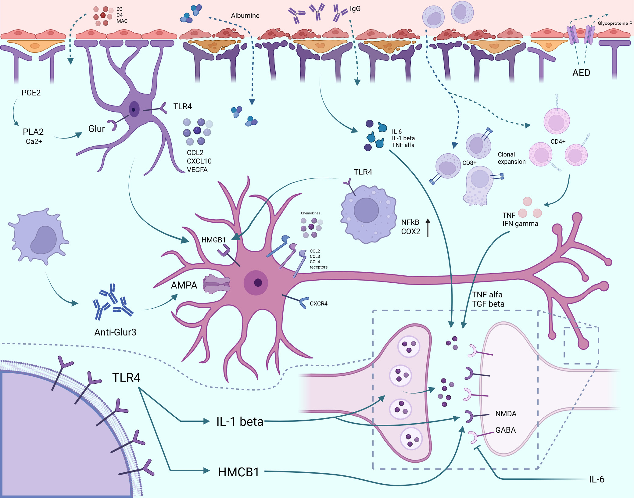
Inflammatory mediators activated during neuroinflammatory processes in DRE contribute to increased blood–brain barrier (BBB) permeability. This is associated with disruption of tight junctions and enhanced transcytosis, leading to vascular leakage within the brain. A key factor in this process is the proinflammatory phenotype of astrocytes, which express increased levels of inflammatory mediators in response to BBB dysfunction. This self-reinforcing cycle may contribute to seizure recurrence. Increased BBB permeability allows albumin to extravasate into the brain parenchyma, which may alter the pharmacokinetics and efficacy of antiseizure medications. Additionally, BBB disruption activates compensatory mechanisms, including upregulation of drug efflux transporters such as P-glycoprotein (P-gp). Increased expression of these transporters in endothelial and perivascular glial cells contributes to reduced drug penetration and pharmacoresistance.

Inflammatory processes may contribute to the etiopathogenesis of DRE, as exemplified by Rasmussen encephalitis (RE), which is characterized by drug-resistant seizures and driven by cytotoxic CD8+ T lymphocytes interacting with neurons and astrocytes. Conversely, inflammation may also arise as a consequence of epilepsy itself, as observed in temporal lobe epilepsy (TLE). Interleukin (IL)-1β appears to play a central role in the development of DRE and is produced by multiple cell types, including endothelial cells of the BBB. It may be particularly relevant during spontaneous seizures. In addition to IL-1β, increased levels of IL-6 and tumor necrosis factor alpha (TNF-α) have been reported in epileptogenic tissue. The role of chemokines and their receptors in the etiopathogenesis of DRE has also been suggested, including CCL2, CCL3, CCL4 (chemokine [C-C motif] ligands), and CXCR4 (C-X-C chemokine receptor type 4). Cyclooxygenase 2 (COX-2) also plays an important role, as its activity may promote seizure recurrence by increasing the expression of P-gp at the BBB, thereby reducing drug penetration from plasma into brain tissue and contributing to pharmacoresistance. Additionally, increased expression of toll-like receptor 4 (TLR4) and high-mobility group box 1 (HMGB1) has been observed in human neural tissue following seizures, suggesting that signaling through these molecules may contribute to seizure generation. The prevalence of drug resistance in epilepsy varies by seizure type. It is more common in patients with frequent seizures and occurs more often in focal epilepsy than in generalized epilepsy. Hippocampal sclerosis is present in 50–75% of cases of drug-resistant temporal lobe epilepsy and is associated with drug resistance in up to 89% of affected patients. A high prevalence of drug resistance is also observed in patients with temporal lobe cortical malformations (76%). When both hippocampal sclerosis and cortical malformations are present, the likelihood of drug resistance increases to 97%, whereas in other conditions it is approx. 65%.19, 30, 31, 32, 33, 34

Differences in the pathomechanisms of autoimmune drug-resistant epilepsy in children and adults

Autoimmune DRE exhibits distinct pathophysiological differences between pediatric and adult populations. In children, innate immune mechanisms appear to predominate and are characterized by sustained microglial activation and cytokine-mediated neuroinflammation. These processes often occur in the absence of identifiable autoantibodies or in the presence of antibodies of uncertain clinical significance. When detected, the most commonly reported autoantibodies include anti-N-methyl-D-aspartate receptor (anti-NMDAR), anti-myelin oligodendrocyte glycoprotein (MOG), and anti-glutamic acid decarboxylase 65 (GAD65). In pediatric patients, epileptogenesis appears to be more closely associated with neuroinflammatory mechanisms than with classical antibody-mediated autoimmunity. Inflammation in the immature, highly plastic brain may result in persistent alterations in neuronal network development and organization, thereby facilitating secondary epileptogenesis and more widespread forms of DRE.

In adults, autoimmune DRE more commonly involves adaptive immune mechanisms, with autoantibodies directed against neuronal surface antigens and ion channels, including the NMDAR, leucine-rich glioma-inactivated protein 1 (LGI1), and contactin-associated protein-like 2 (CASPR2). These antibodies directly disrupt synaptic neurotransmission and are typically associated with focal epilepsy. These age-related differences have important clinical implications, as immunotherapy may be effective in children even in the absence of defined autoantibody specificity, whereas in adults, treatment response is more closely linked to the underlying autoantibody profile.11, 35, 36, 37, 38, 39, 40

Materials and methods

This review was conducted in accordance with the Preferred Reporting Items for Systematic Reviews and Meta-Analyses (PRISMA) guidelines and is based on available data on patients with DRE of presumed or confirmed immunologic etiology. The review also includes case reports, clinical registry data, and studies on the use of immunomodulatory therapies.

Search strategy

The literature search was conducted using the following sources: PubMed, ClinicalTrials.gov, ILAE documents, and relevant literature in neurology and neuroimmunology. Publications available up to February 2025 were included. The following English-language keywords were used: (“drug-resistant epilepsy” OR “refractory epilepsy”) AND (“autoimmune” OR “immune-mediated” OR “autoantibodies”) AND (“focal seizures” OR “temporal lobe epilepsy”) AND (“IVIG” OR “methylprednisolone” OR “immunotherapy” OR “rituximab”). Only publications with abstracts available in English or Polish were included. Additionally, data from clinical trial registries and published case reports were analyzed. The protocol was registered in PROSPERO (ID: 1086558).

Inclusion and exclusion criteria

Inclusion criteria were as follows: 1) case reports, clinical trials (ClinicalTrials.gov), meta-analyses, original studies, and systematic reviews involving patients with DRE of possible autoimmune origin; 2) studies reporting immunomodulatory treatment (intravenous immunoglobulin (IVIG), methylprednisolone, rituximab, plasmapheresis); and 3) studies including clinical assessment of treatment response. Exclusion criteria were as follows: 1) articles not available in full text; 2) studies conducted exclusively in animals; 3) studies addressing other forms of epilepsy (e.g., idiopathic generalized epilepsy); and 4) case reports not related to autoimmune mechanisms.

Data extraction and quality assessment

Data extracted from the included studies comprised patient age and sex, epilepsy type, presence of autoantibodies, type of immunomodulatory therapy, seizure frequency and severity, and clinical response. Publications were manually reviewed for eligibility according to the inclusion criteria. Study selection was performed independently by both authors (K.A.K. and E.D.), with discrepancies resolved through discussion. No formal assessment of risk of bias was performed due to the narrative nature of the review.

Synthesis methods

Data were compiled in tables: Table 137, 41, 42, 43, 44 contains case reports; Table 245, 46, 47, 48 compares clinical trials and provides a qualitative narrative analysis; Table 349, 50, 51, 52 summarizes autoimmune and inflammatory markers in focal epilepsy and DRE; and Table 453, 54, 55, 56, 57, 58, 59, 60, 61, 62, 63, 64, 65, 66 presents key findings in drug-resistant autoimmune epilepsy with focal seizures. Meta-analysis was not performed due to data heterogeneity and the limited number of comparable studies. Data and mechanisms related to the etiology of DRE are also presented graphically in Figure 1.

Data coding and analysis

Treatment response was defined as clinical improvement, including reduction in seizure frequency, improvement in electroencephalography (EEG) findings, neuroimaging results, or neurological status, according to descriptions in the original publications. Changes in EEG recordings, particularly the presence of interictal epileptiform discharges (IEDs), have been associated with the risk of seizure recurrence. A reduction in the frequency of epileptiform discharges is often considered a favorable indicator of therapeutic response; however, it does not always directly correlate with a reduction in seizure frequency. In clinical practice, EEG serves as one of several tools for assessing treatment efficacy and prognosis.67, 68 Data from studies involving patients with antineuronal antibodies (e.g., NMDAR, GluR3) were analyzed to evaluate the efficacy of IVIG, corticosteroids, rituximab, and other immunomodulatory therapies.

Only therapies with quantitative efficacy data were included in the analysis. The most frequently reported interventions were IVIG, steroids, and plasmapheresis. Disease severity and treatment effects were assessed using neurological evaluation, EEG, magnetic resonance imaging (MRI), documentation of seizure frequency before and after treatment, and clinical response as defined in each study. Variation in rating scales across studies limited the possibility of performing meta-analysis; therefore, a descriptive approach was applied. From an etiologic perspective, epilepsy may be classified as cryptogenic (of unknown origin), including cases of DRE. The occurrence of such forms and the still incompletely understood etiopathogenesis suggest that mechanisms beyond currently established concepts may contribute to disease development. These include, for example, imbalances between excitatory postsynaptic potentials (EPSPs) and inhibitory postsynaptic potentials (IPSPs), leading to excessive neuronal depolarization. One proposed hypothesis is that autoimmune processes may underlie the development of certain forms of epilepsy. Early studies focused on alterations in the levels and ratios of nonspecific immunoglobulins, such as immunoglobulin G (IgG) and immunoglobulin M (IgM); however, these changes are now considered more likely to be consequences of seizures rather than their primary cause.45, 69 Rasmussen encephalitis is a prototypical example of an autoimmune disease associated with focal seizures. In this condition, autoantibodies against the α-amino-3-hydroxy-5-methyl-4-isoxazolepropionic acid (AMPA) receptor, particularly the GluR3 subunit, have been described. These autoantibodies may also contribute to complement activation, including components such as C3, C8, and the membrane attack complex (MAC). Under physiologic conditions, IgG antibodies do not cross the BBB; however, disruption of BBB integrity may allow anti-GluR3 antibodies to access neuronal targets and contribute to disease pathogenesis. Recent evidence also highlights the role of cellular immune responses, with increased infiltration of CD8+ T lymphocytes. CD4+ T cells may also undergo clonal expansion and secrete proinflammatory cytokines, including tumor necrosis factor (TNF) and interferon gamma (IFN-γ).45, 70, 71, 72

Some autoimmune diseases are also characterized by an increased frequency of seizures. This is the case with systemic lupus erythematosus (SLE), where there is an increase in the level of antiphospholipid antibodies (aPL), antibodies against cardiolipin (aCL), B2-glycoprotein, dsDNA, and antinuclear antibodies (ANA). The increase in these antibodies correlates with the frequency of seizures in patients with SLE, which is estimated to be 28%, while the occurrence of seizures ranges from 17% to 37%.73, 74

Other conditions potentially associated with autoimmune epileptogenesis include:

− Stiff-person syndrome, characterized by elevated levels of autoantibodies against glutamic acid decarboxylase (GAD), the enzyme responsible for γ-aminobutyric acid (GABA) synthesis;

− West syndrome and Lennox–Gastaut syndrome, in which the etiology remains incompletely understood; however, a more favorable response to immunotherapy than to standard antiseizure medications suggests a potential immune-mediated component;

− Landau–Kleffner syndrome (LKS), in which clinical improvement has been observed following treatment with IVIG, along with evidence of intrathecal IgG synthesis, as indicated by elevated cerebrospinal fluid (CSF) IgG levels;

− Hashimoto encephalopathy;

− Channelopathies.

Some antiseizure medications have immunosuppressive effects, particularly carbamazepine, phenytoin, and valproate. These agents may reduce lymphocyte counts, alter the CD4+/CD8+ ratio, and decrease immunoglobulin levels, including IgG, immunoglobulin A (IgA), and IgM. Their effects on cytokine levels remain less well defined. Evidence supporting the efficacy of IVIG therapy has been reported. In a study by Biliau et al.,58 involving 13 children with DRE, a significant reduction in seizure frequency was observed in 4 patients, a moderate reduction (20% to 50%) in 3 patients, no change in 5 patients, and an increase in seizure frequency in 1 patient. Intravenous immunoglobulig therapy has also been investigated in RE. Hart et al.74 evaluated corticosteroid and IVIG therapy in 9 patients with RE, of whom 8 who received IVIG demonstrated a reduction in seizure frequency.63, 75

The impact of epilepsy on oral health, clinical observations, and implications

Epilepsy in adults is associated with impaired oral health. This is primarily related to difficulties in motor coordination and reduced manual dexterity, which may hinder effective oral hygiene practices. Studies have shown that individuals with epilepsy, compared with healthy controls, exhibit poorer oral health and a higher prevalence of gingival overgrowth. These patients more frequently require dental interventions, including root canal treatment, dental fillings, and tooth extractions. A similar pattern is observed in pediatric populations. Children with epilepsy are more prone to gingivitis, anterior tooth injuries, and increased dental plaque accumulation on permanent teeth compared with their healthy peers.

Children with epilepsy have been reported to exhibit reduced relative abundance of Bacillota and Bacteroidota, along with an increased presence of Actinomycetota in the oral microbiome compared with healthy controls. One contributing factor to poorer oral health in this population is the reduced frequency of dental visits, often related to increased anxiety about dental procedures. Additional factors include impaired motor function affecting oral hygiene, lower socioeconomic status, and oral injuries sustained during seizures, such as mucosal and tongue biting or dental trauma. Seizure-related injuries may also involve the temporomandibular joint (TMJ). Seizures may manifest with involuntary movements affecting this joint, such as oral automatisms and masticatory movements, particularly in focal seizures originating in the frontal or temporal lobes. Such oromandibular automatisms may result in TMJ dislocation. Case reports have described TMJ dislocation occurring during seizure episodes, including in a female patient. Additionally, involuntary facial movements may occur as adverse effects of certain antiseizure medications, manifesting as drug-induced dyskinesias. For example, 1 case report described involuntary facial and limb movements following levetiracetam administration, without corresponding epileptiform discharges on EEG.

Antiseizure medications (ASMs) may exert various effects on oral health. Studies suggest that newer ASMs – such as oxcarbazepine, levetiracetam, ethosuximide, clobazam, lamotrigine, zonisamide, perampanel, topiramate, vigabatrin, and lacosamide – may be associated with slight improvements in oral health indices, including decayed, missing, and filled surfaces (DMFS), plaque index (PI), and decayed, missing, and filled teeth (DMFT), compared with regimens involving older ASMs alone (e.g., phenytoin (PHT), valproate, carbamazepine, and phenobarbital). Notably, a reduced incidence of gingivitis and periodontitis has been observed in patients treated with newer ASMs. Gingival hyperplasia, in particular, is a well-established complication of chronic PHT therapy. The oral cavity may play an important role in the development of psychiatric and neuropsychiatric disorders, including epilepsy. One of the key mechanisms underlying this association involves the oral microbiota. Through the oral microbiota–brain axis and the gut–brain axis, oral microorganisms may influence neurotransmission both directly and indirectly. The microbiota may also access the central nervous system via neural pathways, including the trigeminal and olfactory nerves. Oral diseases may disrupt the mucosal barrier, facilitating the translocation of bacteria and endotoxins into the bloodstream and across the BBB, potentially triggering neuroinflammatory processes associated with epilepsy.

Patients with epilepsy have been reported to exhibit increased oral microbial diversity compared with healthy controls, characterized by higher α-diversity and significant differences in β-diversity between groups. Specifically, reduced abundance of 14 bacterial genera has been observed, including Schaalia, Neisseria, and Peptostreptococcus, along with increased abundance of 26 genera, such as Kluyvera, Granulicatella, and Streptococcus. Importantly, no significant differences in oral microbiota composition have been observed before and after seizure control. Increased abundance of Streptococcus has also been reported in the gut microbiota of patients with epilepsy and correlates with elevated levels of IL-6 and TNF-α, cytokines involved in neuroinflammatory processes. Interleukin-6 and TNF-α are well-established salivary biomarkers, e.g., in the diagnosis of oral and oropharyngeal squamous cell carcinoma, and their elevated levels may also indicate odontogenic infections in the maxillofacial region. Beyond these contexts, increased concentrations of these cytokines may reflect ongoing neuroinflammation. Although not currently used as standard biomarkers for epilepsy in clinical practice, growing evidence suggests their potential utility in monitoring inflammatory states in patients with epilepsy.76, 77, 78, 79, 80, 81, 82, 83, 84, 85, 86, 87, 88, 89, 90, 91

Autoimmune and paraneoplastic antibody-associated encephalitides and blood biomarkers in epilepsy

Autoimmune encephalitis (AE) comprises a heterogeneous group of disorders in which immune responses directed against neuronal antigens result in central nervous system inflammation and a broad spectrum of neurologic and psychiatric manifestations. Target antigens may include neuronal surface or synaptic proteins (e.g., the NMDAR, α-amino-3-hydroxy-5-methyl-4-isoxazolepropionic acid receptor (AMPAR), or components of the voltage-gated potassium channel complex, such as leucine-rich glioma-inactivated protein 1 (LGI1) and CASPR2) as well as intracellular antigens. Antigen localization has important implications for pathophysiology, clinical presentation, and prognosis.

Among these, antibodies against the NMDAR are among the best characterized. They are frequently associated with psychiatric symptoms, movement disorders, autonomic instability, and seizures, with multiple clinical series demonstrating a high prevalence of seizures in this condition. Less common but clinically relevant are antibodies targeting the AMPAR, which have been reported in cases of limbic encephalitis (LE), often accompanied by seizures, although the precise incidence varies across studies. Within the voltage-gated potassium channel (VGKC) complex, antibodies against LGI1 and CASPR2 are associated with distinct clinical phenotypes. LGI1 antibody-associated AE is strongly linked to faciobrachial dystonic seizures (FBDS) – brief dystonic movements involving the face and arm – that often precede LE. In contrast, CASPR2 antibody-associated AE exhibits more heterogeneous neurologic involvement, sometimes including both central and peripheral features. Seizures occur less frequently than in LGI1-associated AE but remain a recognized component of the disease. Antibodies directed against dipeptidyl peptidase-like protein 6 (DPPX) are rare but clinically important causes of AE characterized by neuronal hyperexcitability. Case reports and series indicate that DPPX autoimmunity may present with myoclonus, startle phenomena, gastrointestinal symptoms, and autonomic instability. Seizures have been reported in up to 30–50% of cases, reflecting increased neuronal excitability, particularly within the hippocampus.

Diagnosis of AE relies on an integrated approach combining clinical assessment, MRI, EEG, CSF analysis, and detection of specific antibodies in serum and/or CSF. Although antibody detection supports the diagnosis, results must be interpreted in the appropriate clinical context, as isolated seropositivity may occur without overt autoimmune inflammation. Consensus diagnostic algorithms emphasize stepwise evaluation and timely initiation of immunotherapy. Recent studies highlight the utility of fluid biomarkers of central nervous system injury, including neurofilament light chain (NfL) and glial fibrillary acidic protein (GFAP). Elevated levels of NfL in serum and CSF reflect axonal damage and correlate with disease activity and severity, whereas GFAP serves as a marker of astrogliosis and glial activation. These biomarkers may complement immunologic and neuroimaging findings, providing insight into the extent of neuronal injury and potential response to therapy; however, they are not yet diagnostic substitutes for antibody testing.

In addition to classic AE, paraneoplastic neurologic syndromes (PNS) mediated by onconeural antibodies should be considered. Antibodies such as anti-Hu (ANNA-1), anti-Yo, anti-Ma2, and anti-collapsin response mediator protein 5 (CRMP5; CV2) target intracellular antigens and are associated with cytotoxic T cell-mediated neuronal injury. Their detection should prompt an urgent search for an underlying malignancy, as PNS often precede tumor diagnosis. Current guidelines recommend combining antibody testing with whole-body positron emission tomography/computed tomography (PET/CT) for tumor detection and staging.

Autoimmune and paraneoplastic encephalitides constitute an important and often reversible cause of seizures and encephalopathy. Accurate diagnosis requires integration of clinical, electrophysiologic, imaging, and immunologic data. Novel biomarkers, such as NfL and GFAP, together with traditional paraneoplastic antibody panels, enhance diagnostic precision and prognostic assessment. Early recognition and initiation of immunotherapy or tumor-directed treatment significantly improve outcomes, underscoring the need for clinician awareness and prompt diagnostic evaluation.61, 92, 93, 94, 95, 96, 97, 98, 99, 100, 101, 102, 103, 104, 105, 106, 107, 108, 109 In addition to human studies, animal models of AE provide complementary evidence for the pathogenic role of neuronal autoantibodies. Passive transfer and active immunization models targeting antigens such as the NMDAR, LGI1, and CASPR2 replicate key electrophysiologic and behavioral features observed in patients, including increased seizure susceptibility and background EEG alterations. These findings highlight the translational relevance of preclinical models for understanding antibody-mediated mechanisms in DRE.

Limitations of the study

This review has several limitations that should be considered when interpreting its findings. First, the available studies on autoimmune DRE with focal seizures are limited and often involve small sample sizes, restricting the generalizability of the results. Second, heterogeneity in study design, patient populations, diagnostic criteria, and treatment protocols precluded quantitative synthesis or meta-analysis, necessitating a primarily descriptive approach. Third, many included studies are case reports or observational studies, which are susceptible to reporting bias and lack randomization. Fourth, standardized outcome measures for seizure reduction and response to immunotherapy are often lacking, making comparisons across studies challenging. These limitations underscore the need for larger, well-designed clinical trials and standardized protocols to better assess the role of autoimmune mechanisms and immunotherapies in DRE.

Conclusions

This review demonstrates that autoimmune mechanisms represent a clinically relevant and potentially modifiable contributor to DRE in a distinct subset of patients. Evidence summarized in this article indicates that neuroinflammation, BBB dysfunction, mechanistic mTOR pathway hyperactivation, and neuronal autoantibodies – particularly against the NMDAR and GluR3 – play a significant role in both epileptogenesis and resistance to antiseizure medications. Importantly, clinical data suggest that immunomodulatory therapies, including intravenous immunoglobulin, corticosteroids, rituximab, and plasmapheresis, can lead to meaningful seizure reduction in selected patients, particularly those with identifiable autoimmune markers or inflammatory features.

The main conclusion of this review is that DRE should not be regarded as a purely pharmacologic failure but rather as a heterogeneous condition in which immune-mediated mechanisms may directly contribute to seizure persistence in a subset of cases. Early recognition of an autoimmune background – through targeted antibody testing, inflammatory biomarkers, and clinical phenotyping – may enable timely initiation of immunotherapy and improve seizure control, neurologic outcomes, and quality of life. However, the available evidence remains limited by small cohort sizes, heterogeneous study designs, and a lack of standardized treatment protocols. Therefore, although immunotherapy cannot be universally recommended for all patients with DRE, it should be actively considered in those with focal seizures and features suggestive of an autoimmune etiology. Future prospective studies and randomized controlled trials (RCTs) are needed to establish clear diagnostic criteria, identify reliable predictive biomarkers, and optimize treatment strategies for autoimmune-associated DRE.

Use of AI and AI-assisted technologies

ChatGPT was used in editing and translating sections of the article.

Tables


Table 1. Immunotherapy in drug-resistant epilepsy – case reports

Clinical study

Drug

Mechanism of action (MOA)

Clinical data

Demographic data

Type of seizure

Anti-neuronal autoantibodies in both drug-responsive and resistant focal seizures with unknown cause41

7-day course of i.v. methylprednisolone therapy.

Methylprednisolone and its derivatives, methylprednisolone acetate succinate and methylprednisolone sodium, are intermediate-acting synthetic glucocorticoids. They are typically prescribed as anti-inflammatory or immunosuppressive agents. As an anti-inflammatory, methylprednisolone is 5 times as potent as hydrocortisone (cortisol) while demonstrating minimal mineralocorticoid activity.

He did not have any specific symptoms suggesting genetic epilepsy and experienced seizures with aura. He was admitted to the outpatient clinic with treatment-resistant epilepsy. His EEG showed generalized epileptiform activity, and his cranial MRI was normal. During further treatment, he had 3–4 focal seizures per month while on 4 antiepileptic drugs.

A 34-year-old man, his seizures began at the age of 8.

The first episode was focal status epilepticus, which developed into chronic focal seizures.

7-day course of i.v. methylprednisolone and IVIG (0.4 kg/day for 5 days) treatment.

The precise mechanism of action of intravenous immunoglobulins in refractory epilepsy is still unknown. However, Villani and Avanzini proposed the following possibilities: compensation for IgG2 deficiency, suppression of infection, neutralization of pathogenic autoantibodies, and interference with cytokine production37, 42, 43

The symptoms began a few months ago. His cranial MRI was normal. The seizures persisted despite AED treatment until the detection of anti-neuronal antibodies during the etiological work-up.

76-year-old man.

The patient presented with focal seizures with sensory aura and psychotic features, along with diffuse generalized slow wave activity.

Febrile infection-related epilepsy syndrome (FIRES) treated with immunomodulation in an 8-year-old boy and review of the literature44

A high-dose IVIG therapy was started, which consisted of 3 monthly cycles (0.4 g/kg/day each month over 5 days).

The precise mechanism of action of intravenous immunoglobulins in refractory epilepsy is still unknown. However, Villani and Avanzini proposed the following possibilities: compensation for IgG2 deficiency, suppression of infection, neutralization of pathogenic autoantibodies, and interference with cytokine production.37, 42, 43

His subsequent psychomotor development was normal, and no neurological deficits were apparent. The family history was negative for epilepsy and other neurological disorders. At the age of 15, several weeks after an antibiotic treatment for sinusitis, he developed seizures.

Seizure frequency was completely unaffected by IVIG therapy.

The patient is an 18-year-old left-handed man whose delivery was prolonged and complicated (rupture of the uterus, Apgar score 3/6/9).

At the age of 15, he developed short-lasting CPS with an initial feeling of fear, oral automatisms, speech arrest, left-sided piloerection, and loss of consciousness. Over the next few months, the frequency of these focal seizures increased – at times reaching 30–50 CPS per day. The response to various combinations of high-dose carbamazepine, valproic acid, phenytoin, clonazepam, and levetiracetam was poor.

IVIG – intravenous immunoglobulin; EEG – electroencephalography; MRI – magnetic resonance imaging; AED – anti-epileptic drug; CPS – complex partial seizure; FIRES – febrile infection-related epilepsy syndrome; i.v. – intravenous.
Table 2. Immunotherapy in drug-resistant epilepsy – clinical trials (ClinicalTrials.gov)

Clinical trial

Description

Initial therapy (drug)

Long term therapy (drug)

Target

Therapeutic effects

Type of DRE

The detection of neural autoantibodies in patients with antiepileptic drug-resistant epilepsy predicts response to immunotherapy 45, 46

Immunotherapy was proposed to the 15 patients with AED-resistant epilepsy and neural autoantibodies; 12 of them agreed to receive immunosuppressive treatment or plasmapheresis

6 patients received IVMP (1 g/day for 5 days) followed by IVIG (0.4 g/kg/day for 5 days),

Two patients received IVMP only (1 g/day for 5 days) and 1 (NMDAR-IgG+) with rituximab (375 mg/m2 once weekly for 4 weeks)

3 patients received plasma exchange (5 cycles)

1 patient receive rituximab (375 mg/m2 once weekly for 4 weeks)

prednisone

prednisone + azathioprine

The aim of the study was to assess the response to immunotherapy in patients with DRE and to evaluate the frequency of autoantibodies specific to neuronal antigens.

Nine patients (75%) had an excellent response (>50% reduction in seizure frequency) to the immunotherapy

Two showed a moderate response (20–50% reduction in seizure frequency)

One patient did not respond.

Group 1 included patients with epilepsy due to unknown etiology responding to ≤2 different AEDs associated with at least 1 of the following conditions: psychiatric disturbances (psychosis, delirium); movement disorders (dystonia, chorea, tremor).

Group 2 included patients with AED-resistant epilepsy defined according to the definition of the International League Against Epilepsy Commission on Therapeutic Strategies

(immunotherapy was applied to patients in group 2 (those with AED-resistant epilepsy and neural autoantibodies)).

IVIG treatment for refractory immune-related adult epilepsy47

The purpose of the screening study was to determine if immune problems are an unrecognized cause of epilepsy in some patients, from whom blood samples were taken to identify immune disorders. If such issues were diagnosed, patients with uncontrolled seizures were invited to participate in a study of immune therapy using IVIGs.

IVIGs 2 mg/kg in 2 divided doses with placebo crossover

(IVIG manufactured by Baxter Healthcare Corporation)

The study hypothesis suggested that a significant proportion of patients with young-onset, refractory, image-negative, partial-onset epilepsy have an underlying autoimmune disorder, and many of these patients will respond to immune therapies, including IVIG.

The study was terminated early.

Uncontrolled imaging-negative young adult epilepsy with at least 2 seizures a month for 3 consecutive months.

Refractory to an adequate trial of 2 or more main-line anti-epileptic drugs.

Partial epilepsy

Cryptogenic epilepsy

Seizure disorder

Autoimmune diseases of nervous system

Limbic encephalitis.

Immunotherapy in intractable cryptogenic epilepsy patients with autoimmune antibody48

The purpose of the study is to investigate the effect of immunotherapy in intractable cryptogenic epilepsy patients with autoimmune antibodies.

IVIG (400 mg/kg/day for 5 days) with oral prednisolone (60 mg for 5 days, then decrease by 10 mg every 2nd day)

The aim of the study is to investigate the response to immunotherapy in patients with intractable cryptogenic epilepsy who have been found to have autoimmune antibodies.

Study results have not been submitted. This may be because the study has not been completed yet.

Unspecified, intractable epilepsy.

AED – anti-epileptic drug; DRE – drug-resistant epilepsy; IVIG – intravenous immunoglobulin; IVMP – intravenous methylprednisolone; FIRES – febrile infection-related epilepsy syndrome; NMDAR – N-methyl-D-aspartate receptor.
Table 3. Autoimmune and inflammatory markers in focal and drug-resistant epilepsy – literature-based data

Study/population

Number of patients

Anti-NMDAR (+)

Anti-GAD65 (+)

LGI1 (+)

Inflammatory markers

Neurological autoantibody prevalence in epilepsy of unknown etiology49

112

3.6% (4)

12.5% (14)

3.6% (4)

TPO-Ab: 13.4% (15)

Prevalence of neural autoantibodies in epilepsy of unknown etiology50

1302

1.8%

1.9%

1.1%

onconeural Abs: 0.2%

Neuronal autoantibodies in a sample of Egyptian patients with drug-resistant epilepsy51

25

4% (1)

4% (1)

0% (0)

bot assessed

Immunological markers of drug-resistant epilepsy and its response to immuno­modulatory therapy with ACTH in children52

53

IL-1β, IL-8, MIP-1α, IL-6, IFN-γ, MCP-1 after ACTH

TPO-Ab – thyroid peroxidase antibodies; Abs – antibodies; IL-1β – interleukin-1 beta; IL-6 – interleukin-6; IL-8 – interleukin-8; MIP-1α – macrophage inflammatory protein-1 alpha; IFN-γ – interferon gamma; MCP-1 – monocyte chemoattractant protein-1; ACTH – adrenocorticotropic hormone; anti-NMDAR – antibodies against N-methyl-D-aspartate receptor; anti-GAD65 – antibodies against glutamic acid decarboxylase 65; LGI1 – leucine-rich glioma-inactivated protein 1.
Table 4. The most important discoveries in the field of drug-resistant autoimmune epilepsy with focal seizures

Year

The most important discoveries

1960–1968

Discovery of neuron-specific antibodies – ANNA-1 (1965).53

The first description of a syndrome resembling “limbic encephalitis” (1960).54

The association of limbic syndrome with cancers (paraneoplastic LE) (1968).54

1980–2009

Discovery of ANNA-2, PCA-1, Ma1, PCA-2, MOG, and GAD-65 antibodies.53

Detection of antibodies against the “VGKC-complex” in patients with reversible limbic encephalitis (2001).55

Identification of LGI1 as the actual autoantigen (rather than the potassium channel itself) (2005).56

Identification of “neuropil/neuronal surface” (non-classical) autoantibodies associated with treatment-responsive LE (2005).57

Description and prevalence of the syndrome with antibodies against the NMDA receptor (“anti-NMDAR encephalitis”) (2007).58

Cytokines (IL-1β, IL-6, TNF-α) and chemokines (CCL2–4, CXCL10) contribute to seizure activity (2008).59

2010–2025

Establishment of the definition of drug-resistant epilepsy (2010).60

Description of the impact of the mTOR pathway on epileptogenesis (2013–2025).61

Recategorization of VGKC tests: the significance of LGI1 and CASPR2, and criticism of “VGKC-positive” results without specific antigens (2016).62

Recognition of the role of antibodies against GAD65 and other “intracellular” antibodies in chronic/focal seizures (2000–2010).53

Assessment of the prevalence of autoantibodies in drug-resistant focal epilepsy – epidemiological data and reviews (2010–2024).63

The first RCT evaluating IVIG for seizure frequency reduction (2020).64

Overexpression of P-glycoprotein (Pgp) is responsible for reduced penetration of AEDs into the brain.65

BBB dysfunction is responsible for albumin leakage, which in turn leads to neuroinflammatory processes.59

Some AEDs (carbamazepine, phenytoin, valproate) exhibit mild immunosuppressive effects (2010).66

LE – limbic encephalitis; ANNA-1 – anti-neuronal nuclear antibody type 1; ANNA-2 – anti-neuronal nuclear antibody type 2; PCA-1 – Purkinje cell antibody type 1; PCA-2 – Purkinje cell antibody type 2; GAD-65 – glutamic acid decarboxylase 65-kDa isoform antibody; Ma1 – anti-Ma1 antibody; MOG – myelin oligodendrocyte glycoprotein; VGKC – voltage-gated potassium channel; NMDAR – N-methyl-D-aspartate receptor; IVIG – intravenous immunoglobulin; LGI1 – leucine-rich glioma-inactivated protein 1; IL-1β – interleukin-1 beta; IL-6 – interleukin-6; TNF-α – tumor necrosis factor alpha; CCL2 – C-C motif chemokine ligand 2; CXCL10 – C-X-C motif chemokine ligand 10; mTOR – mechanistic target of rapamycin; CASPR2 – contactin-associated protein-like 2; RCT – randomized controlled trial; P-gp (Pgp) – P-glycoprotein; BBB – blood–brain barrier; AED – antiepileptic drugs.

Figures


Fig. 1. An immunological etiology diagram for drug-resistant epilepsy (DRE) with focal seizures

References (109)

  1. Mesraoua B, Brigo F, Lattanzi S, Abou-Khalil B, Al Hail H, Asadi-Pooya AA. Drug-resistant epilepsy: Definition, pathophysiology, and management. J Neurol Sci. 2023;452:120766. doi:10.1016/j.jns.2023.120766
  2. Sultana B, Panzini MA, Veilleux Carpentier A, et al. Incidence and prevalence of drug-resistant epilepsy: A systematic review and meta-analysis. Neurology. 2021;96(17):805–817. doi:10.1212/WNL.0000000000011839
  3. Serrand C, Rheims S, Faucanié M, et al. Stratifying sudden death risk in adults with drug-resistant focal epilepsy: The SUDEP-CARE score. Eur J Neurol. 2023;30(1):22–31. doi:10.1111/ene.15566
  4. Mecarelli O, Di Gennaro G, Vigevano F. Unmet needs and perspectives in management of drug resistant focal epilepsy: An Italian study. Epilepsy Behav. 2022;137:108950. doi:10.1016/j.yebeh.2022.108950
  5. Janson MT, Bainbridge JL. Continuing burden of refractory epilepsy. Ann Pharmacother. 2021;55(3):406–408. doi:10.1177/1060028020948056
  6. Jang Y, Kim DW, Yang KI, et al. Clinical approach to autoimmune epilepsy. J Clin Neurol. 2020;16(4):519. doi:10.3988/jcn.2020.16.4.519
  7. Gillinder L, Craig D, Powell T, et al. Investigating the frequency of neural autoantibodies in refractory focal epilepsy. Seizure. 2025;125:73–78. doi:10.1016/j.seizure.2025.01.007
  8. Zhao T, Cui X, Zhang X, et al. Hippocampal sclerosis: A review on current research status and its mechanisms. Ageing Res Rev. 2025;108:102716. doi:10.1016/j.arr.2025.102716
  9. Tsai MH, Pardoe HR, Perchyonok Y, et al. Etiology of hippocampal sclerosis: Evidence for a predisposing familial morphologic anomaly. Neurology. 2013;81(2):144–149. doi:10.1212/WNL.0b013e31829a33ac
  10. Thom M. Review: Hippocampal sclerosis in epilepsy. A neuropathology review. Neuropathol Appl Neurobiol. 2014;40(5):520–543. doi:10.1111/nan.12150
  11. Cellucci T, Van Mater H, Graus F, et al. Clinical approach to the diagnosis of autoimmune encephalitis in the pediatric patient. Neurol Neuroimmunol Neuroinflamm. 2020;7(2):e663. doi:10.1212/NXI.0000000000000663
  12. Arcot Jayagopal L, Samson KK, Taraschenko O. Driving with drug-resistant and controlled seizures from a patient’s perspective: Assessment of attitudes and practices. Epilepsy Behav. 2018;81:101–106. doi:10.1016/j.yebeh.2018.01.023
  13. Sun Y, Wang H, Qu T, et al. mTORC2: A multifaceted regulator of auto­phagy. Cell Commun Signal. 2023;21(1):4. doi:10.1186/s12964-022-00859-7
  14. He L, Cho S, Blenis J. mTORC1, the maestro of cell metabolism and growth. Genes Dev. 2024;39(1–2):109–131. doi:10.1101/gad.352084.124
  15. Ragupathi A, Kim C, Jacinto E. The mTORC2 signaling network: Targets and cross-talks. Biochem J. 2024;481(2):45–91. doi:10.1042/BCJ20220325
  16. Gargalionis AN, Papavassiliou KA, Papavassiliou AG. mTOR signaling: Recent progress. Int J Mol Sci. 2024;25(5):2587. doi:10.3390/ijms25052587
  17. Hua H, Kong Q, Zhang H, Wang J, Luo T, Jiang Y. Targeting mTOR for cancer therapy. J Hematol Oncol. 2019;12(1):71. doi:10.1186/s13045-019-0754-1
  18. Panwar V, Singh A, Bhatt M, et al. Multifaceted role of mTOR (mammalian target of rapamycin) signaling pathway in human health and disease. Signal Transduct Target Ther. 2023;8(1):375. doi:10.1038/s41392-023-01608-z
  19. Cao H, Wei P, Huang Y, et al. The alteration of cortical microstructure similarity in drug-resistant epilepsy correlated with mTOR pathway genes. EBioMedicine. 2023;97:104847. doi:10.1016/j.ebiom.2023.104847
  20. Zarogoulidis P, Lampaki S, Turner JF, et al. mTOR pathway: A current, up-to-date mini-review (Review). Oncol Lett. 2014;8(6):2367–2370. doi:10.3892/ol.2014.2608
  21. Mehta D, Rajput K, Jain D, Bajaj A, Dasgupta U. Unveiling the role of mechanistic target of rapamycin kinase (MTOR) signaling in cancer progression and the emergence of MTOR inhibitors as therapeutic strategies. ACS Pharmacol Transl Sci. 2024;7(12):3758–3779. doi:10.1021/acsptsci.4c00530
  22. Dziadkowiak E, Moreira H, Buska-Mach K, et al. Occult autoimmune background for epilepsy: The preliminary study on antibodies against neuronal surface antigens. Front Neurol. 2021;12:660126. doi:10.3389/fneur.2021.660126
  23. Leontariti M, Avgeris M, Katsarou M, et al. Circulating miR-146a and miR-134 in predicting drug-resistant epilepsy in patients with focal impaired awareness seizures. Epilepsia. 2020;61(5):959–970. doi:10.1111/epi.16502
  24. Lai Q, Li Q, Li X, et al. GluR3B antibody was a biomarker for drug-resistant epilepsy in patients with focal to bilateral tonic-clonic seizures. Front Immunol. 2022;13:838389. doi:10.3389/fimmu.2022.838389
  25. Shan W, Mao X, Wang X, Hogan RE, Wang Q. Potential surgical therapies for drug-resistant focal epilepsy. CNS Neurosci Ther. 2021;27(9):994–1011. doi:10.1111/cns.13690
  26. Englot DJ, Birk H, Chang EF. Seizure outcomes in nonresective epilepsy surgery: An update. Neurosurg Rev. 2017;40(2):181–194. doi:10.1007/s10143-016-0725-8
  27. Sheng J, Liu S, Qin H, Li B, Zhang X. Drug-resistant epilepsy and surgery. Curr Neuropharmacol. 2017;16(1):17–28. doi:10.2174/1570159X15666170504123316
  28. Ghosh S, Sinha JK, Khan T, et al. Pharmacological and therapeutic approaches in the treatment of epilepsy. Biomedicines. 2021;9(5):470. doi:10.3390/biomedicines9050470
  29. Nascimento FA, Friedman D, Peters JM, et al. Focal epilepsies: Update on diagnosis and classification. Epileptic Disord. 2023;25(1):1–17. doi:10.1002/epd2.20045
  30. Akassoglou K, Probert L, Kontogeorgos G, Kollias G. Astrocyte-specific but not neuron-specific transmembrane TNF triggers inflammation and degeneration in the central nervous system of transgenic mice. J Immunol. 1997;158(1):438–445. doi:10.4049/jimmunol.158.1.438
  31. Maroso M, Balosso S, Ravizza T, et al. Toll-like receptor 4 and high-mobility group box-1 are involved in ictogenesis and can be targeted to reduce seizures. Nat Med. 2010;16(4):413–419. doi:10.1038/nm.2127
  32. Dubé C, Vezzani A, Behrens M, Bartfai T, Baram TZ. Interleukin-1β contributes to the generation of experimental febrile seizures. Ann Neurol. 2005;57(1):152–155. doi:10.1002/ana.20358
  33. Leary LD. Autoimmune epilepsy in children: Unraveling the mystery. Pediatr Neurol. 2020;112:73–77. doi:10.1016/j.pediatrneurol.2020.03.016
  34. Bauer J, Becker AJ, Elyaman W, et al. Innate and adaptive immunity in human epilepsies. Epilepsia. 2017;58(Suppl 3):57–68. doi:10.1111/epi.13784
  35. Zhang D, Sun H. Immunological factors in pediatric generalized and focal epilepsy: Interplay with anti-seizure medications. BMC Pediatr. 2025;25(1):210. doi:10.1186/s12887-025-05540-9
  36. Smith KM, Budhram A, Geis C, et al. Autoimmune-associated seizure disorders. Epileptic Disord. 2024;26(4):415–434. doi:10.1002/epd2.20231
  37. Ramos S, Novoa V, Aranda C. Link between autoimmunity and epilepsy: Neuronal autoantibodies. Int J Immunol. 2024;12(2):30–37. doi:10.11648/j.iji.20241202.12
  38. Lemoine É, Jemel M, Xu AQ, et al. Prognostic value of interictal epileptiform discharges on routine EEG in adults with epilepsy. Epilepsia. 2025;66(6):2079–2088. doi:10.1111/epi.18318
  39. Baykan B, Dunne J, Wiebe S, et al. Presence of interictal epileptiform EEG discharges implies increased risk of recurrence after the first unprovoked seizure: Report of the International League Against Epilepsy and International Federation of Clinical Neurophysiology. Clin Neurophysiol Pract. 2025;10:380–391. doi:10.1016/j.cnp.2025.07.007
  40. Dantas FG, Yacubian EMT, Jorge CL, Pedreira CC, Bueno JF, Valério RMF. Clinical and EEG analysis of mesial and lateral temporal lobe seizures. Arq Neuropsiquiatr. 1998;56(3A):341–349. doi:10.1590/S0004-282X1998000300001
  41. Zarczuk R, Łukasik D, Jędrych M, Borowicz KK. Immunological aspects of epilepsy. Pharmacol Rep. 2010;62(4):592–607. doi:10.1016/S1734-1140(10)70317-0
  42. Villani F, Avanzini G. The use of immunoglobulins in the treatment of human epilepsy. Neurol Sci. 2002;23(Suppl 1):S33–37. doi:10.1007/s100720200013
  43. Arumugham VB, Rayi A. Intravenous immunoglobulin (IVIG). In: StatPearls. Treasure Island, USA: StatPearls Publishing; 2025:Bookshelf ID: NBK554446. http://www.ncbi.nlm.nih.gov/books/NBK554446. Accessed February 11, 2026.
  44. Alparslan C, Kamit Can F, Anıl AB, Olgaç Dündar N, Çavuşoğlu D, Göç Z. Febrile infection related epilepsy syndrome (FIRES) treated with immunomodulation in an 8 year old boy and review of the literature. Turk J Pediatr. 2017;59(4):463–466. doi:10.24953/turkjped.2017.04.014
  45. Iorio R, Assenza G, Tombini M, et al. The detection of neural autoantibodies in patients with antiepileptic-drug-resistant epilepsy predicts response to immunotherapy. Eur J Neurol. 2015;22(1):70–78. doi:10.1111/ene.12529
  46. Beniczky S, Trinka E, Wirrell E, et al. Updated classification of epileptic seizures: Position paper of the International League Against Epilepsy. Epilepsia. 2025;66(6):1804–1823. doi:10.1111/epi.18338
  47. U.S. National Library of Medicine (NLM). IVIG Treatment for Refractory Immune-Related Adult Epilepsy. ClinicalTrials.gov Identifier: NCT01545518. Bethesda, USA: U.S. National Library of Medicine (NLM); 2014. https://clinicaltrials.gov/study/NCT01545518. Accessed February 17, 2026.
  48. U.S. National Library of Medicine (NLM). Immunotherapy in Intractable Cryptogenic Epilepsy Patients With Autoimmune Antibody. ClinicalTrials.gov Identifier: NCT02695797. Bethesda, USA: U.S. National Library of Medicine (NLM); 2016. https://clinicaltrials.gov/study/NCT02695797. Accessed February 17, 2026.
  49. Dubey D, Alqallaf A, Hays R, et al. Neurological autoantibody prevalence in epilepsy of unknown etiology. JAMA Neurol. 2017;74(4):397. doi:10.1001/jamaneurol.2016.5429
  50. Cabezudo-García P, Mena-Vázquez N, Ciano-Petersen NL, García-Martín G, Estivill-Torrús G, Serrano-Castro PJ. Prevalence of neural autoantibodies in epilepsy of unknown etiology: Systematic review and meta-analysis. Brain Sci. 2021;11(3):392. doi:10.3390/brainsci11030392
  51. Mohamed H, Hemeda M, Gaber A, et al. Neuronal autoantibodies in a sample of Egyptian patients with drug-resistant epilepsy. Egypt J Neurol Psychiatry Neurosurg. 2023;59(1):83. doi:10.1186/s41983-023-00685-9
  52. Kaczorowska M, Czekuć-Kryśkiewicz E, Dądalski M, Kotulska K. Immunological markers of drug resistant epilepsy and its response to immunomodulatory therapy with ACTH in children. Folia Neuropathol. 2023;61(4):360–370. doi:10.5114/fn.2023.131662
  53. Tao K, Yuan Y, Xie Q, Dong Z. Relationship between human oral microbiome dysbiosis and neuropsychiatric diseases: An updated overview. Behav Brain Res. 2024;471:115111. doi:10.1016/j.bbr.2024.115111
  54. Corsellis JA, Goldberg GJ, Norton AR. “Limbic encephalitis” and its association with carcinoma. Brain. 1968;91(3):481–496. doi:10.1093/brain/91.3.481
  55. Buckley C, Oger J, Clover L, et al. Potassium channel antibodies in two patients with reversible limbic encephalitis. Ann Neurol. 2001;50(1):73–78. doi:10.1002/ana.1097
  56. Lai M, Huijbers MG, Lancaster E, et al. Investigation of LGI1 as the antigen in limbic encephalitis previously attributed to potassium channels: A case series. Lancet Neurol. 2010;9(8):776–785. doi:10.1016/S1474-4422(10)70137-X
  57. Ances BM, Vitaliani R, Taylor RA, et al. Treatment-responsive limbic encephalitis identified by neuropil antibodies: MRI and PET correlates. Brain. 2005;128(8):1764–1777. doi:10.1093/brain/awh526
  58. Billiau AD, Witters P, Ceulemans B, Kasran A, Wouters C, Lagae L. Intravenous immunoglobulins in refractory childhood-onset epilepsy: Effects on seizure frequency, EEG activity, and cerebrospinal fluid cytokine profile. Epilepsia. 2007;48(9):1739–1749. doi:10.1111/j.1528-1167.2007.01134.x
  59. Vezzani A, Balosso S, Ravizza T. The role of cytokines in the pathophysiology of epilepsy. Brain Behav Immun. 2008;22(6):797–803. doi:10.1016/j.bbi.2008.03.009
  60. Kwan P, Arzimanoglou A, Berg AT, et al. Definition of drug resistant epilepsy: Consensus proposal by the ad hoc Task Force of the ILAE Commission on Therapeutic Strategies. Epilepsia. 2010;51(6):1069–1077. doi:10.1111/j.1528-1167.2009.02397.x
  61. Meng XF, Yu JT, Song JH, Chi S, Tan L. Role of the mTOR signaling pathway in epilepsy. J Neurol Sci. 2013;332(1–2):4–15. doi:10.1016/j.jns.2013.05.029
  62. Van Sonderen A, Schreurs MWJ, De Bruijn MAAM, et al. The relevance of VGKC positivity in the absence of LGI1 and Caspr2 antibodies. Neurology. 2016;86(18):1692–1699. doi:10.1212/WNL.0000000000002637
  63. Gozubatik-Celik G, Ozkara C, Ulusoy C, et al. Anti-neuronal autoantibodies in both drug responsive and resistant focal seizures with unknown cause. Epilepsy Res. 2017;135:131–136. doi:10.1016/j.eplepsyres.2017.06.008
  64. Dubey D, Britton J, McKeon A, et al. Randomized placebo-controlled trial of intravenous immunoglobulin in autoimmune LGI1/CASPR2 epilepsy. Ann Neurol. 2020;87(2):313–323. doi:10.1002/ana.25655
  65. Potschka H. Transporter hypothesis of drug-resistant epilepsy: Challenges for pharmacogenetic approaches. Pharmacogenomics. 2010;11(10):1427–1438. doi:10.2217/pgs.10.126
  66. Łukawski K, Czuczwar SJ. Understanding mechanisms of drug resistance in epilepsy and strategies for overcoming it. Exp Opin Drug Metab Toxicol. 2021;17(9):1075–1090. doi:10.1080/17425255.2021.1959912
  67. Ocejo A, Correa R. Methylprednisolone. In: StatPearls. Treasure Island, USA: StatPearls Publishing; 2025:Bookshelf ID: NBK544340. http://www.ncbi.nlm.nih.gov/books/NBK544340. Accessed February 16, 2026.
  68. Langhoff E, Ladefoged J. Relative immunosuppressive potency of various corticosteroids measured in vitro. Eur J Clin Pharmacol. 1983;25(4):459–462. doi:10.1007/BF00542111
  69. Cay-Martinez KC, Hickman RA, McKhann Ii GM, Provenzano FA, Sands TT. Rasmussen encephalitis: An update. Semin Neurol. 2020;40(2):201–210. doi:10.1055/s-0040-1708504
  70. Shi J, Xie J, Li Z, et al. The role of neuroinflammation and network anomalies in drug-resistant epilepsy. Neurosci Bull. 2025;41(5):881–905. doi:10.1007/s12264-025-01348-w
  71. Zawadzka M, Pietruszka M, Krygier M, Mazurkiewicz-Bełdzińska M. Role of neuroinflammation factors as potential biomarkers of epilepsy: A narrative review. Neurol Neurochir Pol. 2025;59(3):210–220. doi:10.5603/pjnns.102426
  72. Jennekens FGI. The central nervous system in systemic lupus erythematosus. Part 1. Clinical syndromes: A literature investigation. Rheumatology. 2002;41(6):605–618. doi:10.1093/rheumatology/41.6.605
  73. Mok CC, Lau CS, Wong RW. Neuropsychiatric manifestations and their clinical associations in southern Chinese patients with systemic lupus erythematosus. J Rheumatol. 2001;28(4):766–771. PMID:11327248.
  74. Hart YM, Cortez M, Andermann F, et al. Medical treatment of Rasmussen’s syndrome (chronic encephalitis and epilepsy): Effect of high-dose steroids or immunoglobulins in 19 patients. Neurology. 1994;44(6):1030–1030. doi:10.1212/WNL.44.6.1030
  75. Galas-Zgorzalewicz B, Borysewicz-Lewicka M, Zgorzalewicz M, Borowicz-Andrzejewska E. The effect of chronic carbamazepine, valproic acid and phenytoin medication on the periodontal condition of epileptic children and adolescents. Funct Neurol. 1996;11(4):187–193. PMID:8934150.
  76. Károlyházy K, Vass AF, Csillik A, Schmidt P, Márton K. Is temporomandibular joint involvement more frequent in patients with epilepsy? A clinical study. J Prosthet Dent. 2024;131(4):626–632. doi:10.1016/j.prosdent.2022.03.022
  77. Kubota T, Jin K, Honoki K, et al. Temporomandibular joint dislocation during epileptic seizures in the epilepsy monitoring unit: A case report. Epileptic Disord. 2023;25(6):904–906. doi:10.1002/epd2.20158
  78. Rakotomavo F, Raotoson H, Rasolonjatovo TY, Raveloson N. Temporomandibular joint dislocation during status epilepticus. Oxf Med Case Rep. 2016;2016(8):omw055. doi:10.1093/omcr/omw055
  79. Turner MD, Glickman RS. Epilepsy in the oral and maxillofacial patient: Current therapy. J Oral Maxillofac Surg. 2005;63(7):996–1005. doi:10.1016/j.joms.2004.04.038
  80. Ogunbodede EO, Adamolekun B, Akintomide AO. Oral health and dental treatment needs in Nigerian patients with epilepsy. Epilepsia. 1998;39(6):590–594. doi:10.1111/j.1528-1157.1998.tb01426.x
  81. Wang M, Ding D, Zhang Q, et al. Oral health and dental status in people with epilepsy in rural China. Seizure. 2019;65:42–47. doi:10.1016/j.seizure.2018.12.022
  82. Orzechowska-Wylęgała BE, Wylęgała AA, Zalejska-Fiolka J, Czuba Z, Toborek M. Pro-inflammatory cytokines and antioxidative enzymes as salivary biomarkers of dentofacial infections in children. Dent Med Probl. 2024;62(6):1027–1034. doi:10.17219/dmp/185733
  83. Rengasamy G, Shree H, Veeraraghavan VP, Ramani P, Cervino G, Minervini G. Salivary cytokines as a biomarker for diagnosis, prognosis and treatment of oral squamous cell carcinoma: A systematic review. Dent Med Probl. 2025;62(2):351–359. doi:10.17219/dmp/186664
  84. Lian X, Liu Z, Wu T, et al. Oral microbiome alterations in epilepsy and after seizure control. Front Microbiol. 2023;14:1277022. doi:10.3389/fmicb.2023.1277022
  85. Goswami M, Johar S, Khokhar A. Oral health considerations and dental management for epileptic children in pediatric dental care. Int J Clin Pediatr Dent. 2023;16(1):170–176. doi:10.5005/jp-journals-10005-2516
  86. Borrego-Ruiz A, Borrego JJ, Departamento de Psicología Social y de las Organizaciones, Universidad Nacional de Educación a Distancia (UNED), Madrid, Spain, Departamento de Microbiología, Universidad de Málaga, Málaga, Spain. Human oral microbiome and its influence on mental health and brain disorders. AIMS Microbiol. 2025;11(2):242–294. doi:10.3934/microbiol.2025013
  87. Yoshida K. Movement disorders of the stomatognathic system: A blind spot between dentistry and medicine. Dent Med Probl. 2024;61(4):613–625. doi:10.17219/dmp/185249
  88. Sowmya S, Haripriya A. The influence of epilepsy on oral health outcomes: A retrospective study in South Indian adults. Cureus. 2024;16(8):e66101. doi:10.7759/cureus.66101
  89. Tripodi D, Cacciagrano G, Tieri M, D’Ercole S, Verrotti A. Effects of anti-epileptic drugs on the oral health of paediatric patients. Eur J Paediatr Dent. 2023;24(1):45–48. doi:10.23804/ejpd.2023.24.01.08
  90. Nandana J, George J, Priya L, et al. Are newer antiseizure medications better off in maintaining oral health in persons with epilepsy? Ann Indian Acad Neurol. 2025;28(2):213–219. doi:10.4103/aian.aian_773_24
  91. Husari KS, Dubey D. Autoimmune epilepsy. Neurotherapeutics. 2019;16(3):685–702. doi:10.1007/s13311-019-00750-3
  92. Ansari B, Etemadifar M, Najafi M, Nasri M, Meamar R. Neuronal autoantibodies in focal epilepsy with or without mesial temporal sclerosis. Curr J Neurol. 2019;18(1):1–6. doi:10.18502/ijnl.v18i1.941
  93. Zhou K, Zhang L, Shen S, et al. Neurological autoantibody prevalence in chronic epilepsy: Clinical and neuropathologic findings. Seizure. 2024;115:28–35. doi:10.1016/j.seizure.2023.12.018
  94. Graus F, Titulaer MJ, Balu R, et al. A clinical approach to diagnosis of autoimmune encephalitis. Lancet Neurol. 2016;15(4):391–404. doi:10.1016/S1474-4422(15)00401-9
  95. Dalmau J, Graus F. Diagnostic criteria for autoimmune encephalitis: Utility and pitfalls for antibody-negative disease. Lancet Neurol. 2023;22(6):529–540. doi:10.1016/S1474-4422(23)00083-2
  96. Binks SNM, Klein CJ, Waters P, Pittock SJ, Irani SR. LGI1, CASPR2 and related antibodies: A molecular evolution of the phenotypes. J Neurol Neurosurg Psychiatry. 2018;89(5):526–534. doi:10.1136/jnnp-2017-315720
  97. Li L, Guo Y, Wang J. Detection of paraneoplastic antibodies and their significance in paraneoplastic neurologic syndromes: A narrative review. Ann Transl Med. 2023;11(7):283. doi:10.21037/atm-21-2434
  98. Graus F, Vogrig A, Muñiz-Castrillo S, et al. Updated Diagnostic Criteria for Paraneoplastic Neurologic Syndromes. Neurol Neuroimmunol Neuroinflamm. 2021;8(4):e1014. doi:10.1212/NXI.0000000000001014
  99. Wang S, Hou H, Tang Y, et al. An overview on CV2/CRMP5 antibody-associated paraneoplastic neurological syndromes. Neural Regen Res. 2023;18(11):2357–2364. doi:10.4103/1673-5374.371400
  100. Binks S, Uy C, Honnorat J, Irani SR. Paraneoplastic neurological syndromes: A practical approach to diagnosis and management. Pract Neurol. 2022;22(1):19–31. doi:10.1136/practneurol-2021-003073
  101. Wu X, Shi M, Zhang H, Fang S. Autoimmune encephalitis with CASPR2 antibody: A case series and updated literature review. Int Immunopharmacol. 2025;162:115200. doi:10.1016/j.intimp.2025.115200
  102. Benoit J, Muñiz-Castrillo S, Vogrig A, et al. Early-stage contactin-associated protein-like 2 limbic encephalitis: Clues for diagnosis. Neurol Neuroimmunol Neuroinflamm. 2023;10(1):e200041. doi:10.1212/NXI.0000000000200041
  103. Marksteiner J, Humpel C. Glial fibrillary acidic protein as a biomarker for diagnosis of Alzheimer’s disease in cerebrospinal fluid, plasma and saliva measured with Lumipulse technology: A narrative review. Neuromarkers. 2025;2(1):100038. doi:10.1016/j.neumar.2025.100038
  104. Zheng X, Yang J, Hou Y, Shi X, Liu K. Prediction of clinical progression in nervous system diseases: Plasma glial fibrillary acidic protein (GFAP). Eur J Med Res. 2024;29(1):51. doi:10.1186/s40001-023-01631-4
  105. Khalil M, Teunissen CE, Otto M, et al. Neurofilaments as biomarkers in neurological disorders. Nat Rev Neurol. 2018;14(10):577–589. doi:10.1038/s41582-018-0058-z
  106. Barro C, Chitnis T, Weiner HL. Blood neurofilament light: A critical review of its application to neurologic disease. Ann Clin Transl Neurol. 2020;7(12):2508–2523. doi:10.1002/acn3.51234
  107. Hara M, Ariño H, Petit-Pedrol M, et al. DPPX antibody-associated encephalitis: Main syndrome and antibody effects. Neurology. 2017;88(14):1340–1348. doi:10.1212/WNL.0000000000003796
  108. Boronat A, Gelfand JM, Gresa-Arribas N, et al. Encephalitis and antibodies to dipeptidyl-peptidase-like protein-6, a subunit of Kv4.2 potassium channels. Ann Neurol. 2013;73(1):120–128. doi:10.1002/ana.23756
  109. Irani SR, Michell AW, Lang B, et al. Faciobrachial dystonic seizures precede Lgi1 antibody limbic encephalitis. Ann Neurol. 2011;69(5):892–900. doi:10.1002/ana.22307