Abstract
Background. Thoracic endometriosis syndrome (TES) is the most common extrapelvic manifestation of endometriosis, yet its classification, diagnosis, and treatment remain debated. Thoracic endometriosis syndrome has recently been proposed to be categorized into 3 types: pleural, bronchopulmonary, and diaphragmatic.
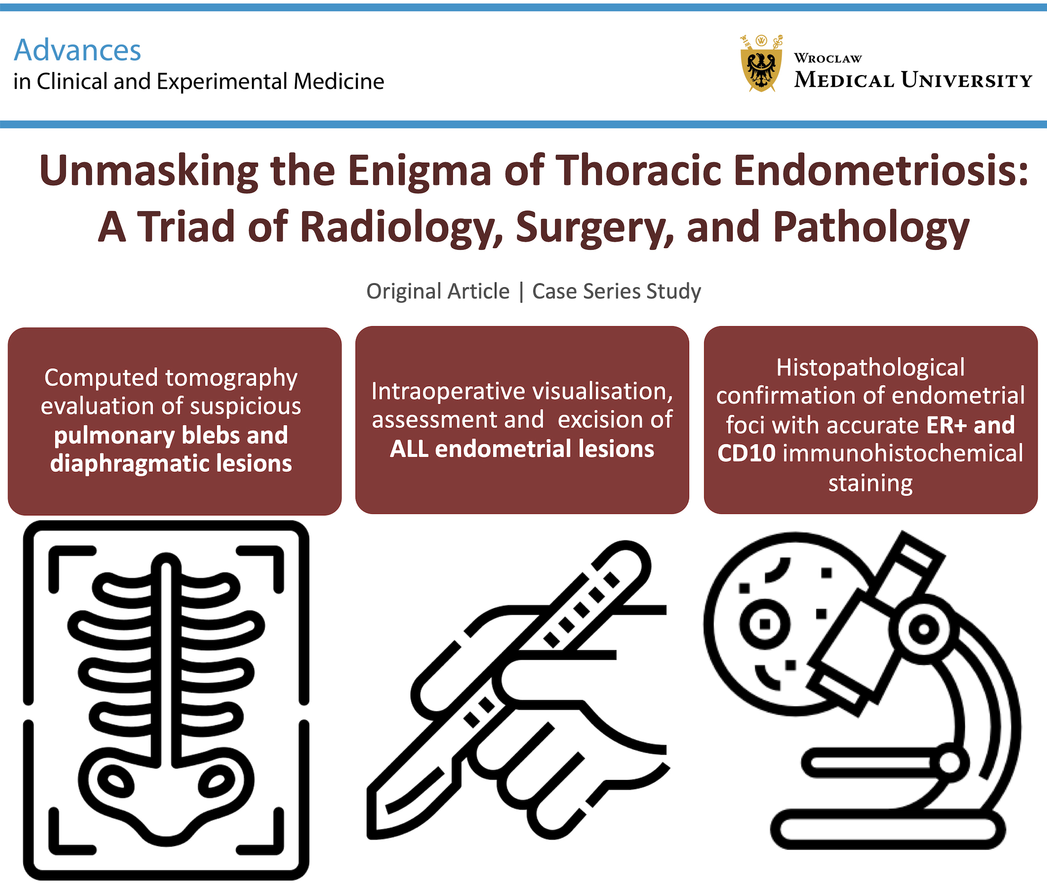
Objectives. Our study aims to propose a 3-step approach – computed tomography (CT) imaging, careful intraoperative assessment, and targeted pathology – to enhance the diagnostic process and optimize the management of TES-related pneumothorax.
Materials and methods. We retrospectively analyzed 3 cases of recurrent right-sided catamenial pneumothorax in female patients with a history of pelvic endometriosis, managed surgically at the Department of Thoracic Surgery, Medical University of Gdansk (Poland), between 2021 and 2024. All included patients underwent video-assisted thoracoscopic surgery (VATS), with comprehensive preoperative CT imaging, intraoperative lesion mapping, and postoperative histopathological examination, improving diagnostic accuracy in TES, with a possible impact on therapeutic results.
Results. Chest CT is essential for detecting bullous lesions, aiding in intraoperative identification and surgical treatment. Immunohistochemistry (IHC), assessing estrogen receptor (ER) and CD10 for stromal identification, improves pathological accuracy.
Conclusions. This study underlines the diagnostic value of integrating imaging, intraoperative assessment, and targeted histopathology, as well as clear communication between surgeons, radiologists, and pathologists to ensure accurate diagnosis and an improved therapeutic process for patients in clinical practice.
Key words: Thoracic endometriosis syndrome, catamenial pneumothorax, video-assisted thoracoscopic surgery (VATS), computed tomography imaging, immunohistochemistry
Background
Thoracic endometriosis syndrome (TES) is the most prevalent form of extrapelvic endometriosis,1 yet it remains surrounded by controversies regarding its classification,2 diagnosis,3 and treatment.4 Recently, it has been proposed that TES be classified into pleural, bronchopulmonary, and diaphragmatic forms.2 The bronchial presentation of TES has rarely been reported, while seeding of endometrial foci on the parietal pleura is exceedingly rare. The most common manifestation of TES, apart from the diaphragmatic form, involves subpleural localization within the pulmonary parenchyma.2, 5 On this basis, distinguishing between the diaphragmatic and bronchopulmonary forms of TES appears justified and allows for a more appropriate classification of this phenomenon. In pulmonary TES, chest computed tomography (CT) reveals bullous lesions5 and solid pulmonary tumors,6, 7 making CT the primary imaging modality. Magnetic resonance imaging (MRI) is valuable for confirming thoracic endometriosis, offering superior contrast resolution and the ability to characterize hemorrhagic lesions, despite its lower spatial resolution. Magnetic resonance imaging is preferred for visualizing diaphragmatic or pleural implants, while CT scans are crucial for analyzing pulmonary parenchyma, facilitating difficult intraoperative identification and effective surgical treatment.3
Catamenial pneumothorax represents a distinct clinical manifestation within the spectrum of TES. Although it is likely not the most prevalent form of TES, it constitutes a clinically significant cause of emergency hospital admissions related to primary pneumothorax and, in particular, recurrent pneumothoraces in women of reproductive age.8 The mean age at diagnosis of TES typically ranges between 34 and 37 years,9 which is generally attributed to the long-standing, often undiagnosed or inadequately treated course of pelvic endometriosis preceding thoracic involvement in 85% of patients.10 Management of patients with catamenial pneumothorax remains challenging, largely due to an exceptionally high recurrence rate, reported to reach or even exceed 40% despite standard surgical interventions.11, 12 These unfavorable outcomes underscore the need to reconsider the conventional approach to such patients, as catamenial pneumothorax should not be managed as a form of spontaneous pneumothorax. Instead, it has been proposed that TES requires treatment within dedicated multidisciplinary teams integrating thoracic surgery, gynecology, radiology, pathology, and hormonal management to optimize diagnostic accuracy and reduce the risk of recurrence.13
Adequate pathological analysis of TES involves immunohistochemistry (IHC) to evaluate the expression of estrogen receptor (ER) and CD10 for stromal identification. Typically, the examination of material resected during pneumothorax surgery does not include ER IHC for endometrial gland visualization or CD10 IHC for stromal visualization.14, 15 Although hematoxylin and eosin (H&E) staining may be indicative in large pelvic endometriomas, it is often insufficient for the minimal lesions typically found in pulmonary TES.
Objectives
We propose a 3-step approach for patients with TES-related pneumothorax: comprehensive CT assessment, meticulous intraoperative visualization, and dedicated pathological evaluation.
Materials and methods
This study is a retrospective analysis of 3 patients who underwent surgery for recurrent right-sided catamenial pneumothorax at the Department of Thoracic Surgery, Medical University of Gdansk, Poland, between February 25, 2021, and January 22, 2024. Each patient had a documented history of pelvic endometriosis treatment. We reanalyzed data from preoperative chest CT imaging, intraoperative findings, and postoperative examination of surgical specimens. The study was approved by the Institutional Review Board of the Medical University of Gdansk (approval No. NKBBN/134/2020-2021 issued on October 15, 2021). The study was carried out in accordance with the Code of Ethics of the World Medical Association (Declaration of Helsinki) for experiments involving humans. The approval covered the retrospective analysis of data routinely collected in the electronic medical records prior to the date it was issued; the data of the 1st patient were therefore included under the approved protocol. Individual patient consent was not required owing to the retrospective, observational design of the study.
Computed tomography scans were performed before the surgical intervention. For patients A and B, preoperative scans were performed 1 month before surgery, while for patient C, the scan was performed 1 week prior. The phase of the menstrual cycle was not recorded at the time of imaging. Patients A, B, and C had experienced 4, 2, and 2 prior recurrences of right-sided catamenial pneumothorax, respectively. All patients subsequently underwent right-sided video-assisted thoracoscopic surgery (VATS).
The standardized VATS approach included positioning the patient in the left lateral decubitus position, utilizing double-lumen intubation with one-lung ventilation, and making incisions as follows: a port in the 7th intercostal space in the anterior axillary line (30° scope), a 3-cm utility incision in the 5th intercostal space with a soft tissue retractor, and a 2nd 10-mm incision in the 8th intercostal space in the posterior axillary line without a port.
During the procedure, a thorough assessment of the parietal and visceral pleura, lung, and diaphragm was conducted. Any bullous lesions were excised, and diaphragmatic defects were either reconstructed or partially excised. The procedure concluded with a complete pleurectomy and placement of a 24-French (F) chest drain. Detailed tissue-processing and immunohistochemical staining protocols are provided in Supplementary File 1.
Microscopic assessment of the slides was performed by 2 pathologists (A.C. and R.P.). A positive ER staining result was indicated by a nuclear reaction, while a positive CD10 staining result was identified by a membrane reaction.
Published case reports emphasize the role of ER and CD10 staining in histopathological confirmation of TES, whereas progesterone receptor (PR) immunostaining appears to be less frequently reported.16 However, recent studies recommend including PR alongside ER and CD10, as it may improve recognition of ectopic endometrial foci in the chest cavity. Given that the PR staining was not performed in our study, we suggest that routine PR assessment may be useful in the diagnosis of suspected TES.
A positive pathological diagnosis of pulmonary endometriosis was retrospectively compared with intraoperative findings and preoperative imaging. A pattern of consecutive preoperative, intraoperative, and postoperative lesions typical of pulmonary endometriosis was presented.
Results
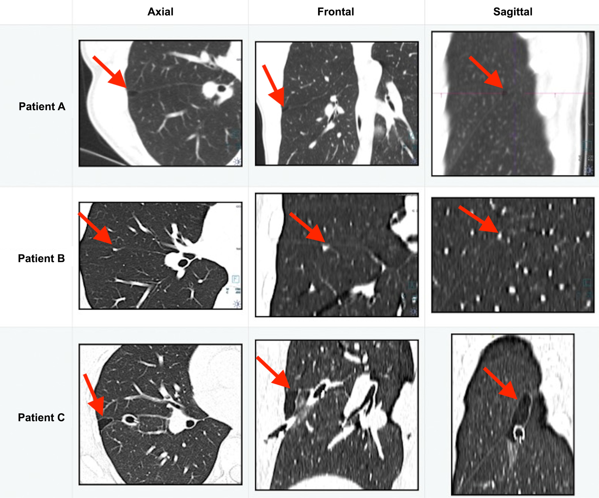
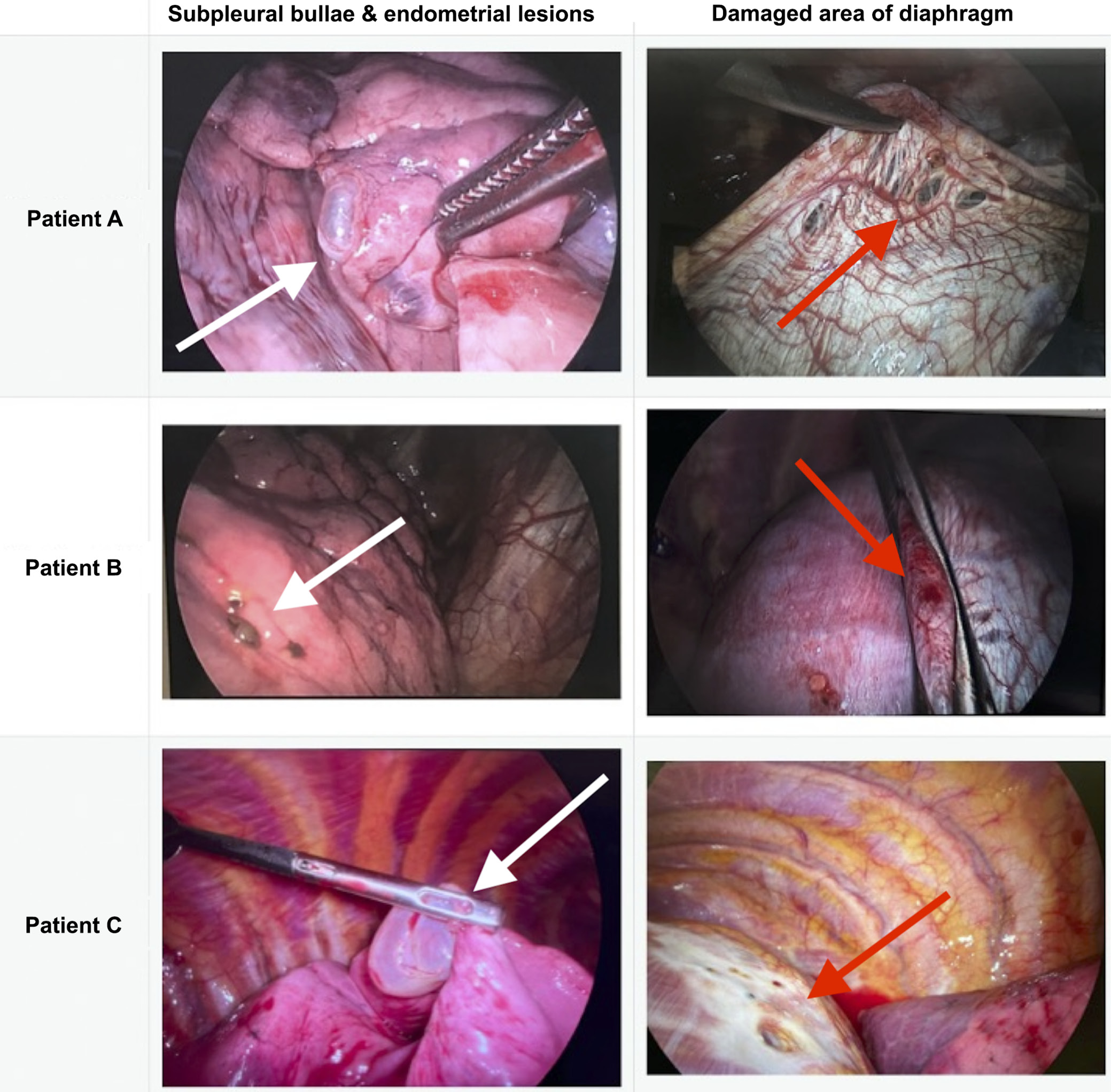
Three female patients of reproductive age (37, 39, and 43 years) with recurrent catamenial right-sided pneumothorax participated in this study. The primary symptom for all patients was pleuritic pain on the right side of the chest. Preoperative CT scans (axial, frontal, and sagittal views) revealed bullous lesions in the middle lobe of the right lung in all patients (Figure 1).17 Every patient presented with solitary, non-clustered small bullous lesions on CT imaging, which were round and subcentimeter in patients A and B and elliptical, measuring 20 mm, in patient C. No additional lesions were observed. Using VATS, we identified bullous lesions in the middle lobe of the right lung in all 3 patients (Figure 2), diaphragmatic defects with nodules in 2 patients, and a porous area without nodules in 1 patient.
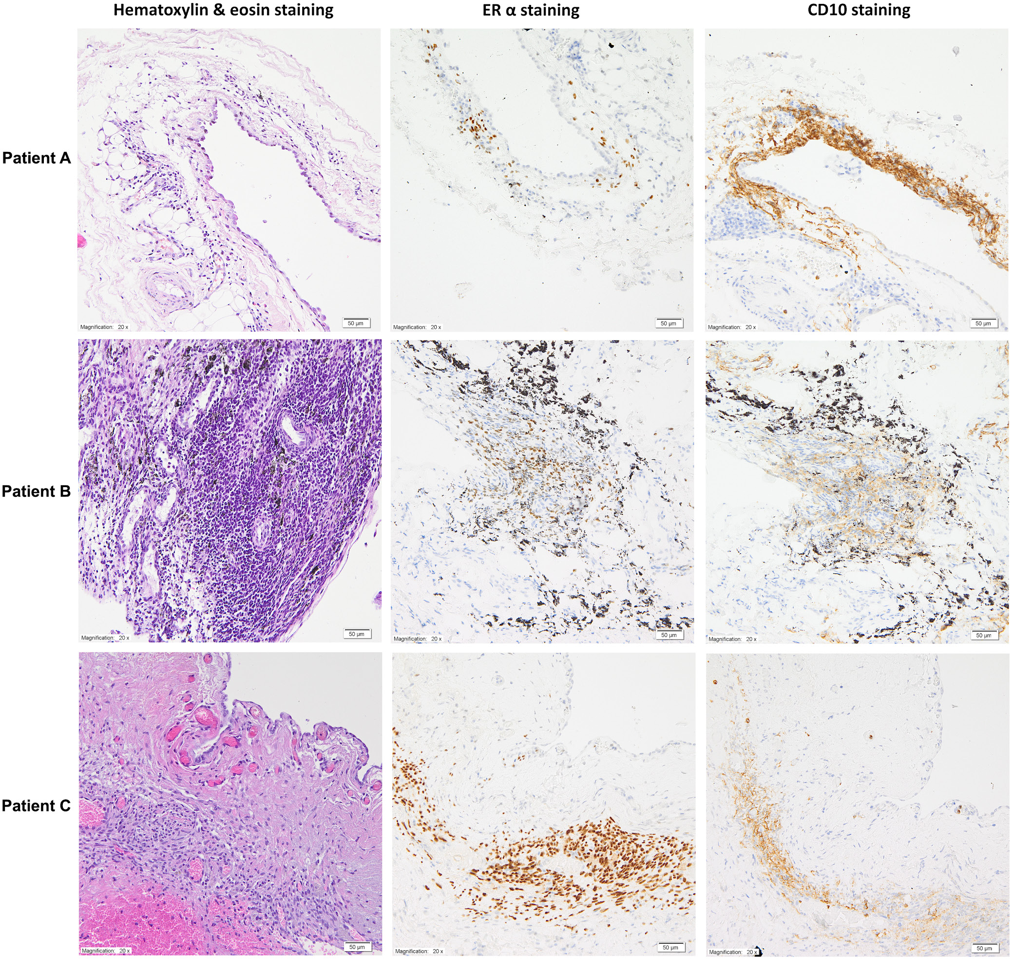
During VATS, all patients underwent wedge excision of pulmonary bullous lesions and complete pleurectomy. Two patients had partial resection of the tendinous part of the diaphragm without mesh, and 1 patient had direct diaphragm reconstruction. Clinical details are summarized below in Table 1. Histopathological examination of tissue samples obtained intraoperatively from the right lung revealed subpleural emphysematous changes. Immunohistochemical evaluation of endometrial lesions used antibodies specific for ERα and CD10, revealing estrogen receptor-positive (ER+) cells characteristic of endometrial glands and CD10+ cells indicating the presence of endometrial stroma (Figure 3).
In patient A, pleural tissue fragments showed 8 subpleural microfoci of endometriosis measuring 0.5–3 mm in diameter, composed of endometrial stromal cells (CD10+, ER+) accompanied by dilated blood vessels and small hemorrhagic foci. In patient B, a peripheral lung parenchyma specimen contained a single subpleural microfocus of endometriosis measuring 1.5 mm, characterized by numerous endometrial stromal cells (CD10+, ER+), dilated blood vessels, and deposits of anthracotic pigment. In patient C, pleural tissue fragments revealed a single subpleural microfocus of endometriosis up to 2.5 mm in size, composed mainly of endometrial stromal cells (CD10+, ER+) and associated with dilated blood vessels.
The operative times for patients A, B, and C were 75, 70, and 139 min. The postoperative course was uncomplicated in all patients, with hospital stays of 4, 6, and 4 days, respectively. Patients A and B had not received hormonal therapy before admission. After hospitalization, patient A received dienogest, and patient B received combined therapy with dienogest and estradiol. Patient C was already on hormonal therapy, receiving dienogest at admission. Postoperative hormonal management was determined individually by the attending physician. Patient A experienced 1 right-sided episode of recurrent catamenial pneumothorax 45 months after surgery, during a 50-month follow-up. Imaging confirmed the recurrence, but no surgical treatment or chest drainage was required, and no pathology was detected on the left side. Patients B and C have not experienced recurrence in 26 and 15 months of follow-up, respectively.
Discussion
This study underscores the necessity for TES-focused preoperative and postoperative assessments to improve the diagnosis of TES. Patients with recurrent TES-related pneumothoraces and underdiagnosed pulmonary TES are common, largely due to low awareness of TES among thoracic care teams. Currently, there is no standardized approach for patients with catamenial, TES-related pneumothorax. Thoracic endometriosis syndrome remains an orphan disease and a rare but probably important cause of recurrent pneumothoraces. Its management warrants a modified and highly coordinated approach, beginning with tailored radiological evaluation, followed by meticulous surgical assessment, comprehensive pathological examination, and, finally, postoperative guidance on hormonal therapy. Although it is not yet established whether this condition should be managed exclusively in high-volume centers, it is clear that patients with endometriosis benefit from a multispecialist model of care involving gynecological and fertility counseling, pain management, general surgical and urological input, specialized physiotherapy, dietary support, psychological and psychiatric care, and access to patient support groups.18 Thoracic endometriosis syndrome represents a rare and distinctive manifestation within this disease spectrum, and in cases of recurrent symptoms, referral to a multidisciplinary center should be strongly considered.13
Catamenial pneumothorax should be considered in ovulating women presenting with spontaneous pneumothorax, even when there is no clinical history suggestive of pelvic endometriosis. Video-assisted thoracoscopic surgery is currently considered the gold standard for the management of TES-related pneumothorax, as it allows thorough inspection of the pleural cavity, lung parenchyma, and diaphragm, together with definitive surgical treatment using a minimally invasive approach.2 In cases with suspected diaphragmatic involvement, the addition of simultaneous laparoscopic visualization may be advantageous,13, 19 since assessment of diaphragmatic lesions from the pleural surface alone may underestimate the true extent of disease affecting the diaphragmatic muscle and its peritoneal aspect.20, 21
Several surgical series emphasize the importance of a meticulous intraoperative assessment, particularly careful inspection of the diaphragmatic surface during video-assisted thoracic procedures.12 Limited diaphragmatic interventions, such as plication of suspicious areas, have been reported to be effective in selected cases, while adjunctive postoperative hormonal suppression has been shown to improve outcomes, especially in more complex or recurrent presentations. Collectively, published data indicate that diaphragmatic involvement is detected in a substantial proportion of patients, and that outcomes vary according to the extent and method of diaphragmatic management. Direct surgical repair involving mesh-based reconstruction has been described, yet analyses expressed in proportional terms consistently suggest superior results when all visible diaphragmatic abnormalities are actively addressed.9 In particular, reconstruction with prosthetic materials, including biological meshes, has been associated with favorable perioperative safety and durable long-term results when compared with more limited approaches.
Large literature-based series further demonstrate that optimal management of catamenial pneumothorax and other thoracic manifestations of endometriosis relies on a combined surgical and hormonal strategy. Surgical treatment commonly encompasses apical lung resection, excision of all suspected visceral and parietal pleural lesions, and partial resection or reconstruction of the diaphragm in the presence of nodules or fenestrations, aiming to eliminate diseased tissue and restore diaphragmatic integrity. Obliteration of the pleural cavity (pleurectomy, surgical abrasion, and chemical or talc pleurodesis) is considered an important component of treatment, intended to reduce the risk of recurrence related to microscopic or newly implanted endometrial foci.22 Despite these observations, no uniform, evidence-based recommendations currently exist regarding the optimal extent of surgery. Nevertheless, based on the cumulative evidence from the literature and our own experience, we advocate a comprehensive approach consisting of removal of all suspected pulmonary lesions, including small or atypical bullous lesions, systematic repair or reconstruction of the diaphragm, and adjuvant hormonal therapy following thoracic surgery.2 Female patients of reproductive age with pneumothorax, particularly right-sided, should be thoroughly evaluated for a TES-related etiology prior to any treatment, including chest drainage.
The risk of TES increases in the presence of coexisting pelvic endometriosis,13, 23 the recurrent nature of the pneumothorax,24, 25 a history of infertility,26, 27 and menses-related arm or chest pain.19 The incidence of TES-related pneumothorax among all patients of childbearing age with pneumothorax ranges from 7% to 37%.28 In such cases, patients should be referred to centers capable of complex interventions, where multidisciplinary care – involving specialists in thoracic surgery, gynecology, radiology, pathology, anesthesia, and pain management – is readily available, and where surgical teams have the expertise to perform combined procedures, including simultaneous thoracoscopic and laparoscopic evaluation of the diaphragm to ensure comprehensive identification and resection of all endometriotic foci.13 The need for dedicated TES care teams remains an open question, though this solution has been discussed by a specialist team organized by the Polish Ministry of Health.29, 30 As a general principle, surgery for endometriosis should be meticulously planned with the aim of identifying and removing all foci of deep infiltrating endometriosis (DIE).31 In thoracic endometriosis, diaphragmatic involvement is present in approx. 84% of cases, and VATS is the preferred surgical approach in the majority of patients, while laparoscopy is performed concomitantly or in a staged fashion in about half of them. Taken together, these observations support the need to plan for complete management of all diaphragmatic endometriotic foci, including direct suturing of defects or ablation of superficial lesions, and excision of diaphragmatic segments in cases of DIE with subsequent reconstruction, preferably with mesh implantation.16, 32
There are various interpretations of the radiological presentation of pulmonary TES.3, 5, 7 In our view, MRI may be beneficial for identifying diaphragmatic implants, whereas CT remains the standard for strictly pulmonary lesions. Computed tomography is useful for diagnosing extensive diaphragmatic defects that can lead to the rarely observed occurrence of hepatic herniation.33 In our series, all patients had diaphragmatic defects identified intraoperatively, despite the absence of a preoperative diagnosis on CT scans. A meticulous preoperative CT assessment with careful attention to small, subpleural bullous lesions is indicated, as these subtle findings may represent early or atypical manifestations of thoracic endometriosis rather than incidental or clinically insignificant small bullous lesions. Although their predominant occurrence in the middle lobe may be coincidental, this pattern could also reflect the inherently unpredictable distribution of subpleural endometriotic foci. Whereas previous studies have primarily described CT and MRI as diagnostic methods for evaluating abdominal and diaphragmatic endometriotic lesions,3 our study highlights the value of CT in identifying endometrial features within the thoracic cavity itself by presenting 3 cases in which pulmonary lesions were delineated on CT, thereby reinforcing the diagnostic value of meticulous thoracic CT evaluation.5 Recognizing these nuances can improve diagnostic accuracy and guide more targeted intraoperative assessment. Unlike previous studies, which associated TES-related pneumothorax with solid tumors,3, 7 our research identified minimal subpleural bullous lesions that correlate with the pathological presentation of TES. These subtle intrapulmonary bullous lesions are adequately visualized with non-contrast CT. Large lung cancer screening trials using CT have improved the understanding of sub-centimeter solid pulmonary nodules. The clinical interpretation of these lesions, found in nearly 25% of screened individuals,34 is challenging. Most subpleural nodules are benign, often attributed to carbon-loaded lymph nodes.35 The CT scans of patients A and B did not reveal significant bullous lesions or other pathological findings at first glance; however, on retrospective review, subtle abnormalities can be appreciated, underscoring the need for meticulous radiologic assessment in premenopausal patients with recurrent pneumothorax, in whom TES should be considered. This radiologic impression was confirmed intraoperatively, when patient C was found to have the most substantial disease burden, whereas patients A and B demonstrated only minor lesions. We strongly advocate for the preoperative identification of even minimal bullous lesions in patients with suspected TES-related pneumothorax, given the high risk of their association with TES.
In many articles addressing IHC in thoracic endometriosis, antibodies for detecting CD10 are predominantly utilized, often in conjunction with antibodies for ER and/or PR detection. While CD10 is generally adequate for identifying endometriosis, its co-expression with ER/PR can be beneficial in diagnosing catamenial pneumothorax.14, 15 Indeed, ER, PR, and CD10 are the most commonly evaluated markers in TES. However, interferon-inducible transmembrane protein 1 (IFITM1) for stromal cells and PAX8 for epithelial cells have also been proposed as sensitive markers in extrapelvic endometriosis.14 In a retrospective study involving 55 women undergoing surgery for pneumothorax, IHC for ER, PR, and CD10 was performed on bland stromal spindle cells of the lung.36 A total of 37 cases that tested positive for both receptors were subsequently classified as exhibiting either “scattered” or “aggregated” patterns. The scattered pattern (33/37) was CD10-negative and associated with a lower 5-year recurrence rate of pneumothorax (16.7%), whereas the aggregated pattern (4/37) was CD10-positive and linked to a higher 5-year recurrence rate (62.5%). Notably, as the scattered pattern was also found in the male control group, a diagnosis of thoracic endometriosis was made only in cases exhibiting the aggregated pattern.36 All patients in our study were ER- and CD10-positive. Although further research is needed, meticulous evaluation of staining patterns may enhance the precision of TES diagnosis. Early identification of even subtle lesions appears to contribute to a more favorable prognosis, reflected in fewer recurrences of TES-related pneumothorax. Close collaboration between surgical and pathological teams remains essential for recognizing lesions most likely associated with TES. Importantly, we believe that TES should be considered a potential etiology of recurrent pneumothorax in every woman of premenopausal age. Diagnosing endometriotic foci within the lung not only establishes the presence of thoracic endometriosis but also enables appropriate systemic treatment of endometriosis as a disease entity. Incorporating routine ER/PR/CD10 immunostaining into the pathological assessment of lung specimens obtained during pneumothorax surgery in premenopausal women may further increase diagnostic yield and improve overall quality of care, ultimately helping to reduce recurrence rates. Although simple pneumothorax surgery can be effectively performed using a uniportal VATS approach, our strategy prioritizes a more comprehensive intraoperative assessment, including meticulous inspection of the lung parenchyma, diaphragmatic evaluation, partial resection, and, when necessary, diaphragmatic reconstruction. While increasingly complex procedures are now performed through uniportal access in high-volume centers of excellence, a utility incision complemented by 1 or 2 additional ports remains the standard technique in many institutions and continues to be widely adopted in routine clinical practice.37 Moreover, we believe that the debate regarding the optimal number of ports in minimally invasive thoracic surgery has largely diminished following the widespread introduction of robot-assisted approaches.
Limitations of the study
This study acknowledges several important limitations. First, it involves a small cohort of patients treated at a single center by 1 general thoracic surgeon. While this may reflect the clinical approach of a single specialist, such scenarios are not uncommon in the context of orphan diseases. Second, the order in which assessments were performed represents a limitation. Ideally, a meticulous radiological assessment should precede intraoperative findings, which are then confirmed by a pathologist’s report. In our study, intraoperative findings, when confirmed by the pathological report, were subsequently compared with preoperative CT scans, which identified minimal yet clinically significant lesions. Third, the follow-up duration was variable among patients A, B, and C (50, 26, and 15 months). Moreover, data on the menstrual cycle phase at the time of chest CT imaging were not collected. Although some recent studies have assessed the radiological features of TES in relation to the menstrual cycle phase, this approach is not routinely implemented. The absence of this information may have limited the interpretative specificity of our imaging findings. We suggest that future studies may benefit from standardized documentation of menstrual cycle phase at the time of imaging. An additional potential limitation is the absence of PR staining, which may have reduced our ability to detect or confirm some TES lesions. Despite all these limitations, we assert that it is both clinically and scientifically justified to emphasize the recommendation not to underestimate the risk of TES in small, subpleural bullous lesions located outside the lung apex.
Conclusions
Small subpleural bullous lesions may represent radiological manifestations of TES. Preoperative CT scans likely facilitate the intraoperative localization of minimal lesions. Effective communication between surgeons, radiologists, and pathologists enhances the diagnostic yield in TES, potentially influencing treatment outcomes.
Supplementary data
The supplementary material is available at https://doi.org/
10.5281/zenodo.17641838. The package contains the following files:
Supplementary File 1. Tissue processing and analysis.
Data Availability Statement
The datasets supporting the findings of the current study are available from the corresponding author on reasonable request. The data are not publicly available due to ethical reasons, by reason of human research participant data presence.
Consent for publication of personal information
Not applicable.
Use of AI and AI-assisted technology
Not applicable.