



Abstract
Background. Percutaneous cryoablation (PCA) is a minimally invasive treatment modality for renal cell carcinoma (RCC) that is often implemented in patients with impaired renal function. However, this procedure inevitably results in the loss of functional renal parenchyma.
Objectives. The objective was to establish the influence of PCA on renal function, presented as a difference in estimated glomerular filtration rate (eGFR).
Materials and methods. This study included patients treated with PCA of renal tumors at 1 institution between June 2023 and February 2025. Creatinine was measured 1 day before the procedure and ≥3 months after. Estimated glomerular filtration rate was calculated using the Modification of Diet in Renal Disease (MDRD) equation. Mean differences in eGFR were assessed for the entire cohort and subgroups. Correlation analyses were performed to assess the relationship between eGFR reduction and tumor volume/diameter. Multiple linear regression analysis was conducted to identify factors independently associated with the change in eGFR.
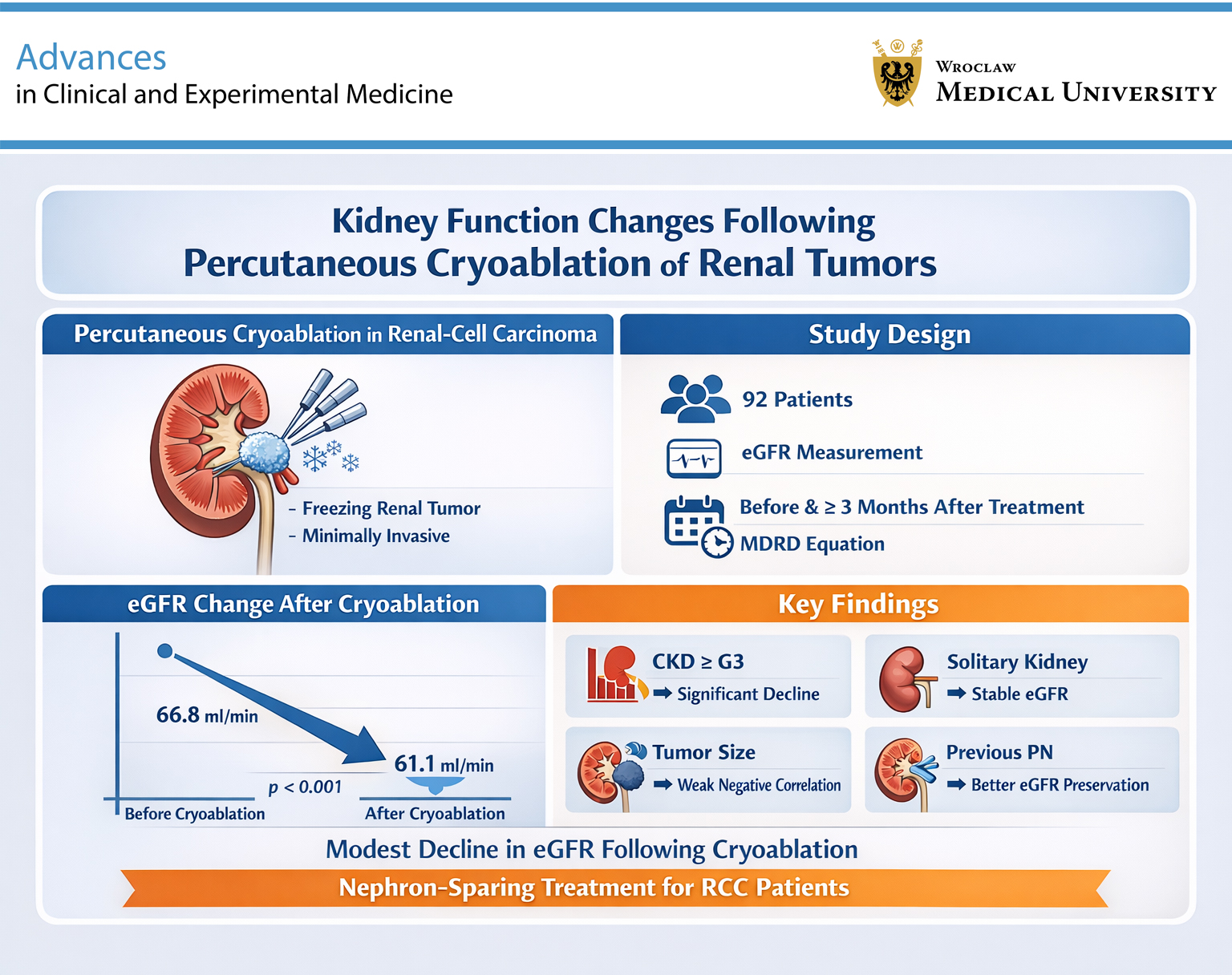
Results. Ninety-two procedures met inclusion criteria. A minor but statistically significant decrease in mean eGFR was observed (66.8–61.1 mL/min/1.73 m2, p < 0.001). Patients with a solitary kidney showed no significant decline in eGFR (56.6–54.5 mL/min/1.73m2, p = 0.200), while those with CKD ≥ G3 experienced a modest but significant reduction (47.2–43.5 mL/min/1.73 m2, p = 0.004). After excluding cases with >10% eGFR improvement, the mean eGFR decline remained statistically significant (68.0–59.9 mL/min/1.73 m2, p < 0.001). Weak negative correlations were identified between eGFR reduction and both tumor volume (r = −0.24, p = 0.024) and tumor diameter (r = −0.21, p = 0.042). Finally, previous partial nephrectomy (PN) was associated with better eGFR preservation after PCA in multiple linear regression analysis (p = 0.014).
Conclusions. Our findings support the nephron-sparing nature of PCA, which is a safe treatment modality of RCC, especially in patients with significant primary comorbidities.
Key words: renal function, cryoablation, glomerular filtration rate, ablation techniques, renal-cell carcinoma
Background
Renal cell carcinoma (RCC) is one of the most prevalent malignancies of the genitourinary system. In addition, the detection of small renal masses (SRMs) has increased with the widespread adoption of advanced imaging techniques, such as computed tomography (CT) and magnetic resonance imaging.1 Partial nephrectomy (PN) is the gold standard treatment for T1 RCC. However, this is an invasive procedure associated with a significant risk of complications. Therefore, in highly comorbid patients with SRMs, percutaneous tumor ablation (TA) can be performed as a minimally invasive alternative to surgery. Despite its advantages, percutaneous cryoablation (PCA) inevitably destroys some functional renal parenchyma, potentially compromising renal function. This is of particular concern in patients with advanced chronic kidney disease (CKD) or a solitary kidney. Most existing studies focus on oncological outcomes, while the functional impact of PCA remains underreported.
Objectives
The objective of this study was to assess the influence of PCA on renal function, measured as the change in estimated glomerular filtration rate (eGFR), with particular attention to patients with a solitary kidney and those with baseline CKD ≥ G3.
Materials and methods
Informed consent was obtained from all patients prior to the PCA procedure. The study was approved by the Institutional Bioethics Committee of Wroclaw Medical University, Poland (approval No. KB/72/2025).
Study design
A prospectively maintained database was used for this retrospective, single-center observational study. The database consisted of all PCA procedures performed at the University Clinical Hospital in Wrocław between June 2023 and February 2025. Percutaneous cryoablation was performed in patients who were either found ineligible for PN or did not consent to surgery/active surveillance. Percutaneous cryoablations were performed by an interventional radiologist skilled in CT interventions (M.G.) and a urologist with extensive experience in ultrasound-guided percutaneous renal procedures (W.K.). All patients had RCC confirmed by prior biopsy. The full methodology of the procedure has been described in detail in another study.2 In the present paper, we selected PCA procedures with available pre- and postoperative serum creatinine concentration measurements obtained after at least 3 months. Patients receiving chronic renal replacement therapy were excluded. For each procedure, data on sex, age at the time of the procedure, Charlson comorbidity index (CCI), body mass index (BMI), tumor size on preoperative CT/MRI, solitary kidney status, and kidney function were collected.
Outcome ascertainment
The primary objective of this study was to assess the influence of PCA on renal function. Estimated glomerular filtration rate was calculated using the Modification of Diet in Renal Disease (MDRD) equation. The mean difference in eGFR before and after the procedure was assessed.
The secondary aim was to explore the influence of PCA on renal function in patients with solitary kidneys and in those with CKD MI (defined as eGFR < 60 mL/min/1.73 m2 on pre- and postoperative measurements). An additional analysis was conducted after excluding patients who exhibited an eGFR increase >10% in a follow-up measurement. Additionally, the relationship between eGFR change and tumor volume/diameter was evaluated. Finally, independent predictors of eGFR change were examined.
Statistical analyses
Continuous variables were reported as means ± standard deviations (SDs) for normally distributed data and as medians with interquartile ranges (IQRs) for non-normally distributed data. Categorical variables were presented as counts and percentages (%). Normality of distribution was assessed using the Shapiro–Wilk test and visual inspection of Q–Q plots (Supplementary Fig. 1).
To assess the statistical significance of differences in eGFR before and after PCA, either the paired Student’s t-test or the Wilcoxon matched-pairs test was used, depending on data distribution. Correlation analyses were performed using Pearson’s correlation coefficient for normally distributed variables and Spearman’s rank correlation coefficient for non-normally distributed variables.
Multiple linear regression was conducted to identify factors independently associated with the change in eGFR. Prior to model fitting, all key regression assumptions were evaluated (Supplementary Table 1 and Supplementary Fig. 2), including linearity (RESET test), normality of residuals (Shapiro–Wilk test), homoscedasticity (Breusch–Pagan test), absence of autocorrelation (Durbin–Watson test), and absence of multicollinearity (variance inflation factor (VIF)). Influential observations were inspected using Cook’s distance.
The final model included tumor volume, solitary kidney status, prior PN, patient age, and sex as covariates. All analyses were two-sided, with p < 0.05 considered statistically significant. Statistica v. 13 (TIBCO Software Inc., Palo Alto, USA), StataNow v. 18.5 (StataCorp LLC, College Station, USA), and R (R Foundation for Statistical Computing, Vienna, Austria) were used to perform all analyses.
Results
Study cohort
A total of 110 PCA procedures (in 102 patients) were performed at our center between June 2023 and February 2025. Of these, 92 cases had both serum creatinine measurements available and were included in the present study. Baseline patient and tumor characteristics of the cohort are presented in Table 1.
Outcomes
Loss of kidney function
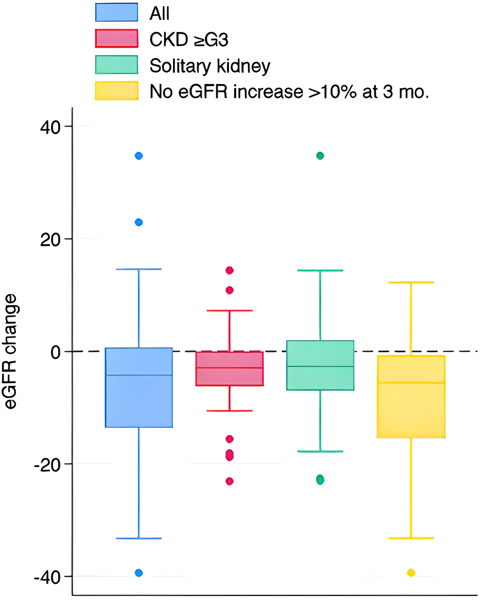
Data regarding the mean change in eGFR after PCA are presented in Figure 1. In the whole cohort, a minor deterioration in renal function was recorded. The mean preprocedural eGFR was 66.8 (±20.6) mL/min/1.73 m2, which was reduced to 61.1 (±19.6) mL/min/1.73 m2 3 months after PCA. The mean loss of eGFR (−5.6 (±12.5) mL/min/1.73 m2) was statistically significant (p < 0.001).
In patients with CKD ≥ G3, PCA resulted in a statistically significant loss of eGFR (−3.7 (±7.8) mL/min/1.73 m2, p = 0.004). The average eGFR decreased from 47.2 (±11.2) mL/min/1.73 m2 before PCA to 43.5 (±11.7) mL/min/1.73 m2 after the intervention.
Finally, patients with a solitary kidney did not experience a statistically significant reduction in eGFR (−2.1 (±13.2) mL/min/1.73 m2, p = 0.200). Nonetheless, in this subgroup, average eGFR before and after the procedure was 56.6 (±19.9) and 54.5 (±18.8) mL/min/1.73 m2, respectively.
Patients with eGFR increase >10%
In 10 cases, the eGFR at 3 months was >10% higher than the preoperative measurement. The characteristics of these patients are presented in Table 2. In this group, we did not find any distinct attributes that could explain the improvement in renal function after PCA. Therefore, an additional analysis was performed, excluding these patients. In 82 cases, a statistically significant reduction in kidney function after PCA was confirmed (−8.1 (±10.5) mL/min/1.73 m2, p < 0.001). Mean pre- and postprocedural eGFR values were 68.0 (±20.5) and 59.9 (±19.4) mL/min/1.73 m2, respectively (Figure 1).
Correlation analyses
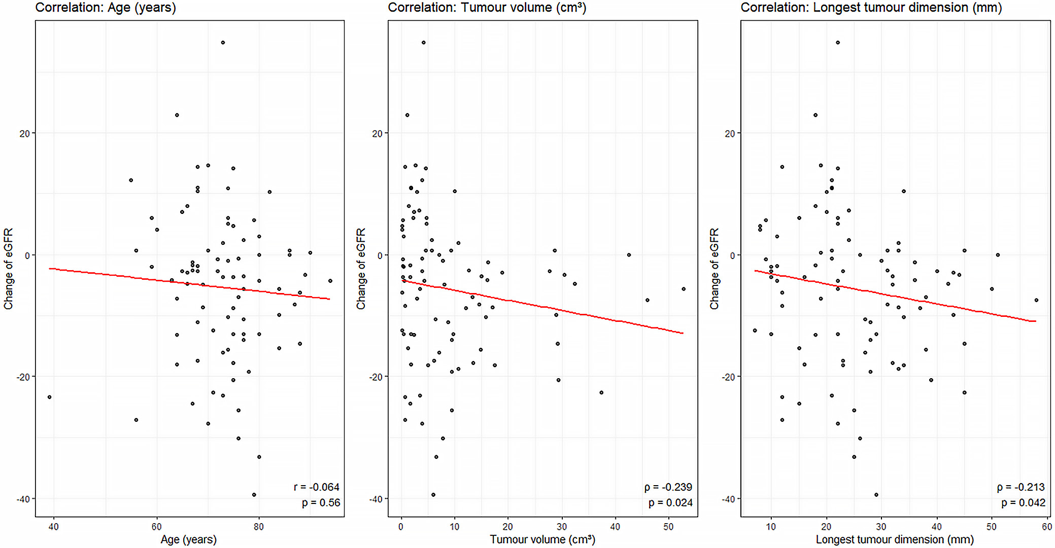
The correlation analyses are presented in Figure 2. A statistically significant weak negative correlation was observed between the change in eGFR and tumor volume (ρ = −0.24, p = 0.024) and between the change in eGFR and the longest tumor dimension (ρ = −0.21, p = 0.042). These monotonic relationships indicate that PCA of larger tumors was associated with a more pronounced postoperative decline in eGFR. No significant linear correlation was found between age and the change in eGFR (r = −0.064, p = 0.560).
Multiple linear regression analysis
The results of the multiple linear regression analysis predicting postoperative eGFR change are presented in Figure 3 and Table 3. All key assumptions of linear regression were met (Supplementary Table 1 and Supplementary Fig. 2).
Tumor volume, solitary kidney status, prior ipsilateral PN, age, and sex were included as predictors. The overall model was statistically significant (F(5,86) = 2.36, p = 0.047) and explained 12.1% of the variance in eGFR change (R2 = 0.121). The intercept term was negative (β = −27.12), suggesting an overall tendency toward a postoperative decline in eGFR in the absence of protective factors.
Previous PN was significantly associated with better preservation of eGFR after PCA. On average, patients with a history of PN had a less pronounced reduction in eGFR (by 7.97 mL/min/1.73 m2) than those without prior PN (95% confidence interval (95% CI): 1.68–14.26; p = 0.014). Other predictors were not significantly associated with eGFR change after PCA.
Discussion
The present study assessed changes in kidney function following PCA of renal tumors in a cohort of 92 procedures, providing insight into the functional consequences of this treatment modality.
Our findings demonstrate a minor yet statistically significant decrease in eGFR at least 3 months after the procedure. The observed eGFR reduction is unlikely to be clinically relevant for patients with preserved baseline kidney function. Nonetheless, such a change can be meaningful in patients with advanced CKD.
Also, patients with CKD ≥ G3 experienced a small but statistically significant decrease in eGFR. The absolute change was smaller than in the whole cohort. This was likely due to the nonlinear relationship between creatinine and eGFR: in advanced CKD, even large changes in serum creatinine levels do not result in significant changes in eGFR. In addition, in patients with primarily reduced kidney function, the procedure was probably performed less aggressively to spare as much renal parenchyma as possible, even at the expense of a thinner surgical margin. This suggests that PCA may be considered a functionally safe option even in this high-risk group, particularly when weighed against the potential risks of PN.
In patients with solitary kidneys, no statistically significant decline in renal function was observed. However, this finding should be interpreted with caution. The lack of significance might be caused by the small sample size, which may have limited statistical power. Again, it may be a consequence of the more conservative approach during PCA.
Interestingly, a subset of patients (n = 10) showed more than a 10% postoperative improvement in eGFR. This was likely due to factors unrelated to PCA, such as falsely low preoperative eGFR, which might have been caused by preoperative dehydration, nonsteroidal anti-inflammatory drug administration, contrast-related acute kidney injury (AKI), etc. Unfortunately, we do not have additional preoperative serum creatinine measurements to determine whether the lower eGFR before PCA was incidental. Nevertheless, no common features were identified to account for this improvement. Given the possibility of erroneous preoperative eGFR results, these outliers were excluded from the analysis, thereby confirming our primary findings. In the group of patients without significant renal function improvement after PCA, the mean eGFR decrease was comparable to that in the whole cohort.
Correlation analyses revealed only weak associations between eGFR change and tumor diameter/volume. The more profound eGFR decrease in larger tumors is logical, as these cases require more cryoprobes and larger ice-ball formation in order to ensure radical PCA. This naturally damages more functional renal parenchyma than in smaller lesions.
Multiple linear regression analysis showed that prior PN was associated with better preservation of renal function following PCA. Although PCA after PN is a safe and feasible procedure associated with minimal loss of eGFR, we are unable to explain this finding.3 Importantly, the other predictors in the model were not statistically significant. Furthermore, the overall model demonstrated limited explanatory power, accounting for only a modest proportion of the variance in eGFR change. These results indicate that reliably predicting eGFR decline using common clinical and tumor variables remains challenging.
The majority of the studies on the topic are in line with our results and report a minor decrease in eGFR after PCA. A 2024 meta-analysis including 3,202 patients found that PCA was associated with a statistically significant eGFR reduction of −3.06 mL/min/1.73 m2.4 Rizzo et al. observed a minor deterioration in eGFR at 1-year follow-up (−2.9 mL/min/1.73 m2).5 A Danish study on 259 patients showed a statistically significant reduction in eGFR after PCA at 12-month follow-up. The authors reported a mean eGFR change of −4 mL/min/1.73 m2.6 Finally, a study by Yamagami et al. included 27 patients who underwent cryoablation. The authors noted a mean decrease in eGFR from 56.5 to 53.8 mL/min/1.73 m2 at 6 months, which was not statistically significant.7
Unfortunately, our analysis lacks a comparative group. In the literature, reported eGFR loss after PN typically ranges from −2 to −9 mL/min/1.73 m2.8, 9, 10, 11 Nonetheless, a large meta-analysis did not find statistically significant differences between PCA and PN in the mean change in eGFR and the percentage of eGFR preservation.12 Also, a recent multicenter analysis did not observe any significant differences in kidney function between microwave, radiofrequency, and cryoablation.13 Another meta-analysis compared radical nephrectomy (RN), PN, and TA. Unsurprisingly, the authors reported that RN was associated with a greater eGFR decrease (by 9.94 mL/min/1.73 m2) than TA.14 However, the difference between PN and TA was statistically insignificant. Finally, stereotactic ablative radiotherapy has recently emerged as a viable treatment for RCC. Siva et al. conducted a phase 2 non-randomized trial on this procedure and reported eGFR deterioration of −10.8 mL/min/1.73 m2 at 12 months and −14.6 mL/min/1.73 m2 at 24 months.15 These trials suggest that stereotactic radiotherapy might be associated with worse preservation of renal function than TA. In addition, radiotherapy affects the entire kidney and may negatively impact endocrine renal function, leading to reduced production of erythropoietin and disturbances in the renin–angiotensin–aldosterone system.16
Limitations of the study
This study has several limitations that should be acknowledged. First, the follow-up period was relatively short, which may have limited our ability to capture long-term changes in renal function. Second, eGFR is influenced by numerous factors that were not fully controlled for in our analysis. Moreover, we did not identify strong correlations or predictive models for a more pronounced decline in eGFR, which may reflect sample size limitations or the multifactorial nature of kidney function decline. Finally, the absence of a comparator group restricts the interpretability of our findings and precludes direct comparisons with standard care or alternative interventions.
Conclusions
This study advances current clinical practice by providing further evidence for the nephron-sparing nature of PCA, a safe treatment modality for RCC, especially in patients with significant primary comorbidities. We found a minor but statistically significant decrease in renal function 3 months after PCA. Nevertheless, close follow-up of postprocedural renal function is crucial.
Supplementary data
The supplementary materials are available at https://doi.org/10.5281/zenodo.18480146. The package contains the following files:
Supplementary Table 1. Diagnostics of the linear regression assumptions for the model predicting change in eGFR.
Supplementary Fig. 1. Diagnostic Q–Q plots for evaluating the normality of residual distributions for all numerical variables. The red line represents the theoretical quantiles of the normal distribution. W statistics and corresponding p-values from the Shapiro–Wilk test are shown for each variable.
Supplementary Fig. 2. Diagnostic plots for the linear regression model. The panel displays the posterior predictive check, assessment of linearity (RESET test), homoscedasticity (Breusch–Pagan test), influential observations (Cook’s distance), multicollinearity (VIF), and normality of residuals (Shapiro–Wilk test).
Data Availability Statement
The datasets supporting the findings of the current study are openly available in Zenodo at https://doi.org/10.5281/zenodo.18480082.
Consent for publication of personal information
Not applicable.
Use of AI and AI-assisted technologies
Not applicable.
.jpg)