Abstract
Background. Complex lung segmentectomy, although effective for early-stage lung cancer, is less widely adopted because of its technical complexity.
Objectives. This study aimed to compare perioperative outcomes, quality of life (QoL), and survival between complex and simple lung segmentectomy performed using single-port thoracoscopy to determine its clinical feasibility.
Materials and methods. This retrospective analysis screened 218 patients; after exclusions, 210 patients underwent complex (n = 100) or simple (n = 110) segmentectomy at Zhanjiang Central Hospital. Propensity score matching resulted in 92 patients per group. Single-port thoracoscopic surgery was performed, and perioperative outcomes, complications, QoL assessed using the 36-Item Short Form Health Survey (SF-36), and survival evaluated using Kaplan–Meier curves were analyzed. Statistical analyses were performed using t-tests, χ2 tests, and log-rank tests (p < 0.05).
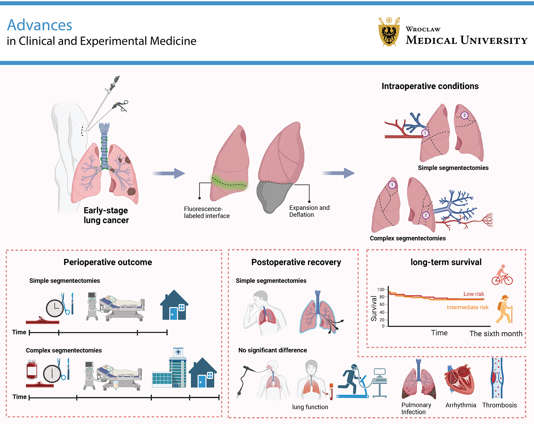
Results. Complex segmentectomy was associated with longer operative time, greater postoperative drainage, and longer hospital stays (p < 0.001). Cough (60.87% vs 32.61%; p = 0.007) and air leakage (54.35% vs 19.57%; p < 0.001) were more common in the complex group. No significant differences were observed in postoperative lung function, other complications, such as infection or arrhythmia, or QoL at 6 months (p = 0.071). Survival curves showed comparable prognosis (p = 0.860).
Conclusions. Despite increased perioperative demands, complex lung segmentectomy achieves lung function, QoL, and survival outcomes similar to those of simple segmentectomy, supporting its role in the treatment of early-stage lung cancer. These findings support its broader application in specialized settings and may inform optimized surgical approaches.
Key words: quality of life, complex lung segmentectomy, simple lung segmentectomy, single-port thoracoscopy, perioperative outcomes
Background
According to the latest cancer incidence and mortality data, lung cancer remains the most prevalent and deadliest cancer in our country.1 Among treatment options for lung cancer, surgical resection remains the most effective approach for resectable pulmonary tumors.2, 3 Based on the nature and location of the lesion, lung segmentectomy is categorized as either simple or complex. Simple lung segmentectomy is suitable for patients with small lesions and clear anatomical structures, and is associated with broad applicability, technical ease, shorter operative time, preservation of lung function, and faster recovery.4, 5, 6, 7 In contrast, complex lung segmentectomy is typically used for lesions that involve multiple segments or adjacent critical anatomical structures. This procedure is more technically complex, carries higher risks, and may result in postoperative air leakage; however, it demonstrates superior outcomes in tumor control and survival compared with simple lung segmentectomy.8, 9, 10 With the development of thoracoscopic technology, particularly single-port thoracoscopy, the invasiveness of lung segmentectomy has been significantly reduced, surgery has become more precise, and postoperative recovery has improved.11, 12, 13 However, direct comparative studies of postoperative outcomes, quality of life (QoL), and survival between complex and simple lung segmentectomy remain lacking, necessitating further investigation.
Existing research primarily focuses on the advantages of single-port thoracoscopy in lung segmentectomy, particularly its application in simple lung segmentectomy, highlighting benefits such as reduced invasiveness, faster recovery, and fewer postoperative complications.14, 15, 16, 17, 18 In contrast, complex lung segmentectomy involves more lung segments and more complex anatomical structures, resulting in higher intraoperative risks and longer operative time. However, it can effectively remove more extensive lesions. Despite this, systematic studies comparing complex with simple lung segmentectomy remain lacking, particularly regarding perioperative outcomes, QoL, and survival. Although some studies suggest slower recovery after complex lung segmentectomy,19 comprehensive discussion of long-term differences in survival and QoL recovery remains limited.
Objectives
The primary objective of this study is to compare complex and simple lung segmentectomy in terms of perioperative outcomes, QoL, and long-term survival in order to assess the clinical suitability and risks of both surgical approaches. Through this comparison, the study aims to provide clinicians with a more scientifically grounded basis for decision-making, helping them choose the most appropriate surgical method for patients. From a scientific perspective, this study will provide data to support individualized treatment strategies for lung segmentectomy and promote the application of minimally invasive techniques in pulmonary surgery. The findings will provide important evidence for surgical selection, postoperative rehabilitation, and QoL assessment in complex cases, further optimizing treatment strategies for lung cancer and other diseases.
Materials and methods
Selection of study subjects and data collection
This was a retrospective study of patients who underwent segmentectomy. Basic information on the study participants, including age, sex, height, weight, and medical history, was collected by reviewing electronic medical records and hospital records. The inclusion criteria comprised all patients who met the indications for segmentectomy. The exclusion criteria included previous thoracic surgery, radiotherapy, chemotherapy, targeted therapy, immunotherapy, or other anticancer treatments; incomplete clinical data; the need for reoperation; and not undergoing segmentectomy.
This study was approved by the Clinical Ethics Committee of Zhanjiang Central Hospital, Guangdong Medical University, Zhanjiang, China (approval No. PJ[IIT-2025026-01]). All procedures involving human participants in this study were conducted in accordance with the ethical standards of the institutional and/or national research committee and with the 1964 Declaration of Helsinki and its later amendments or comparable ethical standards.
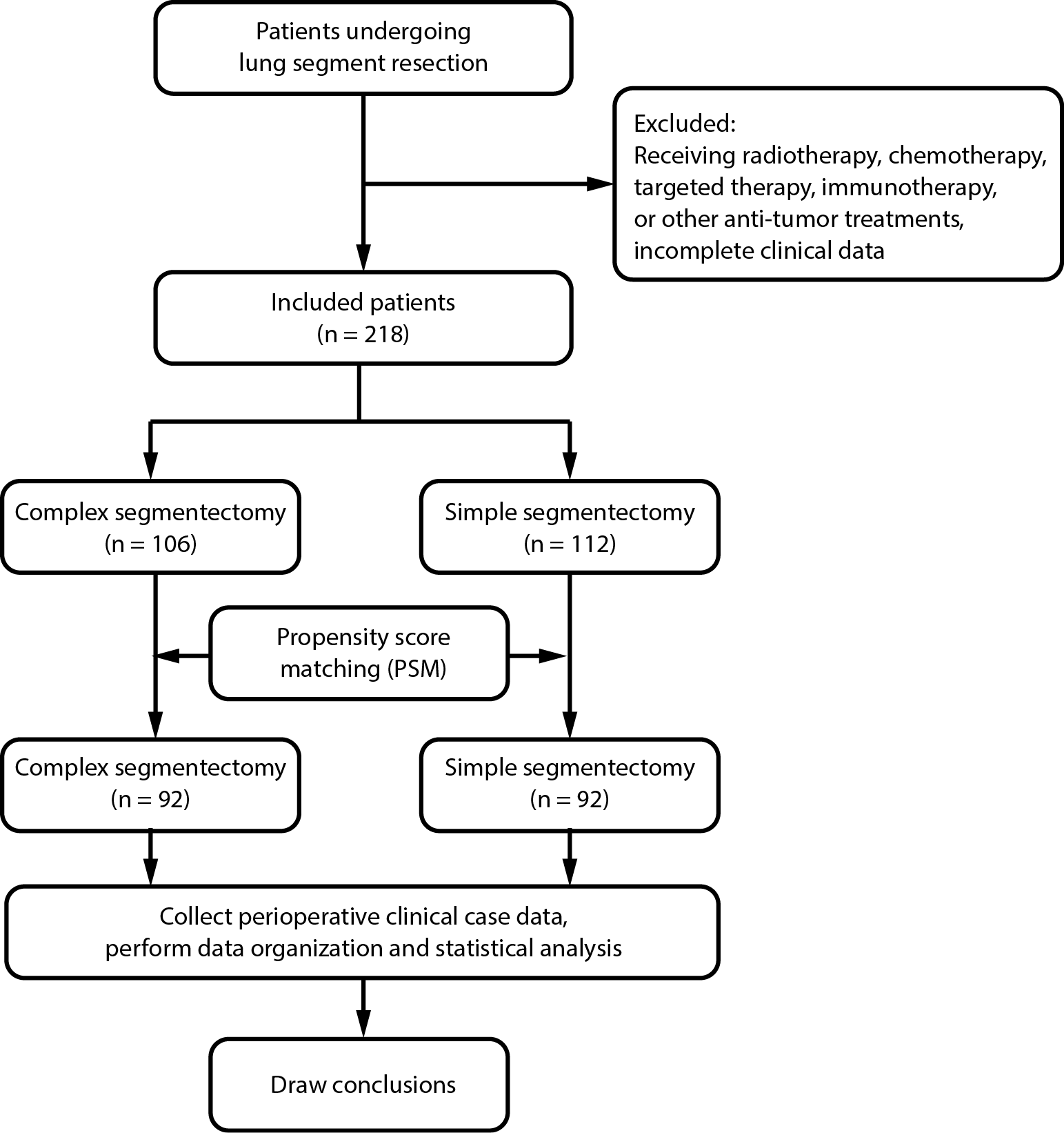
After initial screening, clinical data from a total of 218 patients were included in the analysis. Among them, the complex segmentectomy group consisted of 106 patients (48.6%), and the simple segmentectomy group consisted of 112 patients (51.4%). During the study, 2 patients from the simple segmentectomy group were excluded due to perioperative reoperation; 2 patients from the complex segmentectomy group were excluded due to transfer, and 2 others due to changes in the surgical method (Figure 1).
Sample size calculation
The sample size was estimated based on a power analysis. The primary outcome was a continuous variable, and the effect size was assumed to be Cohen’s d = 0.5, representing a moderate effect and indicating that the mean difference between groups was approximately half of the pooled standard deviation. With a significance level of 0.05 and statistical power of 80%, the minimum required sample size was 64 patients per group. Allowing for a 20% dropout rate, the adjusted required sample size was 77 patients per group. In this study, 100 patients in the complex segmentectomy group and 110 patients in the simple segmentectomy group were included, which was sufficient to meet the statistical power requirement.
Surgical methods
All surgical and clinical data were retrospectively obtained from hospital medical records and operative notes. Preoperative assessment included thin-slice chest computed tomography (CT) and contrast-enhanced CT to evaluate the indications for segmentectomy, with 3-dimensional reconstructions routinely performed and preoperative nodule localization used in selected patients. A multidisciplinary team reviewed the surgical indications and plans, and only patients with early-stage NSCLC (cT1a–cT2aN0M0) were included. All procedures were performed under general anesthesia with single-lung ventilation in the lateral decubitus position (Supplementary Fig. 1). Segmental planes were identified primarily using the inflation–deflation method (95%), with indocyanine green fluorescence used in selected cases (5%). Segmental veins were divided when feasible, and vascular and bronchial structures were managed with staplers, sutures, or energy devices, depending on intraoperative conditions. Mediastinal lymph node sampling or dissection was routinely performed. At the end of surgery, air leakage was tested and, if present, repaired with absorbable sutures and sealants. Chest tubes were placed according to standard practice, and all resected specimens underwent pathological examination, confirming negative surgical margins.
Selection criteria for complex and simple segmentectomy
The surgical approach was determined by segmental anatomical complexity and tumor location. Simple segmentectomy refers to resection of anatomically straightforward segments with clear boundaries, such as right upper lobe S1, S2, and S3 and left upper lobe S1.4 These segments have simple vascular and bronchial distributions, shallow anatomical planes, clear operative fields, and lower technical demands, making them suitable targets for early-stage lung cancer surgery.20
Complex segmentectomy involves multiple or anatomically intricate segments, such as right lower lobe S6 + S7, left lower lobe S8 + S9, and S9 + S10.8, 9 These segments have complex anatomical relationships, multiple vascular and bronchial branches, and close proximity to major structures. The procedures are technically demanding, particularly under uniportal thoracoscopic conditions with limited space and visibility, and require refined dissection and surgical expertise. In selected cases, subsegmentectomy may be performed to preserve lung parenchyma and function, typically in patients with early localized lung cancer or impaired pulmonary reserve.20, 21
Preoperative and postoperative management
All patient data were retrospectively obtained from hospital medical records and operative notes. Preoperative evaluation included brain CT or magnetic resonance imaging (MRI), abdominal ultrasound or contrast-enhanced CT, echocardiography, particularly in patients older than 60 years, and pulmonary function testing. The results were recorded in medical charts and used for surgical decision-making and analysis. Patients routinely underwent preoperative respiratory training, including stair climbing, walking, and balloon-blowing exercises, and surgery was generally scheduled within 3 days of admission.
Postoperative management included fluid restriction to approx. 1,000 mL during the first 3 days, with gradual reduction thereafter. Patients resumed a liquid diet 6 h after surgery, transitioned to a normal diet on the 2nd day, and their dietary and bowel habits were generally maintained as preoperatively. Activity was progressively increased: bedside balloon-blowing on postoperative day 1, short-distance walking on day 2, and stair climbing on day 3. Chest radiographs were performed on postoperative day 2 to evaluate lung re-expansion. Chest tubes were removed when drainage was <100 mL/day for 2 consecutive days without air leakage. Common complications, such as persistent air leakage and cough, were managed with early ambulation, respiratory exercises, nebulization, and medication. Intraoperatively, absorbable sutures and sealants were routinely applied to reinforce the lung surface and reduce air leakage. All patients underwent follow-up 1 month after surgery. Our perioperative respiratory rehabilitation and nursing strategies are consistent with evidence showing that structured nursing interventions can improve functional and psychosocial outcomes in chronic respiratory diseases.22 Discharge criteria included the absence of significant chest pain or dyspnea, resolution or medical control of cough, and removal of all drains. On the day of discharge, patients underwent blood tests and chest radiography and were discharged if no abnormalities were detected.
Quality of life assessment and long-term follow-up analysis
Postoperative QoL was assessed using the 36-Item Short Form Health Survey (SF-36), which has been validated in the local population and demonstrates good reliability and validity (Supplementary Appendix 123). Patients completed the questionnaire 3 and 6 months after surgery to evaluate physical functioning, emotional status, social functioning, vitality, and related domains. Pulmonary function recovery, including forced expiratory volume in 1 s (FEV1) and forced vital capacity (FVC), was assessed retrospectively from follow-up records. Regular chest CT examinations for monitoring tumor recurrence or metastasis were also included in the dataset. Survival status was determined from hospital records and follow-up telephone interviews, including survival, date of death, and cause of death. For deceased patients, causes of death were recorded with particular attention to procedure-related complications, lung cancer recurrence, or metastasis. The median survival follow-up duration was 13 months (range: 6–24 months), whereas QoL was assessed at 3 and 6 months after surgery.
Statistical analyses
To reduce potential selection bias between the complex and simple segmentectomy groups, propensity score matching (PSM) was performed. Propensity scores were calculated using a logistic regression model with covariates including age, sex, body mass index (BMI), preoperative pulmonary function (FEV1/FVC), smoking history, comorbidities (hypertension, diabetes, heart disease, and pulmonary disease), and pathological type. Matching was conducted using 1 : 1 nearest-neighbor matching with a caliper of 0.05. Covariate balance was assessed using standardized mean differences (SMDs), with SMD < 0.1 considered well balanced and SMD < 0.2 considered acceptable. To further control for residual confounding, multivariable regression analyses were performed: linear regression for continuous outcomes (operation time, intraoperative blood loss, postoperative drainage, hospital stay, and postoperative pulmonary function) and multivariable logistic regression for binary outcomes (postoperative air leakage and cough). Results were expressed as regression coefficients (β) or odds ratios (ORs) with 95% confidence intervals (95% CIs).
Continuous variables, including age, BMI, pre- and postoperative FEV1, FEV1/FVC, operation time, blood loss, postoperative drainage, hospital stay, and SF-36 scores, were summarized as means ± standard deviations (SDs). Normality was tested using the Shapiro–Wilk test (Supplementary Table 1), and Levene’s test was used to assess homogeneity of variance. Normally distributed data were compared using independent-samples t-tests for 2 groups and one-way analysis of variance (ANOVA) for multiple groups, with Welch’s correction applied if variances were unequal. Non-normally distributed data were compared using the Mann–Whitney U test or Kruskal–Wallis test, as appropriate. Categorical variables, including sex, comorbidities (hypertension, diabetes, heart disease, and pulmonary disease), smoking history, pathological type, tumor location, and postoperative complications (infection, arrhythmia, thrombosis, cough, atelectasis, air leakage, and conversion to thoracotomy), were presented as counts and percentages and compared using the χ2 test or Fisher’s exact test, as appropriate.
To avoid inflation of type I error due to multiple comparisons, the related tests were grouped into 3 families of hypotheses: 1) perioperative indicators (operation time, intraoperative blood loss, postoperative drainage, hospital stay, and postoperative pulmonary function); 2) postoperative complications (cough, air leakage, infection, arrhythmia, thrombosis, reoperation, atelectasis, etc.); and 3) the 8 dimensions of the SF-36 QoL questionnaire, assessed at 3 and 6 months after surgery. To control the family-wise error rate, Holm–Bonferroni correction was applied within each family. All statistical analyses were performed using IBM SPSS v. 26.0 (IBM Corp., Armonk, USA), and p < 0.05 was considered statistically significant. Unadjusted p-values are reported as the original comparison results, whereas Holm-adjusted p-values represent the corrected values after adjustment within each hypothesis family.
Results
Baseline characteristics of patients
A total of 218 patients were included, with 106 (48.6%) in the complex segmentectomy group and 112 (51.4%) in the simple segmentectomy group. Six patients did not complete the planned segmentectomy due to intraoperative conversion or referral, including 4 in the complex group (3.8%; 2 converted to lobectomy) and 2 in the simple group (1.8%); the difference was not significant (p = 0.412) (Supplementary Table 2). After PSM, 92 patients were retained in each group. Most covariates achieved good balance (SMD < 0.1), while pathological type and tumor location showed mild residual imbalance but remained acceptable (SMD < 0.2) (Supplementary Table 3).
Baseline characteristics are shown in Table 1. No significant differences were observed between groups in age, sex, BMI, preoperative FEV1, or FEV1/FVC (p > 0.05). Comorbidities, including hypertension, diabetes, heart disease, and pulmonary disease, as well as smoking history, were also comparable (p > 0.05). The distribution of pathological types, including adenocarcinoma in situ, invasive adenocarcinoma, minimally invasive adenocarcinoma, squamous cell carcinoma, benign tumors, and other types, was similar between groups (p > 0.05). In contrast, tumor location differed significantly between the complex and simple groups (p < 0.001).
Comparison of perioperative outcomes between complex and simple lung segmentectomy
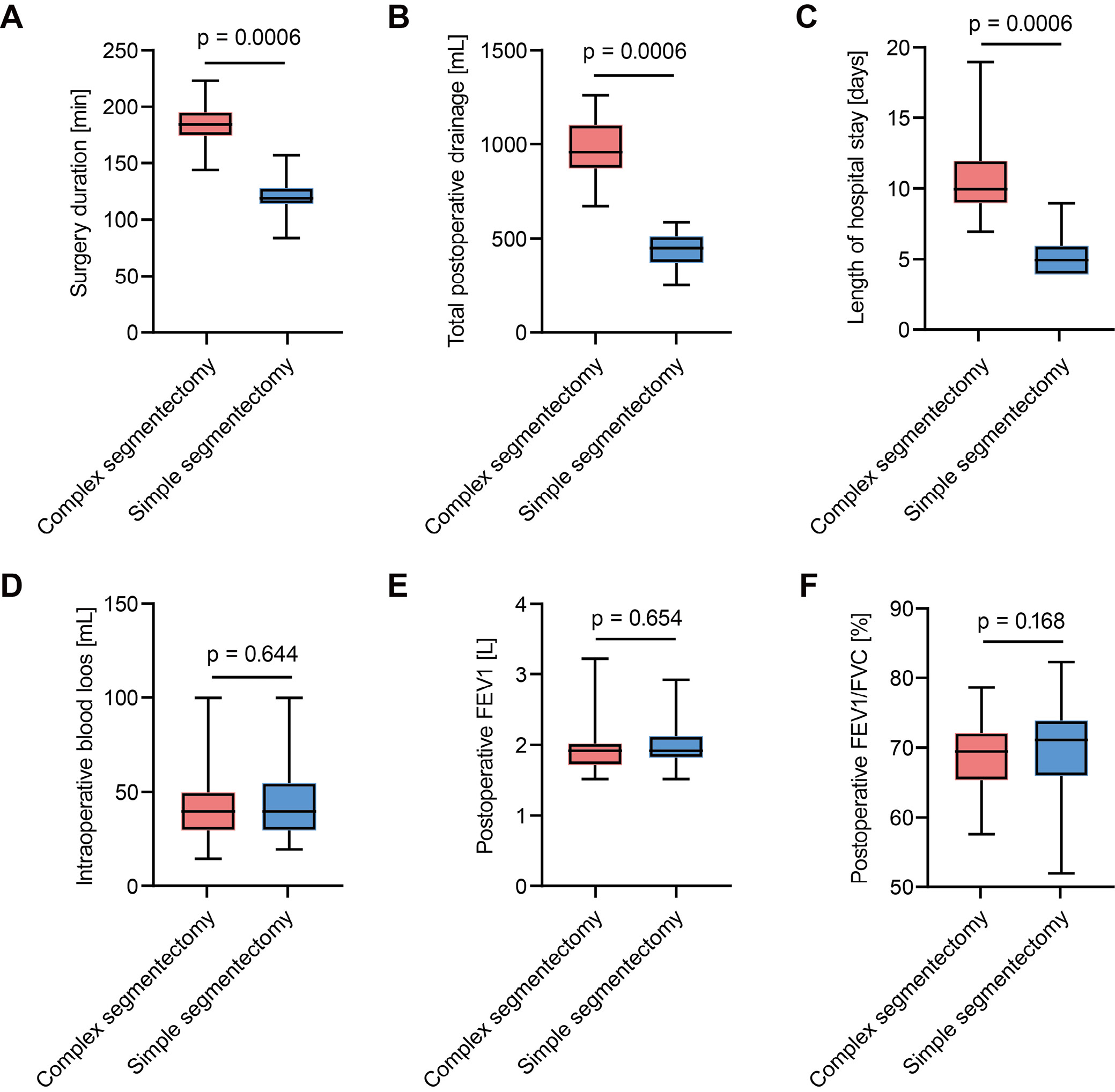
Perioperative outcomes are summarized in Table 2. Operative time was significantly longer in the complex segmentectomy group than in the simple segmentectomy group (p < 0.001) (Figure 2A). Total postoperative drainage was also greater in the complex group (p < 0.001) (Figure 2B), and hospital stay was significantly prolonged (p < 0.001) (Figure 2C), indicating increased resource utilization and longer recovery. In contrast, intraoperative blood loss did not differ between groups (p = 0.644) (Figure 2D). Postoperative pulmonary function, including FEV1 and FEV1/FVC, was comparable between groups (p > 0.05), suggesting no additional short-term functional loss in the complex group (Figure 2E,F). Multivariable regression confirmed these findings: complex segmentectomy was associated with longer operative time (β = +66 min, p < 0.001), greater drainage volume (β = +466 mL, p < 0.001), and longer hospital stay (β = +5.1 days, p < 0.001), while differences in blood loss and postoperative pulmonary function remained non-significant. These results were consistent with the PSM analysis (Supplementary Table 4).
Comparison of postoperative complications between complex and simple lung segmentectomy
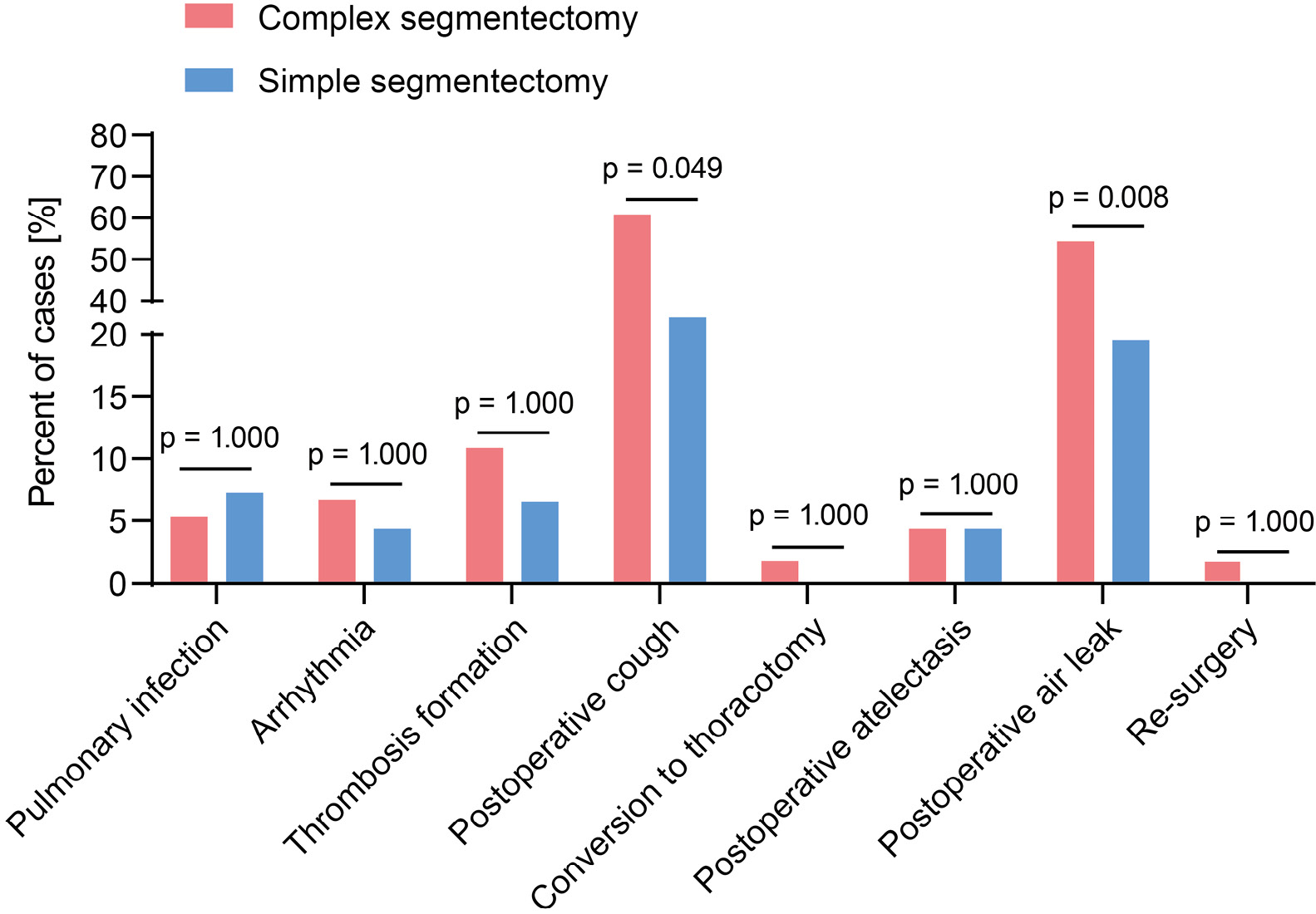
The incidence of postoperative pulmonary infection was 6.52% (6 cases) in the complex segmentectomy group and 10.87% (10 cases) in the simple segmentectomy group, with no significant difference (p = 1.000). Arrhythmia occurred in 6.52% (6 cases) and 4.35% (4 cases), respectively (p = 1.000). Thrombosis occurred in 10.87% (10 cases) in the complex group and 6.52% (6 cases) in the simple group, without statistical significance (p = 1.000). Postoperative cough was significantly more frequent in the complex group (60.87%, 56 cases) than in the simple group (32.61%, 30 cases) (p = 0.049), potentially reflecting greater airway injury and prolonged recovery. Conversion to thoracotomy occurred in 2 patients (2.17%) in the complex group and in none in the simple group (p = 1.000). The incidence of postoperative atelectasis was 4.35% (4 cases) in both groups (p = 1.000). Postoperative air leakage occurred in 54.35% (50 cases) of patients in the complex group compared with 19.57% (18 cases) in the simple group, showing a significant difference (p = 0.008). Reoperation was required in 2 patients (2.17%) in the complex group and in none in the simple group (p = 1.000) (Table 3, Figure 3).
Multivariable logistic regression further confirmed increased risks of postoperative air leakage (OR = 6.14, 95% CI: 2.20–17.10; p < 0.001) and cough (OR = 3.76, 95% CI: 1.50–9.41; p = 0.005) in the complex group. Other complications showed no significant differences because of their low incidence (Supplementary Table 4).
Comparison of perioperative outcomes in complex lung segmentectomy across different lung lobes
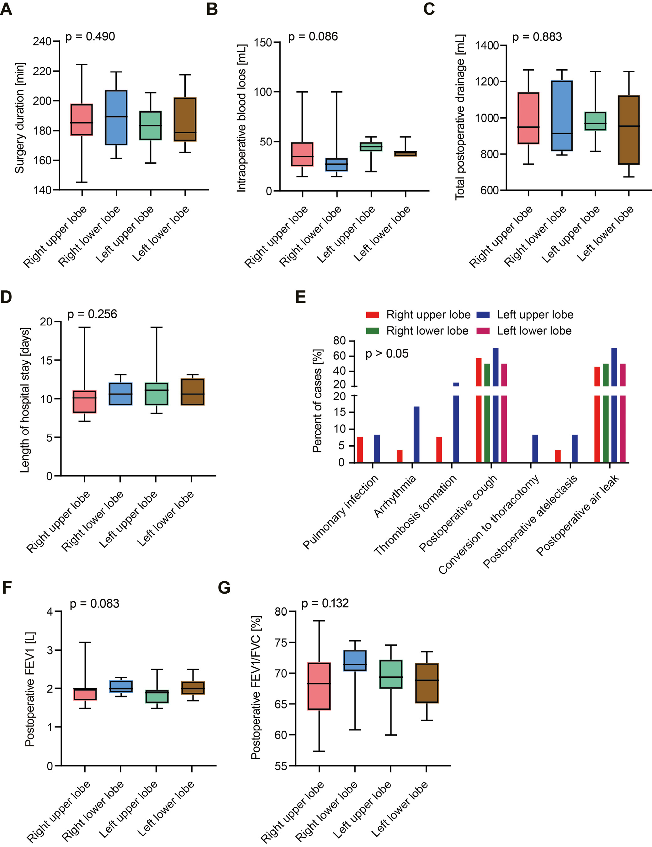
No significant differences were observed among different lobes in operative time, intraoperative blood loss, postoperative drainage, or length of hospital stay (p > 0.05). Postoperative complications, including pulmonary infection, arrhythmia, thrombosis, and cough, also showed no significant differences across lobes (p > 0.05). The incidence of postoperative atelectasis was comparable across groups, while air leakage was more frequent in the left upper lobe group (70.83%) but did not reach statistical significance (p = 0.251). Postoperative pulmonary function, including FEV1 and FEV1/FVC, was similar across groups (p > 0.05) (Table 4, Figure 4).
Impact of complex and simple lung segmentectomy on patients’ QoL
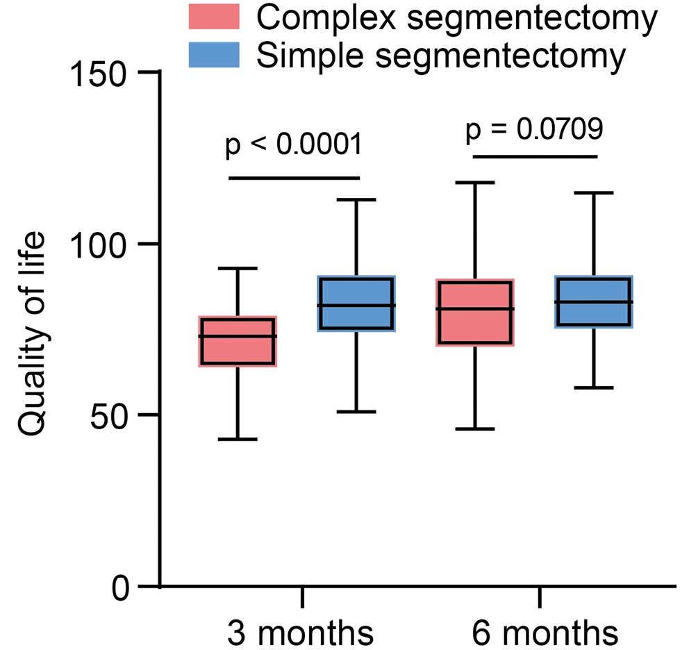
Quality of life was assessed using the SF-36 questionnaire. At 3 months postoperatively, 78 of 92 patients completed the survey (response rate, 84.8%), with 3 excluded because of incomplete data. At 6 months, 72 questionnaires were returned (response rate, 78.3%), with 2 excluded because of incomplete information. At 3 months, the mean SF-36 score was significantly lower in the complex segmentectomy group (70.2 ±10.9) than in the simple segmentectomy group (81.6 ±12.3), reflecting longer recovery and more frequent complications (p < 0.001). By 6 months, scores in the complex group had improved to 79.5 ±11.9, showing no significant difference from the simple group (82.9 ±12.0; p = 0.071) (Table 5, Figure 5).
Analysis of individual SF-36 domains revealed that, at 3 months, the complex group scored significantly lower in physical functioning, role-physical, bodily pain, general health, and vitality (all p < 0.001). Scores for social functioning, role-emotional, and mental health were also lower, but the differences did not reach statistical significance (p > 0.05). By 6 months, scores across all domains had improved in the complex group, and no significant differences were observed between groups (p > 0.05) (Supplementary Table 5).
Impact of complex vs simple segmentectomy on postoperative survival and recurrence
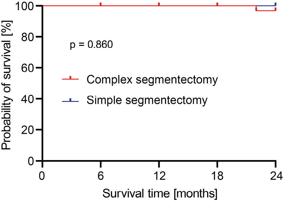
Kaplan–Meier analysis demonstrated nearly overlapping survival curves between the complex and simple segmentectomy groups, with no significant difference (p = 0.860) (Figure 6). Further comparisons of recurrence-free survival (RFS) and local recurrence rates are summarized in Supplementary Table 6. Two-year RFS was similar between groups: 95.7% in the complex group and 96.8% in the simple group (p = 0.782). Local recurrence occurred in 3.3% of patients in the complex group and 2.2% of patients in the simple group, with no significant difference. These findings indicate that complex segmentectomy provides survival and recurrence outcomes comparable to those of simple segmentectomy.
Discussion
Sublobar resection has gained increasing attention because it maximizes the preservation of lung function.24, 25 However, complex segmentectomy remains less widely applied because of anatomical complexity and technical difficulty. A systematic comparison of complex vs simple segmentectomy in terms of perioperative outcomes, QoL, and long-term survival is essential to clarify feasibility and risk and to guide individualized treatment of early-stage lung cancer.
Complex segmentectomy was associated with longer operative time, greater drainage volume, and prolonged hospitalization, whereas postoperative lung function recovery, overall complication rates, and long-term survival were comparable to those of simple segmentectomy.26, 27 This study focused on long-term outcomes and showed that, although patients undergoing complex procedures had lower early postoperative QoL, recovery was comparable to that of the simple segmentectomy group at 6 months.28 Postoperative cough and prolonged air leakage were more frequent after complex resection,29, 30 consistent with previous reports attributing higher complication rates to complex anatomy.31 With standardized perioperative management, including early ambulation, respiratory training, and reinforcement of the lung parenchyma, chest tubes were successfully removed in most patients, and patients were discharged without major adverse events. These findings support the feasibility of complex segmentectomy in terms of long-term outcomes and QoL, indicating its role as an effective surgical option for peripheral early-stage lung cancer.
Previous studies have demonstrated that uniportal thoracoscopic simple segmentectomy provides the advantages of minimal trauma, rapid recovery, and fewer complications.14, 15, 16 In contrast, evidence on complex segmentectomy is limited and mostly addresses technical feasibility and short-term complications.19, 29, 30 This study extends prior work by systematically comparing complex and simple procedures. Although complex surgery increased operative burden and complication incidence, long-term QoL and survival remained comparable.26, 27 Unlike earlier reports, this study used propensity score matching to reduce baseline imbalance and incorporated the SF-36 questionnaire and Kaplan–Meier survival analysis, thereby addressing the lack of long-term follow-up in previous studies.28 The results also confirmed the higher incidence of postoperative cough and prolonged air leakage after complex resections,29, 30, 31 but these complications were manageable with standardized perioperative care, supporting the clinical feasibility of complex segmentectomy in early-stage lung cancer.
The clinical value of complex segmentectomy lies in maximizing the preservation of lung function while maintaining oncological safety. Despite longer operative time, prolonged hospitalization, and a higher complication burden, oncological outcomes were not inferior, highlighting the importance of appropriate patient selection. Evidence suggests that when the tumor diameter is ≤2 cm, the tumor is located at or across a segmental border, presents as ground-glass opacity or has a consolidation-to-tumor ratio (CTR) ≤0.5, and a margin ≥2 cm or ≥ the tumor diameter can be achieved, complex segmentectomy offers advantages over lobectomy by securing adequate margins and systematic nodal staging while preserving lung function.32, 33 In patients with impaired baseline lung function, multiple lesions, or prior resections, complex segmentectomy also prevents unnecessary resection of healthy lung tissue. Compared with wedge resection, it is more advantageous for deep lesions or when margins cannot be guaranteed, particularly when spread through air spaces (STAS) cannot be excluded intraoperatively.34 Lobectomy remains more appropriate for centrally located, solid-dominant tumors >2 cm or those at risk of inadequate margins, whereas wedge resection is sufficient for small, peripheral, GGO-dominant lesions with secure margins. In summary, the essence of complex segmentectomy is “preserving lung function while ensuring complete resection,” and its application should be based on strict patient selection and standardized procedures.
Limitations of the study
This study has important implications for clinical practice, but it also has several limitations. Although 210 patients were included and the overall sample size met the statistical requirements, subgroup numbers, such as the number of patients undergoing complex segmentectomy in different lobes, were small and insufficient to fully assess lobe-specific perioperative risk. QoL follow-up was limited to 6 months, and survival follow-up was relatively short (median, 13 months; range, 6–24 months), which limited assessment of longer-term QoL and survival outcomes. As this was a single-center retrospective study, external validity was limited despite the comprehensive data. All surgeries were performed by a highly experienced thoracic team, with each lead surgeon having performed more than 500 uniportal segmentectomies, ensuring high surgical quality but potentially influencing perioperative outcomes. Previous studies indicate that complex segmentectomy has a steep learning curve, and limited surgical experience may prolong operative time and increase complications.11, 31 Furthermore, lobectomy was not included in the comparison; therefore, this study primarily reflects differences between complex and simple segmentectomy without directly informing the choice between complex segmentectomy and lobectomy.
Future research should involve multicenter, large-sample, prospective studies to enhance generalizability and provide direct comparisons between complex segmentectomy and lobectomy. Follow-up should be extended beyond 5 years to capture long-term outcomes in terms of QoL, recurrence, and survival. Broader clinical data across different populations and regions are needed to strengthen the evidence for treatment decisions. Individualized rehabilitation programs may shorten recovery and improve postoperative QoL. With ongoing advances in uniportal thoracoscopy, further work should focus on new surgical techniques, devices, and optimized perioperative care to enhance the safety and efficacy of complex segmentectomy.
Conclusions
This study compared single-port thoracoscopic complex and simple lung segmentectomy in terms of perioperative outcomes, survival prognosis, postoperative complications, and QoL, leading to the following conclusions.
Perioperative outcomes: Although complex lung segmentectomy was associated with longer operative time, longer hospital stay, higher postoperative drainage volume, and a higher incidence of postoperative cough compared with simple lung segmentectomy, there were no significant differences between the 2 groups in terms of intraoperative blood loss or postoperative complications, including pulmonary infection, arrhythmia, and thrombosis. The perioperative risks of complex lung segmentectomy are manageable and do not significantly increase the postoperative burden on patients.
Survival and recurrence outcomes: This result indicates no significant difference in survival prognosis between the 2 approaches, further validating the clinical feasibility of complex lung segmentectomy.
Quality of life: The QoL score for the complex lung segmentectomy group was lower at 3 months after surgery than that for the simple lung segmentectomy group. However, QoL scores gradually improved over time and approached those of the simple lung segmentectomy group. This suggests that, although complex lung segmentectomy affects short-term QoL, patients’ QoL improves progressively during postoperative recovery. Despite being associated with higher perioperative risks, particularly in non-specialized hospitals, complex lung segmentectomy represents a feasible and promising surgical option for the treatment of early-stage lung cancer. As surgical techniques improve and experience accumulates, complex lung segmentectomy is expected to become a routine option in the treatment of early-stage lung cancer.
Overall, this study provides valuable practical insights into the clinical application of complex lung segmentectomy and lays the foundation for further research.
Supplementary data
The Supplementary materials are available at https://doi.org/10.5281/zenodo.18489943. The package contains the following files:
Supplementary Table 1. Shapiro–Wilk test for normality.
Supplementary Table 2. Incompletion rate of planned segmentectomy.
Supplementary Table 3. Covariate balance before and after PSM.
Supplementary Table 4. Multivariable regression analyses of perioperative outcomes.
Supplementary Table 5. Comparison of SF-36 Dimension Scores.
Supplementary Table 6. Comparison of recurrence outcomes between complex and simple segmentectomy.
Supplementary Fig. 1. Single-port thoracoscopic lung segmentectomy.
Supplementary Appendix 1. SF-36.
Data Availability Statement
The datasets supporting the findings of the current study are openly available in the Zenodo repository at https://doi.org/10.5281/zenodo.18264281.
Consent for publication of personal information
Not applicable.
Use of AI and AI-assisted technologies
Not applicable.