


Abstract
Background. Hemolytic-uremic syndrome (HUS) is a thrombotic microangiopathy that is most commonly caused by infection with Shiga toxin-producing Escherichia coli (STEC-HUS). Atypical HUS (aHUS) is associated with inappropriate activation of the alternative complement pathway caused by infections, including SARS-CoV-2, malignancies, medications, or other triggering factors in genetically predisposed individuals.
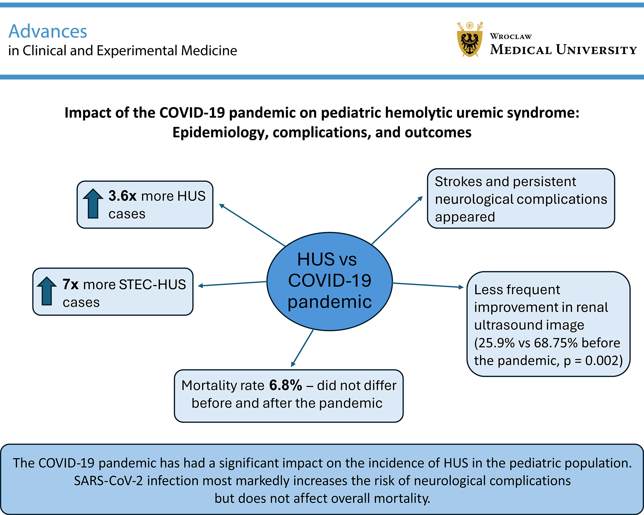
Objectives. Assessment of the impact of the COVID-19 pandemic on epidemiology, course, complications, and outcome of HUS in children in a single tertiary nephrological center in Poland.
Materials and methods. It is a retrospective study, in which a total number of 74 pediatric patients with HUS were analyzed, including 16 cases in 5-year period before the outbreak of the COVID-19 pandemic (HUS-5) and 58 cases in 5-year period after the outbreak of the pandemic (HUS+5).
Results. A more than 3.6-fold increase in HUS cases and a 7-fold higher incidence of STEC-HUS were reported in the HUS+5 population. Strokes and persistent neurological complications were observed only in this group. 91.9% of patients presented with abnormal renal ultrasound (USG) findings at admission, and these findings improved significantly more frequently in the HUS-5 (68.75% vs 25.9%, p = 0.002) group. Mortality did not differ significantly between the 2 analyzed groups and was 6.8% for the entire population.
Conclusions. The COVID-19 pandemic has had a significant impact on the incidence of HUS in the pediatric population. SARS-CoV-2 infection most markedly increases the risk of neurological complications but does not affect overall mortality.
Key words: hemolytic-uremic syndrome, COVID-19, child, Shiga toxin-producing Escherichia coli, neurologic complications
Background
Hemolytic uremic syndrome (HUS) is one of the conditions associated with thrombotic microangiopathy (TMA) and constitutes a life-threatening, rare disease characterized by microangiopathic hemolytic anemia, thrombocytopenia, and acute kidney injury (AKI).
Depending on the triggering factors, HUS can be generally classified as hereditary or acquired.1 Additionally, HUS may be classified as follows: infection-associated HUS (including causes such as Shiga toxin-producing Escherichia coli (STEC), Streptococcus pneumoniae, influenza A, H1N1, HIV, norovirus, Epstein–Barr virus (EBV), parvovirus B19, and COVID-19), HUS secondary to pregnancy, bone marrow or solid organ transplantation, systemic malignancies, radio- or chemotherapy, autoimmune conditions (such as systemic lupus erythematosus (SLE), antiphospholipid antibody syndrome, and scleroderma), some drugs (quinine, calcineurin inhibitors, and a few chemotherapeutic agents), malignant hypertension (HT), cobalamin C disorder, and finally atypical HUS (aHUS) due to alternative complement pathway dysregulation (complement-mediated HUS; CM-HUS) and mutations in the diacylglycerol kinase epsilon (DGKE) gene.2, 3, 4, 5
Shiga toxin-producing Escherichia coli HUS (STEC-HUS), also traditionally called typical HUS, constitutes 90–95% of pediatric HUS cases, while aHUS accounts for 5–10%. The incidence of HUS among individuals infected with STEC ranges from 5% to 15%, predominantly affecting children younger than 5 years.5, 6
Genetic variants in complement regulatory proteins (CRPs, including CFH, CFI, MCP, CFB, and C3) or autoantibodies to factor H (FH) account for 50–60% of all aHUS cases, with approx. 30–50% having no known identifiable mutation.4, 7 While genetic mutations can predispose a person toward developing aHUS, the disease usually requires a trigger event to manifest clinically. Recent studies have suggested that viral infections, including COVID-19, may trigger aHUS in genetically susceptible individuals as severe acute respiratory syndrome coronavirus 2 (SARS-CoV-2) may cause widespread microvascular damage, including TMA, driven by inflammation, cytokine storm, and dysregulated complement system activation.8, 9, 10, 11, 12
Epidemiologically, HUS is a rare disease in children, and the incidence varies considerably depending on the data source and geographical region, but collectively for STEC-HUS and aHUS, it is estimated at an average of 6.3–14.2/million age-related population/year, while the incidence of CM-HUS in children is relatively constant at 0.25–2/million age-related population/year.5, 13, 14, 15
According to the literature, renal involvement in COVID-19 is reported to be 10–15%.16 In a meta-analysis by Gu et al., patients with COVID-19 had a 1.5-fold higher risk of AKI and a more than twofold higher risk of death when AKI occurred, with statistically significant results despite moderate heterogeneity.17 However, in a series of patients with COVID-19 who were biopsied because of AKI or proteinuria, glomerular TMA was a relatively rare finding compared to acute tubular necrosis or collapsing glomerulopathy.18 On the other hand, numerous reports have linked COVID-19 infection in the pediatric population with new episodes or relapses of HUS.9, 19, 20, 21 Therefore, due to the severity and potentially fatal complications of this condition, this study addresses this issue in more detail.
Objectives
There are limited original papers or reports on the effect of SARS-CoV-2 infection on the occurrence of HUS in children. The aim of this project was to assess the impact of the COVID-19 pandemic on the epidemiology, clinical course, complications, and outcomes of HUS in pediatric patients hospitalized in our tertiary pediatric nephrology center.
Materials and methods
The study is a single-center, retrospective chart review of patients who were newly diagnosed with HUS, and were hospitalized at the Department of Pediatric Nephrology and Hypertension University Children’s Hospital of Cracow (Poland), a tertiary reference center, during a 10-year period (from January 1, 2015, to December 31, 2024).
The study population was divided into 2 groups: a subpopulation with HUS before the outbreak of the COVID-19 pandemic, covering the period from January 1, 2015, to December 31, 2019 (HUS-5; 16 patients), and a subpopulation after the outbreak of the pandemic, covering the period from January 1, 2020, to December 31, 2024 (HUS+5; 58 patients).
The ethics committee of Jagiellonian University Medical College, Cracow, Poland, approved the study (approval No. 118.0043.1.429.2024), and informed consent was waived due to its retrospective nature. The study involved a detailed analysis of data from the medical histories and outpatient documentation of each patient included in the study.
STEC-HUS (typical HUS) was diagnosed when genetic material from enteropathogenic Escherichia coli (EPEC), enterohemorrhagic Escherichia coli (EHEC), or STEC (STEC 1 or 2) was identified in the patient’s stool or anal swab using multiplex polymerase chain reaction (PCR) assay (The QIAstat-Dx® Gastrointestinal Panel; Qiagen, Hilden, Germany).22 Atypical HUS was diagnosed when stool testing was negative for these pathogens and ADAMTS13 activity exceeded 10%. This group also included 2 patients with Streptococcus pneumoniae infection who developed HUS (Strep-HUS).
Contact with SARS-CoV-2 was defined as the presence of positive immunoglobulin M or G (IgM or IgG) antibodies against SARS-CoV-2, or a positive antigen or PCR test for SARS-CoV-2 performed upon admission. None of the children had been vaccinated against COVID-19.
Semi-quantitative renal ultrasonography (USG) assessment was performed by evaluating the results of examinations performed on admission and at hospital discharge. The following parameters were assessed: kidney size (normal/enlarged: 0/1 point), parenchymal echogenicity (normal/increased: 0/1 point), and corticomedullary differentiation (normal/reduced/none: 0/1/2 points). The range of possible points was 0–4. After summing up the points, the renal USG findings were categorized as follows: normal (0 points), minor damage (1–2 points), and major damage (3–4 points).
Hypertension was diagnosed according to the European Society of Hypertension (ESH) guidelines for children and adolescents as an average of at least 3 blood pressure measurements greater than or equal to the 95th percentile for age, sex, and height in children aged 0–15 years of age, or ≥140/90 mm Hg for adolescents aged 16 years or older, using an appropriately sized cuff and a validated sphygmomanometer.23
Estimated glomerular filtration rate (eGFR) was calculated using the Schwartz formula.24 When defining the outcome, the basic factors were the eGFR value as well as the presence and severity of proteinuria. The occurrence of HT and the renal USG findings were supplementary factors to the overall assessment. Hence, the outcome of a patient was classified as either:
1) complete recovery, defined as: eGFR > 90 mL/min/1.73 m2 and the absence of proteinuria, the absence of HT, and normal or close to normal renal USG (0–1 points);
2) a) superior partial recovery, defined as: eGFR ≥ 45 mL/min and absence or presence of non-nephrotic range proteinuria (<50 mg/kg/day). Abnormalities including HT and/or abnormal renal USG (2–4 points) may or may not be present; b) inferior partial recovery, defined as: eGFR ≤ 44 mL/min, and/or presence of nephrotic range proteinuria (≥50 mg/kg/day), the presence of non-nephrotic range proteinuria or absence of proteinuria. Abnormalities including HT and/or abnormal renal USG (2–4 points) may or may not be present.
For points 2a and 2b – the final qualification of a child to an outcome group depended on which factor (eGFR or proteinuria) the result was more severe.
3) death.
Statistical analyses
Normality of distribution of variables was assessed using the Shapiro–Wilk test. As most samples deviated from normality, results are reported as medians and interquartile ranges (IQR). Non-normally distributed variables were compared using the nonparametric Wilcoxon rank-sum test (Mann–Whitney U test) with a significance level set at 0.05 (pW). For Gaussian-distributed variables, Student’s t-test (tS) was applied; in cases of heteroscedasticity, Welch’s t-test was used. In cases of significant heterogeneity of variances in Levene’s or Brown–Forsythe test (p < 0.05), Welch’s t-test was applied. Categorical paired data were analyzed using McNemar’s test. The significance of the difference between 2 distributions was assessed using a one-sided Fisher’s exact test. The threshold value of p = 0.05 was adopted when assessing statistical significance. Statistica v. 10 PL package (StatSoft Poland, Cracow, Poland) was used for the analysis.
Results
In this study, we analyzed data from 74 pediatric patients hospitalized for HUS in our center, including 16 cases before the pandemic (HUS-5) and 58 cases after the outbreak of the pandemic (HUS+5). No patient was excluded from the study. In both subgroups, there were boys and girls with almost equal frequency, and the median age was 55.5 months in HUS-5 and 29.3 months in HUS+5.
A more than 3.6-fold increase in the number of HUS cases in the pediatric population was observed after the outbreak of the COVID-19 pandemic compared with the corresponding period before the pandemic (HUS-5 population: 3.2 vs HUS+5 population: 11.6 cases per year).
Interestingly, in the HUS-5 group, the proportion of aHUS was 2.1 times higher than in the post-pandemic period (62.5% vs 29.3%, p = 0.017), with an overall rate of 36.5% during the 10-year observation period. In both subgroups, the mortality rate was comparable and amounted to 6.8% for the entire population. Nineteen patients (32.8%) in the HUS+5 group had confirmed contact with COVID-19 (active – 2, past – 17). The other analyzed parameters, including age, body weight, height, length of stay in the intensive care unit (ICU), circulatory or respiratory failure, and the need for oxygen therapy or intubation, did not differ significantly between the groups.
In the aHUS population, 18 of 27 patients (66.7%) received eculizumab; since 2018, when the drug became available in our country, the proportion was 18 of 20 (90%).
During the summer months (April–September), the incidence of HUS was 1.85 times higher than in the winter months (October–March). Moreover, in the HUS+5 group, there was a significantly higher proportion of children aged 1–5 years compared to the HUS-5 period (Table 1).
Among the analyzed symptoms, diarrhea occurred 1.4 times more frequently in patients in the HUS+5 group compared with the HUS-5 group (81% vs 56.3%, p = 0.046), and its duration, calculated for the entire study population, was also longer in this group (4 days vs 1.5 days, p = 0.03). This is likely due to the fact that this group had a significantly higher proportion of typical HUS cases than in the HUS-5 group (72.4% vs 37.5%). In addition, in this group, the number of schistocytes was 2.3 times higher compared with the HUS-5 group (44.5/µL vs 19/µL, p = 0.03). The remaining symptoms, complications, need for interventions, and their duration did not differ significantly between the studied groups. The most common complaints, besides diarrhea, were abdominal pain (78.4% of the entire study population), nausea and vomiting (75.7%), and oligo/anuria (68.9%). The frequency of HT was approx. 60.8% at admission (similar in both HUS-5 and HUS+5 groups) and 41.9% at discharge (Table 2).
Neurological symptoms occurred in approx. 36.5% of patients, with similar rates in both groups. The severity of these symptoms also did not differ significantly. In the HUS-5 group, no strokes were recorded and no neurological deficits were observed at discharge. In contrast, in the HUS+5 group, strokes (both ischemic and hemorrhagic) occurred in 7 patients (12%). Additionally, at discharge, persistent neurological deficits were observed in 14.8% of patients, the majority of which (5 out of 8; 62.5%) were severe. The overall incidence of strokes in the entire study population did not reach statistical significance, although it showed a trend toward significance (p = 0.067) (Table 3).
The most common neurological symptoms in the entire study group were reduced or absent responsiveness and confusion (39.4% of all symptoms), occurring with similar frequency in both groups. Seizures, stroke, and ophthalmic symptoms were relatively rare. In the HUS+5 group, the spectrum of neurological symptoms was 5 times higher than in the HUS-5 group (83.3% vs 16.7%, p = 0.001), while the type of symptoms did not differ significantly between the groups (Table 4). When considering the distinction between typical and atypical HUS, neurological symptoms were more frequent in the aHUS group than in the typical HUS group (51.9% vs 27.7%, p = 0.03).
In the HUS-5 group, no permanent neurological deficits were observed, while in the HUS+5 group, the most common symptoms at discharge were disturbances in muscle tone and strength, as well as psychomotor slowing (together accounting for 41.2% of all neurological symptoms). Additionally, cranial nerve disorders were observed in 5.2% of these patients, along with single cases of Babinski’s sign, epilepsy, depression, or the need for ventriculoperitoneal shunt placement (Supplementary Table 1).
Full recovery occurred in 17.6% of patients, being slightly more frequent, although not statistically significant, in the HUS-5 group. Partial recovery with mild or moderate kidney damage (eGFR ≥ 45 mL/min) was significantly more common in the HUS+5 group (46.6% vs 18.8%, p = 0.04), while the more severe form (eGFR ≤44 mL/min) occurred with similar frequency in both groups. Nephrotic-range proteinuria and the final eGFR value (at discharge or death) were similar in both groups. No notable differences in mortality were observed between the HUS-5 and HUS+5 groups (6.3% and 6.9%, respectively). This may be due to the small sample size in the HUS-5 group (Table 5).
Neuroimaging was performed in 19 (25.7%) patients (HUS-5, n = 3; HUS+5, n = 16). Computed tomography (CT) was obtained in 10 patients (13.5%), magnetic resonance imaging (MRI) in 8 (10.8%), and USG in 1 (1.4%). In the outcome analysis, normal findings were present in all 3 HUS-5 patients and 4 (25%) HUS+5 patients (p = 0.036). Abnormal findings occurred in 12 of 16 HUS+5 patients (75%), all detected with CT, accounting for 63.2% of the total evaluated group.
Furthermore, no notable differences in complement components were observed between the studied groups. This may indicate a similar level of complement system activation in both analyzed time periods. On the other hand, the HUS-5 group was significantly smaller and therefore may not be representative (Supplementary Table 2).
Most biochemical parameters did not differ significantly between the analyzed groups at any stage of hospitalization. The only significant difference was a clearly higher lactate dehydrogenase (LDH) activity in the HUS-5 group at admission, during hospitalization, and at discharge (p = 0.003, p < 0.001, p = 0.006, respectively) (Supplementary Table 3).
At admission, the USG findings were abnormal in both studied populations, but not statistically significant. Most patients showed minor kidney damage (56.25% in the HUS-5 and 72.4% in the HUS+5), while major kidney damage was observed in 37.5% of the HUS-5 and 19% of the HUS+5 patients. In both groups, improvement in USG findings was noted between the beginning and the end of hospitalization, although this was significantly more frequent in the HUS-5 (semi-quantitative assessment: 68.75% vs 25.9%, p = 0.002; point-based assessment: 68.75% vs 39.7%, p = 0.04). Stable USG findings were more frequently observed in the HUS+5 group. In the overall population, there were no significant differences in the percentage of normal semi-quantitative USG results at admission compared to the endpoint (discharge or death) (6 (8.1%) vs 14 (18.9%), respectively), nor in minor kidney damage (51 (68.9%) vs 52 (70.3%), respectively). However, there was a significant reduction in the percentage of patients with major kidney damage on USG (17 (23%) vs 8 (10.8%); p = 0.04, McNemar’s test) (Table 6, Supplementary Table 4).
Discussion
In this study, we analyzed the clinical course and outcomes of pediatric patients with HUS 5 years before and 5 years after the outbreak of the COVID-19 pandemic. These findings suggest that the pandemic may have a relevant effect on the epidemiology and complications of HUS in children.
Hemolytic uremic syndrome is a rare disease, with an estimated global incidence of 6.3–14.2 cases per million age-related population per year.13, 14 In Poland, approx. 3.9 cases per million age-related population of typical HUS are detected, and an increase in the number of cases has been observed in recent years. In contrast, the incidence of aHUS in Poland has remained stable at around 1.8 cases per million age-related population.25 In our center, the estimated incidence was 4.7 cases per million age-related population for typical HUS and 2.7 cases per million age-related population for aHUS, which appears consistent with national epidemiological data.
In our study, we also observed a significant increase in HUS cases in the 5-year period following the outbreak of the COVID-19 pandemic, with the incidence of typical HUS being 7 times higher than in the corresponding period before the pandemic (42 vs 6 cases). It is worth noting that, according to the European Centre for Disease Prevention and Control (ECDC), the overall notification rate of STEC infection in the European Union was 2.5 cases per 100,000 population, exceeding the pre-pandemic level and representing a 25% increase compared with that in 2021.26
COVID-19 increases the risk of thrombosis and disseminated intravascular coagulation (DIC).12, 27 Additionally, by stimulating the complement system, it may lead to the development of TMA.28, 29, 30 Ziaee and Assari described the occurrence of syndromes similar to HUS, with a positive history of infection or contact with a person infected with COVID-19 within the preceding 1–2 weeks.31 The literature describes the possibility of exacerbation of HUS symptoms or induction of HUS in genetically predisposed individuals through infection with SARS-CoV-2.11, 32, 33 Within our patient population, 1/3 of patients hospitalized for HUS after the outbreak of the COVID-19 pandemic had a documented history of past COVID-19 contact.
Furthermore, the higher percentage of schistocytes observed in the present study in the HUS+5 group may reflect that SARS-CoV-2 acted as an exacerbating factor for TMA through endothelial injury and complement activation, although no significant differences in complement system activity were observed.
Both COVID-19 and HUS share similar pathological mechanisms, such as endothelial injury, cytokine storm, and complement activation, which may result in multi-organ failure.12, 20, 31, 34 Shiga toxin can directly activate the alternative complement pathway, which is particularly evident in severe cases of STEC-HUS.35, 36 Moreover, some studies indicate that neurological symptoms more often arise as a result of other factors, such as electrolyte imbalances, dehydration, or HT.37, 38 SARS-CoV-2 can also reach the brain through hematogenous dissemination.39 Additionally, a cytokine storm could also contribute to neurotoxicity, and the degree of complement component C3 consumption correlates with the severity of renal and neurological complications.39, 40
Furthermore, SARS-CoV-2 uses the angiotensin-converting enzyme 2 (ACE2) receptor to enter cells; this receptor is present not only in respiratory epithelial cells,29, 31, 41 but also in oligodendrocytes and cerebral vascular endothelium, which explains the possibility of neurological complications.39, 42 In addition, ACE2 is found on podocytes, renal tubular cells, and cardiac myocytes, which indicates the renal and cardiac tropism of SARS-CoV-2.41, 43, 44 Excessive angiotensin production due to renin–angiotensin system activation, in both COVID-19 and STEC-HUS, may contribute to the development of TMA, influencing the clinical progression of the disease.45, 46 Thus, the observed increase in STEC-HUS cases after the COVID-19 pandemic may be explained by several pathophysiological mechanisms, including endothelial injury, coagulation abnormalities, and long-lasting immune dysregulation. Moreover, alterations in gut microbiota and increased exposure to STEC following the relaxation of lockdown restrictions may also have contributed to the higher incidence of STEC-HUS.
According to the literature, neurological complications affect 10–33% of patients with STEC-HUS47, 48 and 8–48% of those with aHUS.49, 50 Ylinen et al. reported that the most common severe neurological symptoms in STEC-HUS were seizures (28%) and disturbances of consciousness (17%).48 Zagożdżon et al. observed that neurological complications, such as cerebral edema, seizures, or stroke, were the most common cause of death in the course of STEC-HUS in Poland.25 In our analysis, neurological complications were observed in 36.5% of patients with HUS (including both STEC-HUS and aHUS), and this value did not differ significantly before and after the outbreak of the pandemic. The most common symptoms were impaired responsiveness, drowsiness, and confusion, accounting for almost 40% of all neurological symptoms. Strokes, on the other hand, occurred in 7 patients, only in the HUS+5 group. Four out of 7 had ischemic stroke (with 2 of these patients dying), which occurred only in STEC-HUS, while 3 out of 7 had hemorrhagic stroke, observed only in aHUS. A 5-fold increase in the occurrence of neurological symptoms was also observed after the outbreak of the pandemic. This may result from both direct and indirect effects of the pandemic, such as SARS-CoV-2 co- or post-infection, modification of the immune system by the virus, and changes in the functioning of the healthcare system at the beginning of the pandemic (initially delayed patients’ presentation to hospital and their more severe condition at admission).
The literature describes the incidence of arterial HT at admission, which is a serious complication of HUS, as 34–48.5% in STEC-HUS and 60% in aHUS.25, 48 In our study, HT was observed in approx. 60% of the entire study population, similar in both the HUS-5 and HUS+5 groups. These discrepancies may result from differences in study populations (such as age, sex, and environmental factors), measurement methods, severity of HUS, sample size, or changing guidelines regarding the diagnostic criteria for HT in children over the years.
In our study, USG assessment at admission revealed that only 8.1% of patients presented with normal renal findings, while the vast majority demonstrated minor (68.9%) or major (23%) kidney damage, comparable between the studied populations. During the course of hospitalization, USG findings improved in 35.1% of our patients (based on semi-quantitative assessment), with a statistically significant improvement more frequently observed in the pre-pandemic group. At discharge, the proportion of patients with a normal renal USG increased to 18.9%, with a higher frequency observed in the HUS-5 group. Additionally, the proportion of patients with major damage decreased to 10.8%. These findings are consistent with previous studies, which also demonstrated enlarged kidney size, a notable increase in renal parenchymal echogenicity, and cortical thickening at disease onset in both STEC-HUS and aHUS. Moreover, the published data indicate that abnormalities on USG at disease onset tend to normalize as clinical improvement occurs.51, 52
In the present study, after the acute phase of HUS, less than 1/5 of patients achieved complete recovery, and the mortality rate was 6.8%, with both outcomes being comparable between the 2 analyzed groups. However, the HUS-5 group was substantially smaller, which may have influenced the statistical analysis. It is also worth emphasizing that eculizumab has been available in Poland since 2018. According to the literature, in the pre-eculizumab era, patients were treated with fresh frozen plasma and plasma exchange, and the mortality at the first episode of aHUS was 6.7–15%, while 16–50% of patients developed end-stage chronic kidney disease (CKD stage 5, CKD5).25, 53 In a recent study from Poland, the total mortality rate was 2% for STEC-HUS and 3.7% for aHUS, with no deaths observed among those treated with eculizumab, and most fatalities linked to neurological complications,25 whereas in the Finnish study by Ylinen et al., no deaths were reported during the acute phase of HUS.48 Moreover, the literature shows that several years after an episode of HUS, complete recovery occurs in approx. 70–74% of patients. However, with longer follow-up, an increased risk of progression to CKD is observed in these patients.25, 48
Limitations of the study
Our study is a retrospective analysis, which may result in missing data or patient information. Furthermore, it is a single-center study and therefore cannot be fully generalized to the entire population. The relatively small study group in the HUS-5 group increases the risk of statistical error. It should also be noted that one of the possible reasons for the increased recognition of STEC-HUS after the COVID-19 pandemic is that PCR-based detection of intestinal pathogens was only implemented in our center in 2021. Prior to that, diagnostics relied on stool bacterial cultures and enzymatic assays for Shiga toxin detection, which may have produced false-negative results and consequently led to the misclassification of patients as atypical HUS. Nevertheless, the present study covers an extensive 10-year observation period and adopts an innovative approach aimed at a detailed comparison of pediatric HUS populations from the periods before and after the outbreak of the COVID-19 pandemic. To the best of the authors’ knowledge, at the time of manuscript preparation, no such study has yet been published in the literature.
Conclusions
The COVID-19 pandemic significantly increased the number of HUS cases hospitalized in our center. It appears that the greatest impact was on STEC-HUS cases and neurological complications. Among these children, a severe neurological complication was stroke, which was not previously observed in patients with HUS. Additionally, prior to the pandemic, no neurological deficits were observed at discharge, whereas after the outbreak, such deficits (often severe) were noted. To better understand the impact of the COVID-19 pandemic, further multi-center studies involving a larger group of patients are needed.
Supplementary data
The supplementary materials are available at https://doi.org/10.5281/zenodo.17803156. The package contains the following files:
Supplementary Table 1. Detailed assessment of neurological status at discharge.
Supplementary Table 2. Complement system parameters in the study population at admission.
Supplementary Table 3. Biochemical parameters at hospital admission, minimum/maximum values during hospitalization, and at endpoint (discharge/death) in the study population.
Supplementary Table 4. Ultrasound findings in the study population at hospital admission and at discharge/death.
Supplementary Table 5. Results of the assessment of test assumptions (normality of distributions and homogeneity of variances).
Data Availability Statement
The datasets supporting the findings of the current study are openly available in Zenodo at https://doi.org/10.5281/zenodo.17798989.
Consent for publication
Not applicable.
Use of AI and AI-assisted technologies
Not applicable.