Advances in Clinical and Experimental Medicine

Title abbreviation: Adv Clin Exp Med
5-Year IF – 2.0, IF – 1.9, JCI (2024) – 0.43
Scopus CiteScore – 4.3
Q1 in SJR 2024, SJR score – 0.598, H-index: 49 (SJR)
ICV – 161.00; MNiSW – 70 pts
Initial editorial assessment and first decision within 24 h

ISSN 1899–5276 (print), ISSN 2451-2680 (online)
Periodicity – monthly

Download original text (EN)

Advances in Clinical and Experimental Medicine

Ahead of print

doi: 10.17219/acem/213945

Publication type: editorial

Language: English

License: Creative Commons Attribution 3.0 Unported (CC BY 3.0)

Download citation:

  • BIBTEX (JabRef, Mendeley)
  • RIS (Papers, Reference Manager, RefWorks, Zotero)

Cite as:


Di Gregorio F, Battaglia S. Insights into brain oscillations and connectivity in neuropsychiatric disorders [published online as ahead of print on November 20, 2025]. Adv Clin Exp Med. 2026. doi:10.17219/acem/213945

Insights into brain oscillations and connectivity in neuropsychiatric disorders

Francesco Di Gregorio1,A,D,E,F, Simone Battaglia1,A,D,E,F

1 Faculty of Psychology, eCampus University, Novedrate (Como), Italy

Graphical abstract


Graphical abstracts

Highlights


• EEG connectivity and oscillatory biomarkers reveal rhythm-specific network dysfunctions across neurological and psychiatric disorders.
• Alpha oscillations provide a unifying framework linking local neural activity with large-scale network coordination via thalamocortical circuits.
• Combining cross-frequency coupling, dynamic connectivity and graph-theoretical analyses enhances understanding of brain network dynamics.
• Multimodal and longitudinal EEG approaches enable precision diagnostics and personalized neuromodulation therapies.

Abstract

Electroencephalography has advanced from spectral analyses to integrate functional-connectivity and oscillatory metrics, offering mechanistic insights into network dysfunction across neurological and psychiatric disorders. Methodological advances, such as source reconstruction and brain modelling, enhance spatial precision and mitigate volume conduction. Empirical studies show that oscillatory brain activity and functional connectivity serve human cognition and their disruptions underlie symptoms in a variety of neuropsychiatric disorders. The study of the relation between brain oscillations and connectivity is pivotal for the advances in cognitive and clinical neuroscience. Crucially, integrating these biomarkers into machine-learning frameworks and closed-loop neuromodulation holds promise for personalized diagnostics and interventions.

Key words: neuropsychiatric disorders, electroencephalography, brain connectivity, brain oscillations, clinical neuroscience

Introduction

Electroencephalography (EEG) remains an irreplaceable neuroscientific technique for probing the temporal dynamics of neural activity, combining high temporal resolution with noninvasive, cost-effective implementation at the bedside.1, 2, 3 Over the past 2 decades, EEG has evolved from simple spectral analyses to sophisticated functional connectivity metrics that capture interdependencies among cortical regions in both sensor and source spaces.3, 4, 5, 6, 7, 8, 9, 10, 11, 12, 13 These advances have enabled the detection of subtle network dysfunctions that correlate with clinical phenotypes in neurological and psychiatric disorders, including emerging evidence for rhythm-specific network aberrations.4, 14, 15, 16, 17, 18, 19, 20, 21, 22, 23 These approaches have promoted new insights into the neurophysiological substrates of cognition and the pathophysiology of psychiatric and neurological disorders, yet they also introduce methodological complexities that must be carefully managed.24, 25, 26, 27 In this editorial, we aim to synthesize the converging evidence on the link between brain oscillatory activity and connectivity into a focused perspective to explore pathophysiology in neurological and psychiatric disorders.

A primary challenge in EEG-based connectivity analysis arises from volume conduction and low spatial resolution at the scalp level: Signals recorded at nearby electrodes may reflect the same underlying neural source, inflating estimates of connectivity if uncorrected.9, 10, 28, 29, 30 Source-reconstruction techniques (inverse solutions) can mitigate this issue by estimating activity within cortical nodes, but they demand accurate head models to solve the inverse problem reliably,8, 31, 32, 33, 34, 35 and recent reviews further underscore their broad antidepressant potential.36, 37 Consequently, the quality of the forward model and the choice of regularization parameters critically influence the accuracy of source-level connectivity estimates. Researchers must balance sensor-level convenience against the spatial precision afforded by source analyses, selecting methods that align with their hypotheses and experimental constraints. Standardized reporting of montage density, head model parameters and preprocessing pipelines is vital for reproducibility and cross-study comparisons.

Bridging connectivity and oscillatory frameworks

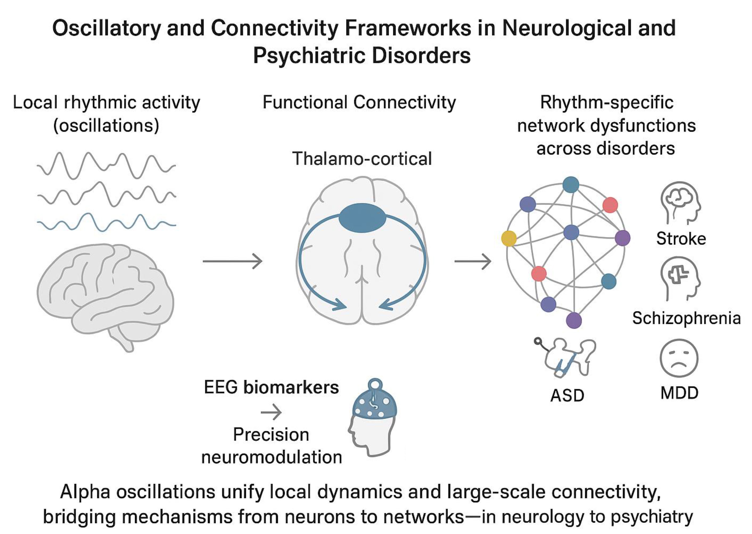
From a computational point of view, functional connectivity metrics and oscillatory dynamics are 2 sides of the same coin: While connectivity indices quantify the coordination between regions, oscillatory measures reflect the underlying rhythmic modes that facilitate or obstruct such coordination.38, 39, 40, 41, 42, 43 Phase-based, amplitude-based and information-theoretic metrics each interrogate these interactions from complementary angles: 1) Phase-lag index (PLI) and its variants assess the consistency of phase differences between signals, isolating genuine interregional coupling by minimizing zero-lag artifacts10, 31; 2) Coherence metrics capture co-modulation of band-limited activities, reflecting how oscillatory fluctuations co-occur across the cortex40, 44, 45; 3) Mutual information (MI) and related entropy-based measures provide a nonlinear gauge of shared signal content, potentially revealing higher-order dependencies that linear metrics miss.46, 47 Finally, by integrating these classes of metrics, researchers can form a unified picture in which oscillations at distinct frequencies (e.g., alpha, theta) serve as communication channels whose efficacy is quantified by connectivity indices. This perspective allows for the computational integration between local oscillations and distributed functional connectivity, identifying oscillatory-specific brain functional networks. In neuroscience, the concept of metastability has been proposed as a signature that balance between these local and distributed activities in the brain. Specifically, metastability captures the brain’s ability to balance functional segregation (specialized processing in localized regions) and integration (coordinated activity across distributed networks).40, 48, 49 Metastable dynamics are thought to underpin key cognitive functions such as attention, working memory and consciousness by allowing the brain to flexibly reconfigure its network topology in response to internal and external demands.50 Theoretical models often describe metastability using nonlinear dynamical systems and coupled oscillatory activities across brain areas, demonstrating how functional integration requires segregated brain areas to influence each other in a way that facilitates integration and serve cognition and behavior.51, 52, 53 Empirical evidence from electrophysiological (EEG/MEG) studies has shown that metastable patterns characterize both resting-state and task-related brain activity,54 with alterations observed in neuropsychiatric disorders.55, 56, 57

Oscillations and connectivity: from neurology to psychiatry

Empirically, EEG-derived connectivity measures have elucidated network alterations across a spectrum of clinical conditions, revealing both shared and disorder-specific patterns for several health conditions, including neurodegenerative diseases.58 In patients with the acute phase of ischemic and hemorrhagic stroke, a shift in spectral power characterized by reduced activity in higher frequency bands (alpha/mu: 8–13 Hz and beta: 14–30 Hz) alongside increased power in lower frequencies (delta: 1–3 Hz and theta: 4–7 Hz) has consistently been associated with unfavorable clinical outcomes.13, 19, 59, 60, 61, 62, 63, 64, 65, 66 In parallel, evidence suggests that stronger connectivity within the fronto-parietal motor network during the early post-stroke period correlates with more favorable motor recovery trajectories.19, 60 Conversely, increases in connectivity occurring at later stages have been linked to less optimal functional outcomes.18, 19, 67, 68, 69, 70, 71, 72

In disorders of consciousness (DoC), patients with unresponsive wakefulness syndrome (UWS) or minimally conscious state (MCS) show reductions in functional connectivity and oscillatory brain dynamics within large-scale default mode and frontoparietal networks correlating with clinical severity and outcomes.2, 17, 20, 73, 74, 75, 76, 77, 78, 79, 80, 81, 82, 83 Mutual information and coherence metrics recorded in the acute post-injury phase can predict functional outcomes with up to ~83% accuracy, highlighting their prognostic utility.2, 17, 20 Furthermore, combining multimodal connectivity metrics with complexity measures (e.g., permutation entropy) and local oscillatory activity can enhance predictive models,84, 85, 86, 87 reflecting the link between recovery trajectories and complex neural dynamics.

In patients with schizophrenia-spectrum disorders (SSD), alpha-band synchrony deficits in SSD manifest as both reduced posterior alpha power and weakened fronto-parietal connectivity, correlating with positive symptom severity and cognitive disorganization.22, 88, 89 These aberrations align with a failure of pulsed inhibition mechanisms, leading to excessive cortical noise and impaired signal-to-noise gating.90, 91, 92, 93, 94 Similarly, beta and gamma band connectivity alterations have been linked to negative symptoms and working-memory deficits, suggesting frequency-specific network dysfunction.95, 96, 97 Integrative models propose that alterations in local processing and large-scale coordination underlie both perceptual distortions and executive dysfunction in SSD.96

In individuals with autism spectrum disorder (ASD), EEG studies have reported long-range underconnectivity in the alpha and beta frequency bands, accompanied by local hyperconnectivity, often manifesting as elevated short-range coherence, particularly in occipital, temporal and parietal regions.98, 99, 100 Abnormal connectivity patterns, including alpha and theta coherence over frontal areas, have been implicated in atypical social perception and sensory integration.98, 101, 102, 103 Within the framework of predictive coding, these aberrant patterns are thought to reflect a deficiency in top-down predictive signaling and an overrepresentation of bottom-up error signals,104 as a consequence of a core deficit in the flexibility.

In patients with attention-deficit/hyperactivity disorder (ADHD), EEG recordings typically show reduced posterior alpha power desynchronization and abnormal alpha and beta-based functional connectivity during sustained attention and cognitive tasks.105, 106, 107, 108, 109 These alterations co-occur with weakened fronto-parietal connectivity, suggesting a functional breakdown in large-scale attentional and executive function networks.110, 111 Combined interventions using behavioral training and alpha-frequency transcranial alternating current stimulation (alpha-tACS) have shown preliminary success in enhancing oscillatory activity and improving executive function and attention performance.112, 113, 114, 115

In major depressive disorder (MDD), elevated frontal alpha power, particularly in the left hemisphere, and a right-to-left alpha asymmetry are frequently reported EEG features (i.e., frontal alpha asymmetry, FAA), suggesting altered interhemispheric connectivity within the prefrontal cortex.108, 109 Although limited diagnostic value of FAA in MDD,116, 117, 118 neuromodulation approaches targeting FAA have shown promising results in reducing depressive symptoms, especially those linked to motivational deficits.119, 120

Taken together, this evidence supports and demonstrates the utility of EEG-derived connectivity and spectral metrics as robust biomarkers across diverse neuropsychiatric conditions (Table 1). Future work should focus on longitudinal, multimodal studies to validate these biomarkers and explore their relationship in cognition and in the diagnosis of neuropsychiatric disorders.121

Alpha activity as a unifying lens

Based on empirical evidence and computations needed to calculate functional connectivity measures, it is plausible to hypothesize a relation between local oscillatory metrics and oscillatory-based functional connectivity. Alpha oscillations (8–13 Hz) may represent the unifying lens to understand this relation. Alpha oscillations are the dominant rhythm in the human EEG and modulate perception, attention and memory. Translational evidence consistently report both disrupted local alpha-band activity and connectivity across several neuropsychiatric disorders.21 For instance, in stroke and severe acquired brain injury, pronounced reductions in posterior alpha power, slowing of the individual alpha frequency (IAF) and weakened fronto-parietal connectivity correlate with attentional deficits and poor functional outcomes,2, 17, 18, 19, 20, 60 whereas in SSD, abnormal alpha-based connectivity is associated with psychotic symptoms and perceptual impairments.95, 96, 97 The most probable neural substrate underlying the cross-diagnostic link between alpha oscillations and functional connectivity measures is the thalamus-cortical circuitry.122, 123, 124 This brain network has been considered the generator of alpha oscillatory activity125, 126, 127, 128 and its dysfunctions yield disorder-specific connectivity signatures, ranging from hypoconnectivity in stroke to hyperconnectivity in ASD.18, 19, 98, 101, 102, 103 The hypothesis of the thalamus-cortical role in alpha generation forges the theoretical bridge between connectivity metrics and oscillatory measures, suggesting that optimized neuromodulation should aim to restore both rhythmic power and interregional synchrony. Clinically, integrating spectral and connectivity biomarkers into unified machine-learning frameworks can refine differential diagnosis, stratify patients for targeted interventions and monitor treatment response.129, 130 Establishing normative databases and open, multicenter repositories with standardized pipelines will be critical for validating hypothesis testing and improving treatment efficacy. Moreover, advances in real-time EEG processing and closed-loop neuromodulation can leverage these biomarkers for personalized treatments, such as alpha-modulation adapted to individual IAF.

Future directions

To enhance the translational impact of EEG connectivity and oscillatory research, future studies should pursue multimodal integration with structural and functional magnetic resonance imaging (MRI), as well as genomic and phenotypic data.131 This integrative approach will enable the identification of neurogenetic factors that modulate rhythmic connectivity, facilitating the development of targeted interventions.132, 133, 134 Longitudinal cohorts that track EEG connectivity trajectories across developmental stages and aging, alongside biomarkers of metabolites that modulate signals, will clarify how network dynamics confer vulnerability or resilience in neuropsychiatric conditions.135

Future research should prioritize integrative, mechanism-oriented approaches that leverage cross-frequency coupling (CFC), dynamic functional connectivity (dFC) and graph-theoretical metrics to resolve how multiscale oscillatory interactions map onto clinical phenotypes and treatment response. Cross-frequency coupling (particularly phase–amplitude coupling) has emerged as a reproducible index of mesoscale coordination and is differentially altered across psychiatric syndromes, suggesting both diagnostic and mechanistic utility.136 Complementing CFC, dFC methods allow the characterization of transient network states and state-switching dynamics that are obscured by static connectivity estimates; these transient states are likely to index symptom-relevant fluctuations in cognition and arousal but require harmonized analytic standards to ensure reproducibility.137 Graph-theoretical measures computed on EEG/MEG/fMRI networks can then quantify topology (e.g., hubness, modularity, efficiency) of both static and dynamic networks, providing compact biomarkers that are amenable to longitudinal tracking and to integration within predictive models.138 We therefore advocate for: 1) multimodal pipelines that combine source-resolved electrophysiology with hemodynamic imaging, 2) standardized connectivity analysis workflows (including surrogate testing and cross-validation), and 3) translational studies that link topology and cross-frequency interactions to cellular and circuit-level mechanisms and to neuromodulatory interventions. Collectively, these convergent approaches will improve mechanistic specificity and accelerate the translation of network biomarkers into personalized diagnostics and closed-loop therapeutics. In conclusion, by recognizing that oscillations and connectivity are inherently interdependent,22, 139 the field can accelerate the translation of EEG-derived metrics into precision diagnostics and personalized neuromodulation protocols.140, 141 This integrative framework not only deepens our mechanistic understanding of brain network dysfunction but also paves the way for novel therapeutic avenues that restore healthy rhythmic coordination across distributed neural circuits.142, 143

Use of AI and AI-assisted technologies

Not applicable.

Tables


Table 1. Summary of the brain oscillatory pattern in the main neuropsychiatric disorders

Disorder

Spectral/connectivity alterations

Network involvement

Clinical/prognostic implications

Major depressive disorder (MDD)

Increased frontal alpha power (left > right); frontal alpha asymmetry (FAA).

Prefrontal interhemispheric connectivity.

FAA has limited diagnostic specificity but is a promising target for neuromodulation, particularly for motivational symptoms.

Schizophrenia-spectrum disorders (SSD)

Reduced posterior alpha power;  fronto-parietal alpha connectivity; beta/gamma connectivity disruptions.

Large-scale fronto-parietal temporal and occipital oscillatory networks.

Alpha deficits correlate with positive symptoms; beta/gamma alterations correlate with negative symptoms and working-memory deficits; overall dysfunction reflects impaired local–global coordination.

Autism spectrum disorder (ASD)

Long-range underconnectivity (alpha, beta); local hyperconnectivity (occipito-temporo-parietal); abnormal frontal alpha/theta coherence.

Imbalance between local vs distributed networks.

Linked to atypical social perception and sensory integration; consistent with predictive coding deficits (↓ top-down,  bottom-up error signaling).

Attention-deficit/hyperactivity disorder (ADHD)

Reduced posterior alpha desynchronization; altered alpha/beta connectivity during attention tasks.

Fronto-parietal attentional/executive networks.

Weakened large-scale connectivity underlies attentional/executive dysfunction; behavioral training + alpha-tACS enhances oscillatory activity and improves attention/executive performance.

Disorders of consciousness (UWS/MCS)

Global reductions in connectivity and oscillatory dynamics.

Default Mode Network (DMN), fronto-parietal networks.

Reduced connectivity correlates with clinical severity; mutual information/coherence metrics predict outcomes (~83% accuracy); multimodal indices enhance prognostic value.

References (143)

  1. Bareham CA, Roberts N, Allanson J, et al. Bedside EEG predicts longitudinal behavioural changes in disorders of consciousness. NeuroImage Clin. 2020;28:102372. doi:10.1016/j.nicl.2020.102372
  2. Di Gregorio F, La Porta F, Petrone V, et al. Accuracy of EEG biomarkers in the detection of clinical outcome in disorders of consciousness after severe acquired brain injury: Preliminary results of a pilot study using a machine learning approach. Biomedicines. 2022;10(8):1897. doi:10.3390/biomedicines10081897
  3. Di Gregorio F, Battaglia S. Advances in EEG-based functional connectivity approaches to the study of the central nervous system in health and disease. Adv Clin Exp Med. 2023;32(6):607–612. doi:10.17219/acem/166476
  4. Baldassarre A, Ramsey LE, Siegel JS, Shulman GL, Corbetta M. Brain connectivity and neurological disorders after stroke. Curr Opin Neurol. 2016;29(6):706–713. doi:10.1097/WCO.0000000000000396
  5. Bastos AM, Schoffelen JM. A tutorial review of functional connectivity analysis methods and their interpretational pitfalls. Front Syst Neuro-sci. 2015;9:175. doi:10.3389/fnsys.2015.00175
  6. Cao J, Zhao Y, Shan X, et al. Brain functional and effective connectivity based on electroencephalography recordings: A review. Hum Brain Mapp. 2022;43(2):860–879. doi:10.1002/hbm.25683
  7. Hallett M, De Haan W, Deco G, et al. Human brain connectivity: Clinical applications for clinical neurophysiology. Clin Neurophysiol. 2020;131(7):1621–1651. doi:10.1016/j.clinph.2020.03.031
  8. Mahjoory K, Nikulin VV, Botrel L, Linkenkaer-Hansen K, Fato MM, Haufe S. Consistency of EEG source localization and connectivity estimates. NeuroImage. 2017;152:590–601. doi:10.1016/j.neuroimage.2017.02.076
  9. Marinazzo D, Riera JJ, Marzetti L, Astolfi L, Yao D, Valdés Sosa PA. Controversies in EEG source imaging and connectivity: Modeling, validation, benchmarking. Brain Topogr. 2019;32(4):527–529. doi:10.1007/s10548-019-00709-9
  10. Stam CJ, Nolte G, Daffertshofer A. Phase lag index: Assessment of functional connectivity from multichannel EEG and MEG with diminished bias from common sources. Hum Brain Mapp. 2007;28(11):1178–1193. doi:10.1002/hbm.20346
  11. Vecchio F, Tomino C, Miraglia F, et al. Cortical connectivity from EEG data in acute stroke: A study via graph theory as a potential biomarker for functional recovery. Int J Psychophysiol. 2019;146:133–138. doi:10.1016/j.ijpsycho.2019.09.012
  12. Di Biase L, Ricci L, Caminiti ML, Pecoraro PM, Carbone SP, Di Lazzaro V. Quantitative high density EEG brain connectivity evaluation in Parkinson’s disease: The phase locking value (PLV). J Clin Med. 2023;12(4):1450. doi:10.3390/jcm12041450
  13. Bentes C, Peralta AR, Viana P, et al. Quantitative EEG and functional outcome following acute ischemic stroke. Clin Neurophysiol. 2018;129(8):1680–1687. doi:10.1016/j.clinph.2018.05.021
  14. Baldassarre A, Ramsey L, Rengachary J, et al. Dissociated functional connectivity profiles for motor and attention deficits in acute right-hemisphere stroke. Brain. 2016;139(7):2024–2038. doi:10.1093/brain/aww107
  15. Bolwig TG. EEG and psychiatry: Time for a resurrection. Acta Psychiatr Scand. 2008;117(4):241–243. doi:10.1111/j.1600-0447.2008.01172.x
  16. Cao KX, Ma ML, Wang CZ, et al. TMS-EEG: An emerging tool to study the neurophysiologic biomarkers of psychiatric disorders. Neurophar-macology. 2021;197:108574. doi:10.1016/j.neuropharm.2021.108574
  17. Chennu S, Annen J, Wannez S, et al. Brain networks predict metabolism, diagnosis and prognosis at the bedside in disorders of consciousness. Brain. 2017;140(8):2120–2132. doi:10.1093/brain/awx163
  18. Di Gregorio F, Petrone V, Casanova E, et al. Hierarchical psychophysiological pathways subtend perceptual asymmetries in neglect. Neu-roImage. 2023;270:119942. doi:10.1016/j.neuroimage.2023.119942
  19. Di Gregorio F, Lullini G, Orlandi S, et al. Clinical and neurophysiological predictors of the functional outcome in right-hemisphere stroke. NeuroImage. 2025;308:121059. doi:10.1016/j.neuroimage.2025.121059
  20. Engemann DA, Raimondo F, King JR, et al. Robust EEG-based cross-site and cross-protocol classification of states of consciousness. Brain. 2018;141(11):3179–3192. doi:10.1093/brain/awy251
  21. Ippolito G, Bertaccini R, Tarasi L, et al. The role of alpha oscillations among the main neuropsychiatric disorders in the adult and developing human brain: Evidence from the last 10 years of research. Biomedicines. 2022;10(12):3189. doi:10.3390/biomedicines10123189
  22. Trajkovic J, Ricci G, Pirazzini G, et al. Aberrant functional connectivity and brain network organization in high-schizotypy individuals: An electroencephalography study. Schizophr Bull. 2025;51(5):1266–1281. doi:10.1093/schbul/sbaf004
  23. Young MJ, Koch C, Claassen J, et al. An ethical framework to assess covert consciousness. Lancet Neurol. 2025;24(3):195–196. doi:10.1016/S1474-4422(24)00432-0
  24. Battaglia S, Servajean P, Friston KJ. The paradox of the self-studying brain. Phys Life Rev. 2025;52:197–204. doi:10.1016/j.plrev.2024.12.009
  25. Battaglia S, Nazzi C, Thayer JF. Genetic differences associated with dopamine and serotonin release mediate fear-induced bradycardia in the human brain. Transl Psychiatry. 2024;14(1):24. doi:10.1038/s41398-024-02737-x
  26. Battaglia S, Nazzi C, Lonsdorf TB, Thayer JF. Neuropsychobiology of fear-induced bradycardia in humans: Progress and pitfalls. Mol Psychiatry. 2024;29(12):3826–3840. doi:10.1038/s41380-024-02600-x
  27. Di Gregorio F, Battaglia S. The intricate brain–body interaction in psychiatric and neurological diseases. Adv Clin Exp Med. 2024;33(4):321–326. doi:10.17219/acem/185689
  28. Nolte G, Bai O, Wheaton L, Mari Z, Vorbach S, Hallett M. Identifying true brain interaction from EEG data using the imaginary part of coherency. Clin Neurophysiol. 2004;115(10):2292–2307. doi:10.1016/j.clinph.2004.04.029
  29. Nolte G, Ziehe A, Nikulin VV, et al. Robustly estimating the flow direction of information in complex physical systems. Phys Rev Lett. 2008;100(23):234101. doi:10.1103/PhysRevLett.100.234101
  30. Vinck M, Oostenveld R, Van Wingerden M, Battaglia F, Pennartz CMA. An improved index of phase-synchronization for electrophysiological data in the presence of volume-conduction, noise and sample-size bias. NeuroImage. 2011;55(4):1548–1565. doi:10.1016/j.neuroimage.2011.01.055
  31. Hardmeier M, Hatz F, Bousleiman H, Schindler C, Stam CJ, Fuhr P. Reproducibility of functional connectivity and graph measures based on the Phase Lag Index (PLI) and Weighted Phase Lag Index (wPLI) derived from high resolution EEG. PLoS One. 2014;9(10):e108648. doi:10.1371/journal.pone.0108648
  32. Lehmann D, Faber PL, Gianotti LRR, Kochi K, Pascual-Marqui RD. Coherence and phase locking in the scalp EEG and between LORETA model sources, and microstates as putative mechanisms of brain temporo-spatial functional organization. J Physiol (Paris). 2006;99(1):29–36. doi:10.1016/j.jphysparis.2005.06.005
  33. Makeig S, Bell AJ, Tzyy-Ping J, Sejnowski TJ. Independent component analysis of electroencephalographic data. In: Touretzky DS, Mozer MC, Hasselmo ME, eds. Advances in Neural Information Processing Systems 8: Proceedings of the 1995 Conference. Cambridge, USA: MIT Press; 1996. https://papers.nips.cc/paper_files/paper/1995/hash/754dda4b1ba34c6fa89716b85d68532b-Abstract.html.
  34. Mantini D, Franciotti R, Romani GL, Pizzella V. Improving MEG source localizations: An automated method for complete artifact removal based on independent component analysis. NeuroImage. 2008;40(1):160–173. doi:10.1016/j.neuroimage.2007.11.022
  35. Zhukov L, Weinstein D, Johnson C. Independent component analysis for EEG source localization. IEEE Eng Med Biol Mag. 2000;19(3):87–96. doi:10.1109/51.844386
  36. Barbalho SM, Leme Boaro B, Da Silva Camarinha Oliveira J, et al. Molecular mechanisms underlying neuroinflammation intervention with medicinal plants: A critical and narrative review of the current literature. Pharmaceuticals (Basel). 2025;18(1):133. doi:10.3390/ph18010133
  37. Figueiredo Godoy AC, Frota FF, Araújo LP, et al. Neuroinflammation and natural antidepressants: Balancing fire with flora. Biomedicines. 2025;13(5):1129. doi:10.3390/biomedicines13051129
  38. Cabral J, Hugues E, Sporns O, Deco G. Role of local network oscillations in resting-state functional connectivity. NeuroImage. 2011;57(1):130–139. doi:10.1016/j.neuroimage.2011.04.010
  39. Cohen MX. Multivariate cross-frequency coupling via generalized eigendecomposition. eLife. 2017;6:e21792. doi:10.7554/eLife.21792
  40. Deco G, Kringelbach ML. Metastability and coherence: Extending the communication through coherence hypothesis using a whole-brain computational perspective. Trends Neurosci. 2016;39(3):125–135. doi:10.1016/j.tins.2016.01.001
  41. Forrester M, Crofts JJ, Sotiropoulos SN, Coombes S, O’Dea RD. The role of node dynamics in shaping emergent functional connectivity pat-terns in the brain. Netw Neurosci. 2020;4(2):467–483. doi:10.1162/netn_a_00130
  42. Modolo J, Hassan M, Wendling F, Benquet P. Decoding the circuitry of consciousness: From local microcircuits to brain-scale networks. Netw Neurosci. 2020;4(2):315–337. doi:10.1162/netn_a_00119
  43. Tewarie P, Hunt BAE, O’Neill GC, et al. Relationships between neuronal oscillatory amplitude and dynamic functional connectivity. Cereb Cortex. 2019;29(6):2668–2681. doi:10.1093/cercor/bhy136
  44. Fries P. A mechanism for cognitive dynamics: Neuronal communication through neuronal coherence. Trend Cogn Sci. 2005;9(10):474–480. doi:10.1016/j.tics.2005.08.011
  45. Fries P. Rhythms for cognition: Communication through coherence. Neuron. 2015;88(1):220–235. doi:10.1016/j.neuron.2015.09.034
  46. King JR, Sitt JD, Faugeras F, et al. Information sharing in the brain indexes consciousness in noncommunicative patients. Curr Biol. 2013;23(19):1914–1919. doi:10.1016/j.cub.2013.07.075
  47. Riedl M, Müller A, Wessel N. Practical considerations of permutation entropy: A tutorial review. Eur Phys J Spec Top. 2013;222(2):249–262. doi:10.1140/epjst/e2013-01862-7
  48. Deco G, Jirsa VK, McIntosh AR. Emerging concepts for the dynamical organization of resting-state activity in the brain. Nat Rev Neurosci. 2011;12(1):43–56. doi:10.1038/nrn2961
  49. Hancock F, Rosas FE, Luppi AI, et al. Metastability demystified: The foundational past, the pragmatic present and the promising future. Nat Rev Neurosci. 2025;26(2):82–100. doi:10.1038/s41583-024-00883-1
  50. Tognoli E, Kelso JAS. The metastable brain. Neuron. 2014;81(1):35–48. doi:10.1016/j.neuron.2013.12.022
  51. Friston KJ. Transients, metastability, and neuronal dynamics. NeuroImage. 1997;5(2):164–171. doi:10.1006/nimg.1997.0259
  52. Friston KJ. Functional and effective connectivity: A review. Brain Connect. 2011;1(1):13–36. doi:10.1089/brain.2011.0008
  53. Tononi G. Consciousness and complexity. Science. 1998;282(5395):1846–1851. doi:10.1126/science.282.5395.1846
  54. Cabral J, Vidaurre D, Marques P, et al. Cognitive performance in healthy older adults relates to spontaneous switching between states of functional connectivity during rest. Sci Rep. 2017;7(1):5135. doi:10.1038/s41598-017-05425-7
  55. Alderson TH, Bokde ALW, Kelso JAS, Maguire L, Coyle D. Metastable neural dynamics in Alzheimer’s disease are disrupted by lesions to the structural connectome. NeuroImage. 2018;183:438–455. doi:10.1016/j.neuroimage.2018.08.033
  56. Hellyer PJ, Scott G, Shanahan M, Sharp DJ, Leech R. Cognitive flexibility through metastable neural dynamics is disrupted by damage to the structural connectome. J Neurosci. 2015;35(24):9050–9063. doi:10.1523/JNEUROSCI.4648-14.2015
  57. Lee WH, Doucet GE, Leibu E, Frangou S. Resting-state network connectivity and metastability predict clinical symptoms in schizophrenia. Schizophr Res. 2018;201:208–216. doi:10.1016/j.schres.2018.04.029
  58. Tanaka M, Battaglia S, Liloia D. Navigating neurodegeneration: Integrating biomarkers, neuroinflammation, and imaging in Parkinson’s, Alzheimer’s and motor neuron disorders. Biomedicines. 2025;13(5):1045. doi:10.3390/biomedicines13051045
  59. Cassidy JM, Wodeyar A, Wu J, et al. Low-frequency oscillations are a biomarker of injury and recovery after stroke. Stroke. 2020;51(5):1442–1450. doi:10.1161/STROKEAHA.120.028932
  60. Cassidy JM, Wodeyar A, Srinivasan R, Cramer SC. Coherent neural oscillations inform early stroke motor recovery. Hum Brain Mapp. 2021;42(17):5636–5647. doi:10.1002/hbm.25643
  61. Finnigan SP, Walsh M, Rose SE, Chalk JB. Quantitative EEG indices of sub-acute ischaemic stroke correlate with clinical outcomes. Clin Neuro-physiol. 2007;118(11):2525–2532. doi:10.1016/j.clinph.2007.07.021
  62. Finnigan S, Van Putten MJAM. EEG in ischaemic stroke: Quantitative EEG can uniquely inform (sub-)acute prognoses and clinical manage-ment. Clin Neurophysiol. 2013;124(1):10–19. doi:10.1016/j.clinph.2012.07.003
  63. Gallina J, Pietrelli M, Zanon M, Bertini C. Hemispheric differences in altered reactivity of brain oscillations at rest after posterior lesions. Brain Struct Funct. 2022;227(2):709–723. doi:10.1007/s00429-021-02279-8
  64. Leon-Carrion J, Martin-Rodriguez JF, Damas-Lopez J, Barroso Y Martin JM, Dominguez-Morales MR. Delta–alpha ratio correlates with level of recovery after neurorehabilitation in patients with acquired brain injury. Clin Neurophysiol. 2009;120(6):1039–1045. doi:10.1016/j.clinph.2009.01.021
  65. Pietrelli M, Zanon M, Làdavas E, Grasso PA, Romei V, Bertini C. Posterior brain lesions selectively alter alpha oscillatory activity and predict visual performance in hemianopic patients. Cortex. 2019;121:347–361. doi:10.1016/j.cortex.2019.09.008
  66. Sheorajpanday RVA, Nagels G, Weeren AJTM, Van Putten MJAM, De Deyn PP. Quantitative EEG in ischemic stroke: Correlation with function-al status after 6 months. Clin Neurophysiol. 2011;122(5):874–883. doi:10.1016/j.clinph.2010.07.028
  67. Gale SD, Pearson CM. Neuroimaging predictors of stroke outcome: Implications for neurorehabilitation. NeuroRehabilitation. 2012;31(3):331–344. doi:10.3233/NRE-2012-0800
  68. Hoshino T, Oguchi K, Inoue K, Hoshino A, Hoshiyama M. Relationship between lower limb function and functional connectivity assessed by EEG among motor-related areas after stroke. Top Stroke Rehabil. 2021;28(8):614–623. doi:10.1080/10749357.2020.1864986
  69. Kim B, Winstein C. Can neurological biomarkers of brain impairment be used to predict poststroke motor recovery? A systematic review. Neurorehabil Neural Repair. 2017;31(1):3–24. doi:10.1177/1545968316662708
  70. Lim JS, Lee JJ, Woo CW. Post-stroke cognitive impairment: Pathophysiological insights into brain disconnectome from advanced neuroim-aging analysis techniques. J Stroke. 2021;23(3):297–311. doi:10.5853/jos.2021.02376
  71. Min YS, Park JW, Park E, et al. Interhemispheric functional connectivity in the primary motor cortex assessed by resting-state functional magnetic resonance imaging aids long-term recovery prediction among subacute stroke patients with severe hand weakness. J Clin Med. 2020;9(4):975. doi:10.3390/jcm9040975
  72. Nicolo P, Rizk S, Magnin C, Pietro MD, Schnider A, Guggisberg AG. Coherent neural oscillations predict future motor and language im-provement after stroke. Brain. 2015;138(10):3048–3060. doi:10.1093/brain/awv200
  73. Bagnato S, Boccagni C, Prestandrea C, Sant’Angelo A, Castiglione A, Galardi G. Prognostic value of standard EEG in traumatic and non-traumatic disorders of consciousness following coma. Clin Neurophysiol. 2010;121(3):274–280. doi:10.1016/j.clinph.2009.11.008
  74. Claassen J, Velazquez A, Meyers E, et al. Bedside quantitative electroencephalography improves assessment of consciousness in comatose subarachnoid hemorrhage patients. Ann Neurol. 2016;80(4):541–553. doi:10.1002/ana.24752
  75. Estraneo A, Loreto V, Guarino I, et al. Standard EEG in diagnostic process of prolonged disorders of consciousness. Clin Neurophysiol. 2016;127(6):2379–2385. doi:10.1016/j.clinph.2016.03.021
  76. Gosseries O, Schnakers C, Ledoux D, et al. Automated EEG entropy measurements in coma, vegetative state/unresponsive wakefulness syndrome and minimally conscious state. Funct Neurol. 2011;26(1):25–30. PMID:21693085. PMCID:PMC3814509.
  77. O’Donnell A, Pauli R, Banellis L, et al. The prognostic value of resting-state EEG in acute post-traumatic unresponsive states. Brain Commun. 2021;3(2):fcab017. doi:10.1093/braincomms/fcab017
  78. Rosanova M, Gosseries O, Casarotto S, et al. Recovery of cortical effective connectivity and recovery of consciousness in vegetative patients. Brain. 2012;135(4):1308–1320. doi:10.1093/brain/awr340
  79. Sarasso S, Rosanova M, Casali AG, et al. Quantifying cortical EEG responses to TMS in (un)consciousness. Clin EEG Neurosci. 2014;45(1):40–49. doi:10.1177/1550059413513723
  80. Scarpino M, Lolli F, Hakiki B, et al. EEG and Coma Recovery Scale-Revised prediction of neurological outcome in disorder of consciousness patients. Acta Neurol Scand. 2020;142(3):221–228. doi:10.1111/ane.13247
  81. Sitt JD, King JR, El Karoui I, et al. Large scale screening of neural signatures of consciousness in patients in a vegetative or minimally con-scious state. Brain. 2014;137(8):2258–2270. doi:10.1093/brain/awu141
  82. Stefan S, Schorr B, Lopez-Rolon A, et al. Consciousness indexing and outcome prediction with resting-state EEG in severe disorders of consciousness. Brain Topogr. 2018;31(5):848–862. doi:10.1007/s10548-018-0643-x
  83. Tanaka M, Szabó Á, Vécsei L. Redefining roles: A paradigm shift in tryptophan–kynurenine metabolism for innovative clinical applications. Int J Mol Sci. 2024;25(23):12767. doi:10.3390/ijms252312767
  84. Amiri M, Fisher PM, Raimondo F, et al. Multimodal prediction of residual consciousness in the intensive care unit: The CONNECT-ME study. Brain. 2023;146(1):50–64. doi:10.1093/brain/awac335
  85. Coleman MR, Bekinschtein T, Monti MM, Owen AM, Pickard JD. A multimodal approach to the assessment of patients with disorders of consciousness. Prog Brain Res. 2009;177:231–248. doi:10.1016/S0079-6123(09)17716-6
  86. Di Perri C, Bahri MA, Amico E, et al. Neural correlates of consciousness in patients who have emerged from a minimally conscious state: A cross-sectional multimodal imaging study. Lancet Neurol. 2016;15(8):830–842. doi:10.1016/S1474-4422(16)00111-3
  87. Gallucci A, Varoli E, Del Mauro L, et al. Multimodal approaches supporting the diagnosis, prognosis and investigation of neural correlates of disorders of consciousness: A systematic review. Eur J Neurosci. 2024;59(5):874–933. doi:10.1111/ejn.16149
  88. Hinkley LBN, Vinogradov S, Guggisberg AG, Fisher M, Findlay AM, Nagarajan SS. Clinical symptoms and alpha band resting-state functional connectivity imaging in patients with schizophrenia: Implications for novel approaches to treatment. Biol Psychiatry. 2011;70(12):1134–1142. doi:10.1016/j.biopsych.2011.06.029
  89. Trajkovic J, Di Gregorio F, Ferri F, Marzi C, Diciotti S, Romei V. Resting state alpha oscillatory activity is a valid and reliable marker of schizotypy. Sci Rep. 2021;11(1):10379. doi:10.1038/s41598-021-89690-7
  90. Braff DL. Prepulse inhibition of the startle reflex: A window on the brain in schizophrenia. Curr Top Behav Neurosci. 2010;4:349–371. doi:10.1007/7854_2010_61
  91. Kumari V, Sharma T. Effects of typical and atypical antipsychotics on prepulse inhibition in schizophrenia: A critical evaluation of current evidence and directions for future research. Psychopharmacology. 2002;162(2):97–101. doi:10.1007/s00213-002-1099-x
  92. Mena A, Ruiz-Salas JC, Puentes A, Dorado I, Ruiz-Veguilla M, De La Casa LG. Reduced prepulse inhibition as a biomarker of schizophrenia. Front Behav Neurosci. 2016;10:202. doi:10.3389/fnbeh.2016.00202
  93. Radhu N, Garcia Dominguez L, Farzan F, et al. Evidence for inhibitory deficits in the prefrontal cortex in schizophrenia. Brain. 2015;138(2):483–497. doi:10.1093/brain/awu360
  94. Swerdlow NR, Weber M, Qu Y, Light GA, Braff DL. Realistic expectations of prepulse inhibition in translational models for schizophrenia research. Psychopharmacology. 2008;199(3):331–388. doi:10.1007/s00213-008-1072-4
  95. Lynn PA, Sponheim SR. Disturbed theta and gamma coupling as a potential mechanism for visuospatial working memory dysfunction in people with schizophrenia. Neuropsychiatr Electrophysiol. 2016;2(1):7. doi:10.1186/s40810-016-0022-3
  96. Pittman-Polletta BR, Kocsis B, Vijayan S, Whittington MA, Kopell NJ. Brain rhythms connect impaired inhibition to altered cognition in schizophrenia. Biol Psychiatry. 2015;77(12):1020–1030. doi:10.1016/j.biopsych.2015.02.005
  97. Senkowski D, Gallinat J. Dysfunctional prefrontal gamma-band oscillations reflect working memory and other cognitive deficits in schizophrenia. Biol Psychiatry. 2015;77(12):1010–1019. doi:10.1016/j.biopsych.2015.02.034
  98. Ghuman AS, Van Den Honert RN, Huppert TJ, Wallace GL, Martin A. Aberrant oscillatory synchrony is biased toward specific frequencies and processing domains in the autistic brain. Biol Psychiatry Cogn Neurosci Neuroimaging. 2017;2(3):245–252. doi:10.1016/j.bpsc.2016.07.006
  99. Kessler K, Seymour RA, Rippon G. Brain oscillations and connectivity in autism spectrum disorders (ASD): New approaches to methodology, measurement and modelling. Neurosci Biobehav Rev. 2016;71:601–620. doi:10.1016/j.neubiorev.2016.10.002
  100. O’Reilly C, Lewis JD, Elsabbagh M. Is functional brain connectivity atypical in autism? A systematic review of EEG and MEG studies. PLoS One. 2017;12(5):e0175870. doi:10.1371/journal.pone.0175870
  101. Barnes SJK, Thomas M, McClintock PVE, Stefanovska A. Theta and alpha connectivity in children with autism spectrum disorder. Brain Com-mun. 2025;7(2):fcaf084. doi:10.1093/braincomms/fcaf084
  102. Belmonte MK, Allen G, Beckel-Mitchener A, Boulanger LM, Carper RA, Webb SJ. Autism and abnormal development of brain connectivity. J Neurosci. 2004;24(42):9228–9231. doi:10.1523/JNEUROSCI.3340-04.2004
  103. Uhlhaas PJ, Singer W. Neural synchrony in brain disorders: Relevance for cognitive dysfunctions and pathophysiology. Neuron. 2006;52(1):155–168. doi:10.1016/j.neuron.2006.09.020
  104. Van De Cruys S, Evers K, Van Der Hallen R, et al. Precise minds in uncertain worlds: Predictive coding in autism. Psychol Rev. 2014;121(4):649–675. doi:10.1037/a0037665
  105. Alba G, Pereda E, Mañas S, et al. The variability of EEG functional connectivity of young ADHD subjects in different resting states. Clin Neu-rophysiol. 2016;127(2):1321–1330. doi:10.1016/j.clinph.2015.09.134
  106. Debnath R, Miller NV, Morales S, Seddio KR, Fox NA. Investigating brain electrical activity and functional connectivity in adolescents with clinically elevated levels of ADHD symptoms in alpha frequency band. Brain Res. 2021;1750:147142. doi:10.1016/j.brainres.2020.147142
  107. Lenartowicz A, Lu S, Rodriguez C, et al. Alpha desynchronization and fronto­parietal connectivity during spatial working memory encoding deficits in ADHD: A simultaneous EEG­fMRI study. Neuroimage Clin. 2016;11:210–223. doi:10.1016/j.nicl.2016.01.023
  108. Lenartowicz A, Mazaheri A, Jensen O, Loo SK. Aberrant modulation of brain oscillatory activity and attentional impairment in attention-deficit/hyperactivity disorder. Biol Psychiatry Cogn Neurosci Neuroimaging. 2018;3(1):19–29. doi:10.1016/j.bpsc.2017.09.009
  109. Murias M, Swanson JM, Srinivasan R. Functional connectivity of frontal cortex in healthy and ADHD children reflected in EEG coherence. Cereb Cortex. 2007;17(8):1788–1799. doi:10.1093/cercor/bhl089
  110. Dipnall LM, Hourani D, Darling S, Anderson V, Sciberras E, Silk TJ. Fronto-parietal white matter microstructure associated with working memory performance in children with ADHD. Cortex. 2023;166:243–257. doi:10.1016/j.cortex.2023.03.015
  111. Parlatini V, Radua J, Robertsson N, et al. Asymmetry of attentive networks contributes to adult attention-deficit/hyperactivity disorder (ADHD) pathophysiology [published online as ahead of print on November 2, 2024]. Eur Arch Psychiatry Clin Neurosci. 2024. doi:10.1007/s00406-024-01927-4
  112. Kannen K, Rasbach J, Fantazi A, et al. Alpha modulation via transcranial alternating current stimulation in adults with attention-deficit hyperactivity disorder. Front Psychol. 2024;14:1280397. doi:10.3389/fpsyg.2023.1280397
  113. Yin Y, Wang X, Feng T. Noninvasive brain stimulation for improving cognitive deficits and clinical symptoms in attention-deficit/hyperactivity disorder: A systematic review and meta-analysis. Brain Sci. 2024;14(12):1237. doi:10.3390/brainsci14121237
  114. Trajkovic J, Di Gregorio F, Marcantoni E, Thut G, Romei V. A TMS/EEG protocol for the causal assessment of the functions of the oscillatory brain rhythms in perceptual and cognitive processes. STAR Protoc. 2022;3(2):101435. doi:10.1016/j.xpro.2022.101435
  115. Trajkovic J, Di Gregorio F, Thut G, Romei V. Transcranial magnetic stimulation effects support an oscillatory model of ERP genesis. Curr Biol. 2024;34(5):1048–1058.e4. doi:10.1016/j.cub.2024.01.069
  116. Debener S, Beauducel A, Nessler D, Brocke B, Heilemann H, Kayser J. Is resting anterior EEG alpha asymmetry a trait marker for depression? Neuropsychobiology. 2000;41(1):31–37. doi:10.1159/000026630
  117. Goldstein BL, Shankman SA, Kujawa A, Torpey-Newman DC, Olino TM, Klein DN. Developmental changes in electroencephalographic frontal asymmetry in young children at risk for depression. Child Psychol Psychiatry. 2016;57(9):1075–1082. doi:10.1111/jcpp.12567
  118. Van Der Vinne N, Vollebregt MA, Van Putten MJAM, Arns M. Frontal alpha asymmetry as a diagnostic marker in depression: Fact or fiction? A meta-analysis. Neuroimage Clin. 2017;16:79–87. doi:10.1016/j.nicl.2017.07.006
  119. Sun L, Peräkylä J, Hartikainen KM. Frontal alpha asymmetry, a potential biomarker for the effect of neuromodulation on brain’s affective circuitry: Preliminary evidence from a deep brain stimulation study. Front Hum Neurosci. 2017;11:584. doi:10.3389/fnhum.2017.00584
  120. Juhász L, Spisák K, Szolnoki BZ, et al. The power struggle: Kynurenine pathway enzyme knockouts and brain mitochondrial respiration. J Neurochem. 2025;169(5):e70075. doi:10.1111/jnc.70075
  121. Szabó Á, Galla Z, Spekker E, et al. Oxidative and excitatory neurotoxic stresses in CRISPR/Cas9-induced kynurenine aminotransferase knockout mice: A novel model for despair-based depression and post-traumatic stress disorder. Front Biosci (Landmark Ed). 2025;30(1):25706. doi:10.31083/FBL25706
  122. Redinbaugh MJ, Phillips JM, Kambi NA, et al. Thalamus modulates consciousness via layer-specific control of cortex. Neuron. 2020;106(1):66–75.e12. doi:10.1016/j.neuron.2020.01.005
  123. Saalmann YB, Pinsk MA, Wang L, Li X, Kastner S. The pulvinar regulates information transmission between cortical areas based on attention demands. Science. 2012;337(6095):753–756. doi:10.1126/science.1223082
  124. Saalmann YB, Kastner S. Cognitive and perceptual functions of the visual thalamus. Neuron. 2011;71(2):209–223. doi:10.1016/j.neuron.2011.06.027
  125. Halgren M, Ulbert I, Bastuji H, et al. The generation and propagation of the human alpha rhythm. Proc Natl Acad Sci U S A. 2019;116(47):23772–23782. doi:10.1073/pnas.1913092116
  126. Schreckenberger M, Lange-Asschenfeldt C, Lochmann M, et al. The thalamus as the generator and modulator of EEG alpha rhythm: A com-bined PET/EEG study with lorazepam challenge in humans [published correction appers in: Neuroimage. 2006;32(1):485 doi:10.1016/j.neuroimage.2006.03.007]. Neuroimage. 2004;22(2):637–644. doi:10.1016/j.neuroimage.2004.01.047
  127. Vijayan S, Kopell NJ. Thalamic model of awake alpha oscillations and implications for stimulus processing. Proc Natl Acad Sci U S A. 2012;109(45):18553–18558. doi:10.1073/pnas.1215385109
  128. Tortora F, Hadipour AL, Battaglia S, Falzone A, Avenanti A, Vicario CM. The role of serotonin in fear learning and memory: A systematic review of human studies. Brain Sci. 2023;13(8):1197. doi:10.3390/brainsci13081197
  129. Battaglia S, Fazio CD, Borgomaneri S, Avenanti A. Cortisol imbalance and fear learning in PTSD: Therapeutic approaches to control abnormal fear responses. Curr Neuropharmacol. 2025;23(7):835–846. doi:10.2174/1570159X23666250123142526
  130. Battaglia S, Di Fazio C, Mazzà M, Tamietto M, Avenanti A. Targeting human glucocorticoid receptors in fear learning: A multiscale inte-grated approach to study functional connectivity. Int J Mol Sci. 2024;25(2):864. doi:10.3390/ijms25020864
  131. Tanaka M, Szabó Á, Vécsei L, Giménez-Llort L. Emerging translational research in neurological and psychiatric diseases: From in vitro to in vivo models. Int J Mol Sci. 2023;24(21):15739. doi:10.3390/ijms242115739
  132. Hernandez-Pavon JC, Schneider-Garces N, Begnoche JP, Miller LE, Raij T. Targeted modulation of human brain interregional effective con-nectivity with spike-timing dependent plasticity. Neuromodulation. 2023;26(4):745–754. doi:10.1016/j.neurom.2022.10.045
  133. Di Gregorio F, La Porta F, Casanova E, et al. Efficacy of repetitive transcranial magnetic stimulation combined with visual scanning treat-ment on cognitive and behavioral symptoms of left hemispatial neglect in right hemispheric stroke patients: Study protocol for a randomized controlled trial. Trials. 2021;22(1):24. doi:10.1186/s13063-020-04943-6
  134. Di Gregorio F, La Porta F, Lullini G, et al. Efficacy of repetitive transcranial magnetic stimulation combined with visual scanning treatment on cognitive-behavioral symptoms of unilateral spatial neglect in patients with traumatic brain injury: Study protocol for a randomized controlled trial. Front Neurol. 2021;12:702649. doi:10.3389/fneur.2021.702649
  135. Tanaka M, Szatmári I, Vécsei L. Quinoline quest: Kynurenic acid strategies for next-generation therapeutics via rational drug design. Phar-maceuticals (Basel). 2025;18(5):607. doi:10.3390/ph18050607
  136. Yakubov B, Das S, Zomorrodi R, et al. Cross-frequency coupling in psychiatric disorders: A systematic review. Neurosci Biobehav Rev. 2022;138:104690. doi:10.1016/j.neubiorev.2022.104690
  137. Hutchison RM, Womelsdorf T, Allen EA, et al. Dynamic functional connectivity: Promise, issues, and interpretations. NeuroImage. 2013;80:360–378. doi:10.1016/j.neuroimage.2013.05.079
  138. Hallquist MN, Hillary FG. Graph theory approaches to functional network organization in brain disorders: A critique for a brave new small-world. Netw Neurosci. 2019;3(1):1–26. doi:10.1162/netn_a_00054
  139. Jaeger C, Nuttall R, Zimmermann J, et al. Targeted rhythmic visual stimulation at individual participants’ intrinsic alpha frequency causes selective increase of occipitoparietal BOLD-fMRI and EEG functional connectivity. NeuroImage. 2023;270:119981. doi:10.1016/j.neuroimage.2023.119981
  140. Battaglia S, Nazzi C, Fullana MA, Di Pellegrino G, Borgomaneri S. ‘Nip it in the bud’: Low-frequency rTMS of the prefrontal cortex disrupts threat memory consolidation in humans. Behav Res Ther. 2024;178:104548. doi:10.1016/j.brat.2024.104548
  141. Battaglia S, Nazzi C, Di Fazio C, Borgomaneri S. The role of pre-supplementary motor cortex in action control with emotional stimuli: A repetitive transcranial magnetic stimulation study. Ann N Y Acad Sci. 2024;1536(1):151–166. doi:10.1111/nyas.15145
  142. Tanaka M. Beyond the boundaries: Transitioning from categorical to dimensional paradigms in mental health diagnostics. Adv Clin Exp Med. 2024;33(12):1295–1301. doi:10.17219/acem/197425
  143. Tanaka M. From serendipity to precision: Integrating AI, multi-omics, and human-specific models for personalized neuropsychiatric care. Biomedicines. 2025;13(1):167. doi:10.3390/biomedicines13010167