Abstract
Background. Posterior capsular opacification (PCO) is a common complication of cataract surgery and is strongly associated with the epithelial–mesenchymal transition (EMT) of lens epithelial cells (LECs). Integrin β3 (ITGB3) has been implicated in various pathological processes; however, its role in PCO remains insufficiently understood.
Objectives. To investigate the regulatory role of ITGB3 in PCO progression and determine whether ITGB3 modulates transforming growth factor-β2 (TGF-β2)-induced migration and EMT in human lens epithelial cells.
Materials and methods. Human lens epithelial HLE-B3 cells were treated with transforming growth factor-β2 (TGF-β2; 10 ng/mL) to establish an in vitro model of PCO. ITGB3 expression at the mRNA and protein levels was assessed using reverse transcription quantitative polymerase chain reaction (RT-qPCR) and western blotting. Cell proliferation was evaluated using the 5-ethynyl-2′-deoxyuridine (EdU) assay, while cell migration was assessed using transwell and wound-healing assays.
Results. TGF-β2 stimulation significantly increased ITGB3 expression in HLE-B3 cells. Silencing of ITGB3 significantly reduced the proliferation of TGF-β2-treated cells and inhibited their migratory capacity, as demonstrated by both transwell and wound-healing assays. Exposure to TGF-β2 promoted EMT, whereas ITGB3 knockdown reversed this effect. Furthermore, activation of the TGF-β/Smad2/3 signaling pathway induced by TGF-β2 was attenuated following ITGB3 silencing.
Conclusions. Knockdown of ITGB3 alleviates TGF-β2-induced migration and EMT in HLE-B3 cells by suppressing the TGF-β/Smad2/3 signaling pathway, supporting ITGB3 as a potential therapeutic target for the prevention and treatment of PCO.
Key words: integrin beta 3, transforming growth factor beta 2, epithelial–mesenchymal transition, lens epithelial cells, posterior capsular opacification
Background
Posterior capsular opacification (PCO) is one of the most common complications following cataract surgery and remains a leading cause of visual impairment and secondary blindness in affected patients.1 Following cataract extraction, wound-healing responses are activated within the eye, inducing the proliferation, migration, and epithelial–mesenchymal transition (EMT) of residual lens epithelial cells (LECs), thereby contributing to the development of PCO.2 At present, no drugs or molecular targets are available that can be safely and effectively applied in clinical practice for the treatment or prevention of PCO. Therefore, identifying novel therapeutic targets to prevent or ameliorate PCO has become an important research priority.3
Integrins are heterodimeric adhesion receptors that physically link cells to the extracellular matrix (ECM) and play essential roles not only in cellular anchorage but also in intracellular signaling.4 Through these combined functions, integrins influence numerous physiological and pathological processes.5 Among them, integrin β3 (ITGB3) has been shown to exert important regulatory effects in a variety of diseases. For example, platelet-derived extracellular vesicles upregulate ITGB3 expression, promote distant metastasis of nasopharyngeal carcinoma, and inhibit ferroptosis.6 In osteosarcoma, ITGB3 activation influences the MAPK and VEGF signaling pathways, enhancing cisplatin resistance and contributing to disease progression.7 Furthermore, miR-25-3p regulates ITGB3 in osteoporosis and suppresses the osteogenic differentiation of bone marrow stromal cells (BMSCs).8 The ITGB3–PKM2 axis has also been implicated in alleviating aerobic glycolysis and exerting protective effects against mechanical ventilation-induced pulmonary fibrosis.9 In addition, downregulation of ITGB3 by isoliquiritigenin has been reported to reduce renal fibrosis and protect against tubular cell senescence.10 Importantly, ITGB3 has also been associated with enhanced transforming growth factor-beta (TGF-β) secretion, a key driver of EMT.10, 11 Despite these findings, the regulatory role of ITGB3 and its associated signaling pathways in the progression of PCO remains poorly understood.
Objectives
This study was designed to investigate the regulatory role of ITGB3 in PCO progression and to elucidate the involvement of its associated signaling pathways, with the aim of identifying potential molecular targets for the prevention and treatment of PCO.
Materials and methods
Cell lines and treatment
Human lens epithelial B3 cells (HLE-B3; AC340494) were purchased from the American Type Culture Collection (ATCC; Manassas, USA). The cells were cultured in Dulbecco’s modified Eagle’s medium (DMEM; Gibco, Waltham, USA) supplemented with 10% fetal bovine serum (FBS; Gibco) and maintained at 37°C in a humidified incubator containing 5% CO2. To establish an in vitro model of PCO, HLE-B3 cells were treated with transforming growth factor-β2 (TGF-β2; 10 ng/mL; Cell Signaling Technology (CST), Danvers, USA) for 48 h.
Cell transfection
For gene silencing, HLE-B3 cells were transfected with small interfering RNAs targeting ITGB3 (si-ITGB3; sequence: GAAAAUCCGUUCUAAAGUA) or a negative control siRNA (si-NC; GenePharma, Shanghai, China) using Lipofectamine 2000 (Invitrogen, Waltham, USA). Transfection was performed according to the manufacturer’s instructions, and the cells were incubated for 48 h before subsequent experiments.
RT-qPCR
Total RNA was extracted from HLE-B3 cells using TRIzol reagent (Invitrogen). Complementary DNA (cDNA) was synthesized using the PrimeScript™ RT Master Mix Kit (Takara, Dalian, China), and reverse transcription quantitative polymerase chain reaction (RT-qPCR) was performed using the SYBR Premix Ex Taq™ Kit (Takara). Gene expression was analyzed using the 2−ΔΔCt method, with GAPDH serving as the internal control.
The primer sequences were as follows:
ITGB3
Forward: 5′-CCAGCACCATCTCTTTAC-3′;
Reverse: 5′-CTTATTCCCAGCCAACTC-3′.
GAPDH
Forward: 5′-CCTCTTGACTTCAACAGCGACCAC-3′;
Reverse: 5′-TGGTCCAGGGGTCTTACTCC-3′.
Western blotting
Total protein was extracted from HLE-B3 cells using radioimmunoprecipitation assay (RIPA) lysis buffer. Equal amounts of protein were separated using 10% sodium dodecyl sulfate–polyacrylamide gel electrophoresis (SDS–PAGE) and subsequently transferred onto polyvinylidene fluoride (PVDF) membranes (Beyotime, Shanghai, China). After blocking, the membranes were incubated overnight at 4°C with the following primary antibodies: ITGB3 (ab179473; rabbit monoclonal; 1:1,000), Smad3 (ab40854; rabbit monoclonal; 1:1,000), phospho-Smad3 (ab63403; rabbit polyclonal; 1:500), Smad2 (ab40855; rabbit monoclonal; 1:2,000), phospho-Smad2 (ab280888; rabbit monoclonal; 1:1,000), alpha-smooth muscle actin (α-SMA; ab5831; mouse monoclonal; 1:1,000), vimentin (ab92547; rabbit monoclonal; 1:1,000), E-cadherin (ab40772; rabbit monoclonal; 1:1,000), N-cadherin (ab76011; rabbit monoclonal; 1:5,000), and β-actin (ab9485; mouse monoclonal; 1:1,000) (all from Abcam, Shanghai, China). The membranes were then incubated for 2 h at room temperature with horseradish peroxidase (HPA)-conjugated secondary antibodies (ab7090; goat anti-rabbit; 1:2,000; Abcam). Protein bands were visualized using an enhanced chemiluminescence detection kit (Thermo Fisher Scientific, Waltham, USA) and quantified using ImageJ v. 2.2.0 (National Institutes of Health (NIH), Bethesda, USA).
5-ethynyl-2′-deoxyuridine cell proliferation assay
Cell proliferation was evaluated using the 5-ethynyl-2′-deoxyuridine (EdU) assay (50 μM; RiboBio, Guangzhou, China). HLE-B3 cells were incubated with EdU for 2 h, fixed with 4% paraformaldehyde, and permeabilized using 0.5% Triton X-100. The cells were then stained with Apollo dye solution and counterstained with DAPI (4′,6-diamidino-2-phenylindole). EdU-positive cells were visualized under a fluorescence microscope Leica DM2500; Leica Microsystems, Wetzlar, Germany). The percentage of proliferating cells was calculated using the following formula:
EdU-positive cells (%) =
(EdU-positive cells/DAPI-positive cells) × 100%
Transwell migration assay
Cell migration was assessed using Transwell chambers (Corning Life Sciences, Corning, USA). A suspension of HLE-B3 cells (1 × 10⁵ cells in 200 μL of DMEM) was seeded into the upper chamber, while the lower chamber was filled with 600 μL of medium containing 20% FBS as a chemoattractant. After 24 h, migrated cells on the lower surface of the membrane were fixed with 90% ethanol, stained with 0.1% crystal violet, and counted in 5 randomly selected fields under a light microscope (IX73; Olympus Corp., Tokyo, Japan).
Wound healing assay
HLE-B3 cells (1 × 10⁵ cells) were seeded into 6-well plates and grown to confluence. A linear scratch was created across the cell monolayer using a sterile pipette tip, and cell migration into the wound area was observed at 0 h and 24 h. Images were captured using a light microscope (IX73; Olympus Corp.), and wound closure was quantified using ImageJ software.
Statistical analyses
Each experimental group consisted of 3 independently treated biological replicates. The expression of collagen 1A1, fibronectin, vimentin, and cell migration in human lens epithelial cells (HLECs) showed a normal distribution when n = 3, and comparisons among multiple groups or between 2 groups were conducted using one-way analysis of variance (ANOVA) or Student’s t-test.12 Protein expression, autophagy, and EMT-related experiments also showed a normal distribution when the experiments were repeated 3 times.13 Comparisons between 2 groups were analyzed using Student’s t-test, whereas comparisons among multiple groups were analyzed using ANOVA. Therefore, according to the statistical guidelines by Kujawa et al.,14 data in this study are presented as the mean ± standard deviation (SD). Comparisons between 2 groups were performed using an unpaired Student’s t-test, while comparisons among multiple groups were performed using one-way ANOVA followed by Tukey’s post hoc test. Statistical analyses and graphical presentations were generated using GraphPad Prism v. 9.0 (GraphPad Software, San Diego, USA), and p < 0.050 was considered statistically significant.
Results
ITGB3 expression is upregulated in HLE-B3 cells following TGF-β2 stimulation
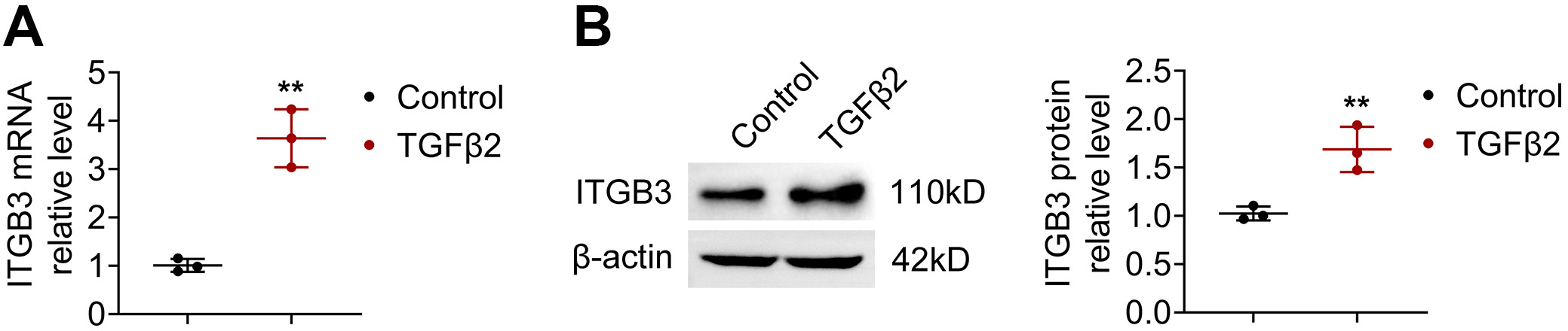
Reverse transcription-quantitative polymerase chain reaction analysis revealed that ITGB3 mRNA expression was significantly increased following transforming growth factor (TGF)-β2 treatment (Figure 1A). Consistently, western blotting demonstrated elevated ITGB3 protein levels in HLE-B3 cells exposed to TGF-β2 (Figure 1B). These findings confirm that TGF-β2 stimulation induces increased ITGB3 expression at both the mRNA and protein levels in HLE-B3 cells.
Inhibition of ITGB3 attenuates TGF-β2-induced proliferation of HLE-B3 cells
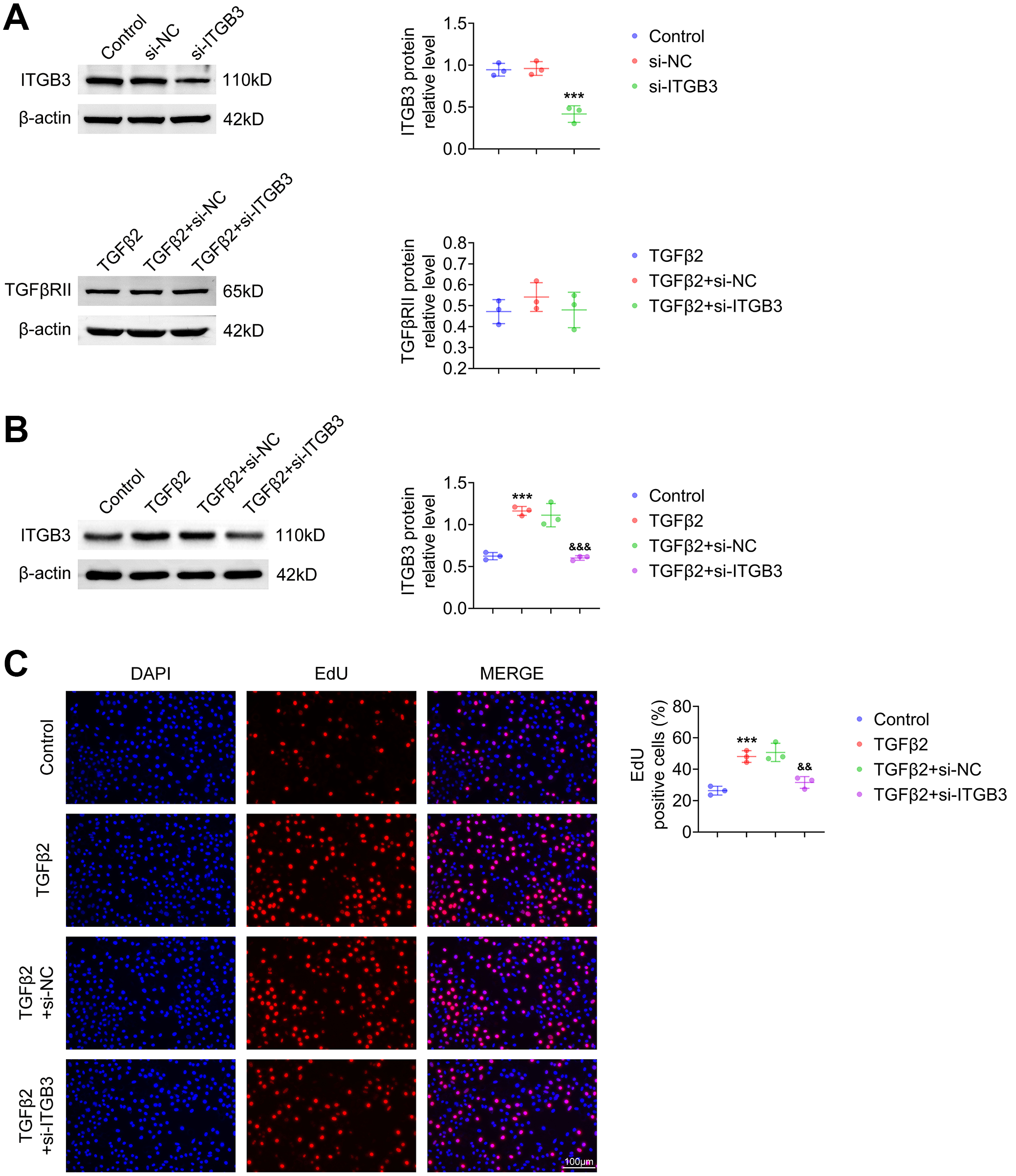
The transfection efficiency of si-ITGB3 was verified (Figure 2A), and TGFβRII protein expression remained unchanged across all groups, indicating the specificity of the knockdown. ITGB3 protein expression, which was elevated following TGF-β2 stimulation, was markedly reduced after si-ITGB3 transfection (Figure 2B). Functionally, TGF-β2 enhanced the proliferation of HLE-B3 cells; however, this effect was significantly attenuated following ITGB3 inhibition (Figure 2C). Collectively, these results suggest that ITGB3 silencing suppresses TGF-β2-induced proliferation in HLE-B3 cells.
Knockdown of ITGB3 suppresses cell migration
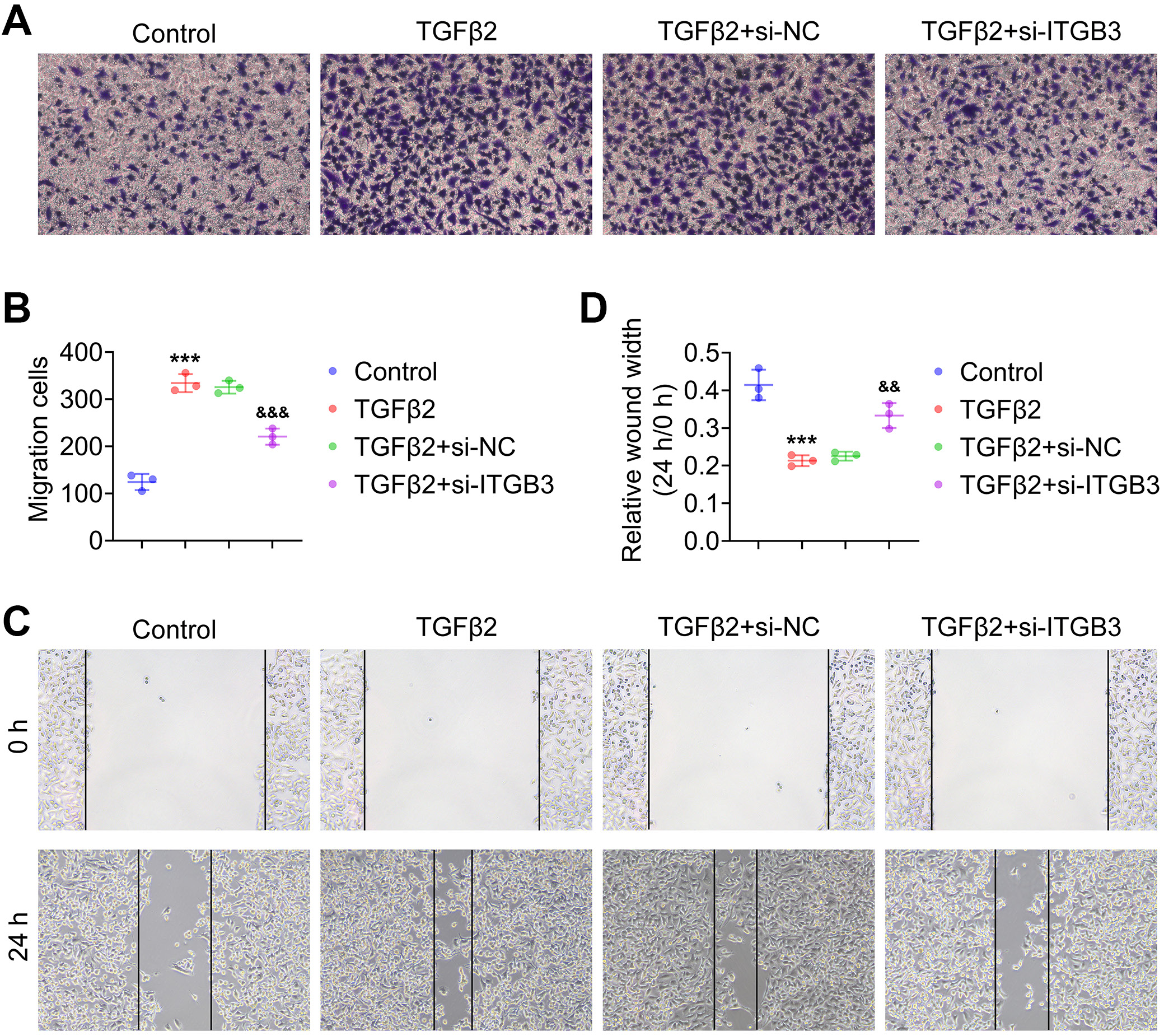
The transwell assay showed that TGF-β2 stimulation significantly increased the migration of HLE-B3 cells, whereas ITGB3 knockdown counteracted this effect (Figure 3A,B). A similar trend was observed in the wound-healing assay, in which the enhanced migratory capacity induced by TGF-β2 was reversed following ITGB3 silencing (Figure 3C,D). These results demonstrate that ITGB3 knockdown effectively restrains TGF-β2-induced migration of HLE-B3 cells.
Suppression of ITGB3 attenuates EMT progression
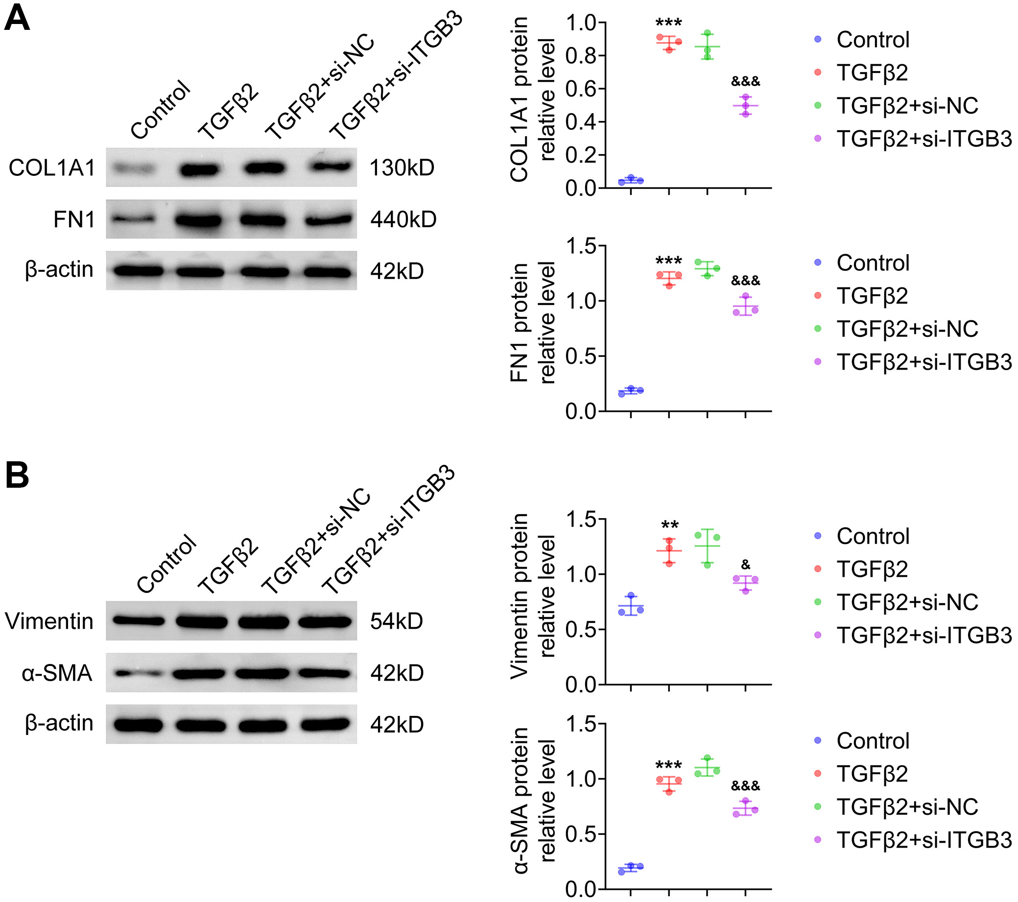
Western blotting demonstrated that TGF-β2 stimulation reduced E-cadherin expression and increased N-cadherin expression, consistent with the induction of EMT. These changes were reversed following ITGB3 suppression (Figure 4A). Moreover, the protein levels of α-SMA and vimentin, both markers of EMT, were elevated by TGF-β2 but decreased following ITGB3 knockdown (Figure 4B). Together, these findings indicate that ITGB3 silencing attenuates EMT progression in HLE-B3 cells.
ITGB3 knockdown inhibits activation of the TGF-β/Smad2/3 pathway
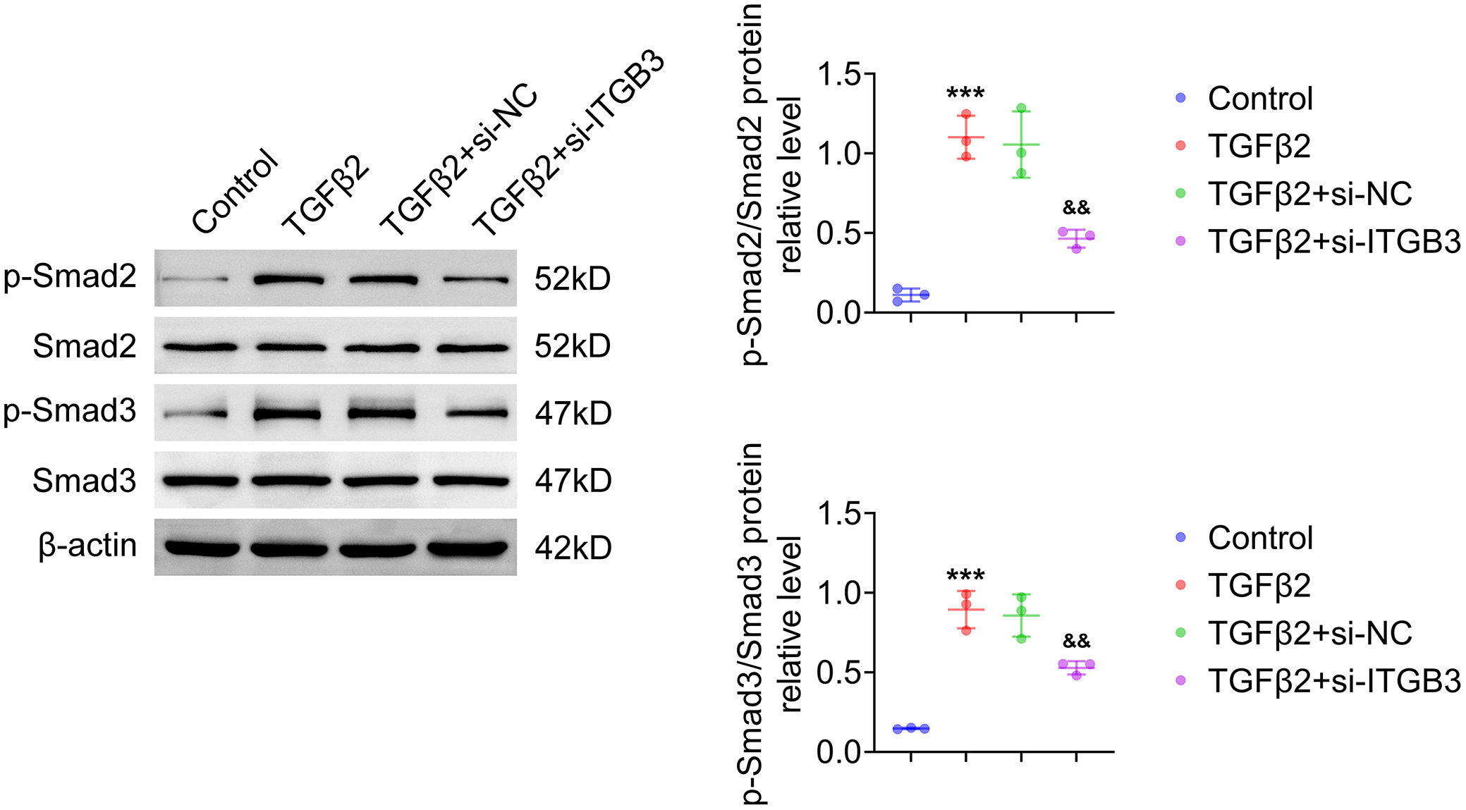
TGF-β2 treatment enhanced the phosphorylation of Smad2 and Smad3, as indicated by increased p-Smad2/Smad2 and p-Smad3/Smad3 ratios. Silencing ITGB3 significantly reduced these phosphorylation events (Figure 5). Overall, these results indicate that ITGB3 knockdown may impair activation of the TGF-β/Smad2/3 signaling pathway in TGF-β2-treated HLE-B3 cells.
Discussion
The development of PCO is a complex process involving multiple cytokines.15 Among these, TGF-β has been identified as one of the most potent cytokines driving the transdifferentiation and fibrosis of LECs.16 TGF-β promotes the proliferation, migration, and EMT of LECs; therefore, TGF-β2-treated LECs are widely used as an in vitro model for PCO research.17, 18 In the present study, this model was established by stimulating HLE-B3 cells with TGF-β2 at a concentration of 10 ng/mL.
ITGB3 has been shown to exert important regulatory functions in various pathological contexts.6, 7, 8, 9, 10 However, its role in PCO progression remains unclear. Our results demonstrated that ITGB3 expression was significantly upregulated in HLE-B3 cells following TGF-β2 treatment. Furthermore, inhibition of ITGB3 reduced the proliferative response of these cells, suggesting that ITGB3 may contribute to TGF-β2-induced cell proliferation.
Both cell migration and EMT are central to the progression of PCO. Considerable attention has been directed toward identifying the molecular mechanisms that regulate these processes. For instance, miR-30a targets Smad2 to attenuate EMT and cell migration in PCO,19 TP53INP2 enhances autophagy to accelerate EMT and migration,13 and REPS2 regulates FAK/Cdc42 signaling to modulate adhesion, EMT, and migration in PCO.20 In addition, silibinin has been reported to inhibit TGF-β2-mediated EMT and migration of LECs, thereby reducing PCO progression.21
Importantly, ITGB3 has been shown to enhance EMT in several diseases.22, 23, 24 Consistent with these findings, our study demonstrated that ITGB3 knockdown reduced the migration of HLE-B3 cells, as shown by transwell and wound-healing assays. Moreover, the EMT process enhanced by TGF-β2 stimulation was effectively reversed following ITGB3 suppression.
The TGF-β/Smad2/3 signaling pathway plays a pivotal role in the progression of PCO. For instance, metformin has been shown to alleviate EMT in LECs through the AMPK/TGF-β/Smad2/3 pathway,25 while FGF-2 regulates TGF-β-induced EMT in LECs by modulating the same pathway, thereby affecting the behavior of LECs in PCO.26 Similarly, capsaicin has been shown to suppress EMT in TGF-β2-stimulated LECs through inhibition of Smad2/3 activation.27
Importantly, ITGB3 has been shown to enhance TGF-β secretion.10, 11 TGF-β binds to its receptors and promotes the activation of downstream Smad proteins, particularly Smad2 and Smad3.28, 29 In this context, our findings revealed that ITGB3 knockdown attenuated TGF-β2-induced phosphorylation of Smad2 and Smad3, indicating that ITGB3 is involved in the regulation of the TGF-β/Smad2/3 pathway during PCO progression.
Limitations of the study
This study has several limitations. First, no human PCO tissue samples were included for validation. Second, the absence of in vivo experiments limits the translational value of the findings. Third, clinical investigations and more detailed phenotypic analyses remain necessary to confirm the role of ITGB3 in the development of PCO. Fourth, although we followed previous studies and applied parametric tests (e.g., t-tests), our small sample size may compromise statistical reliability, resulting in variability in p-values, confidence intervals, and sensitivity to outliers, thereby potentially affecting our findings. Therefore, future studies with larger sample sizes are needed for further validation.
Conclusions
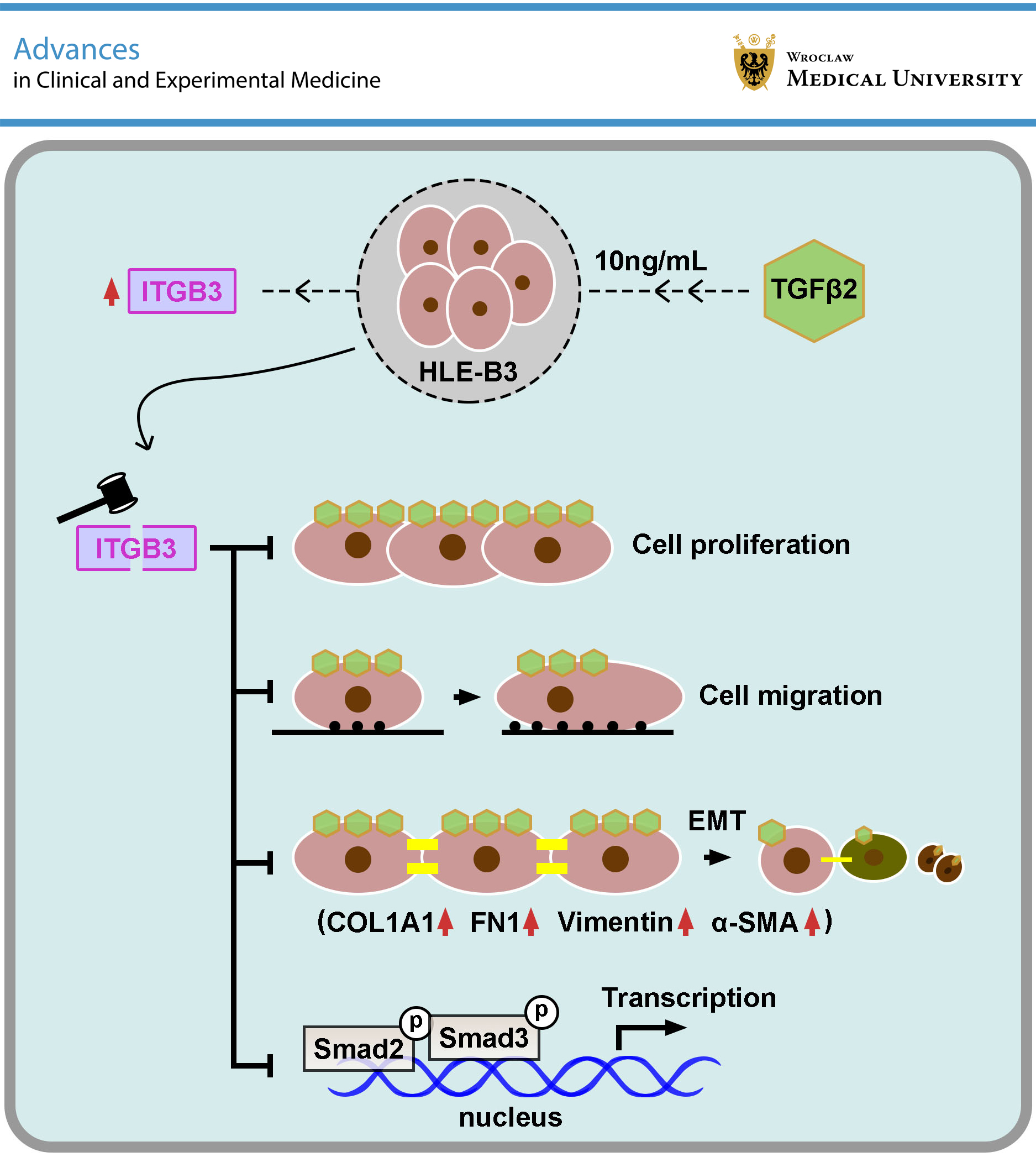
This study demonstrated that ITGB3 knockdown alleviates TGF-β2-induced proliferation, migration, and EMT in HLE-B3 cells. Furthermore, ITGB3 silencing suppressed activation of the TGF-β/Smad2/3 signaling pathway. These findings support ITGB3 as a promising molecular target for the prevention and treatment of PCO and may contribute to the development of future translational strategies aimed at improving postoperative outcomes in patients undergoing cataract surgery.
Data Availability Statement
The datasets supporting the findings of the current study are openly available in Zenodo at https://doi.org/10.5281/zenodo.17607776.
Consent for publication of personal information
Not applicable.
Use of AI and AI-assisted technologies
Not applicable.