Abstract
Background. The adenoma detection rate (ADR) is a key quality metric in colonoscopy. However, it may not accurately reflect the true number of adenomas and requires pathological evaluation of all detected polyps. The adenoma miss rate (AMR), which quantifies adenomas missed during colonoscopy, may provide a more comprehensive measure of colonoscopy quality.
Objectives. To evaluate the effectiveness of ADR and selected surrogate quality indicators (SQIs) in reflecting AMR.
Materials and methods. In this prospective, cross-sectional, multicenter study, we analyzed asymptomatic individuals aged 50–72 years who underwent tandem colonoscopies using a split-dose bowel preparation regimen. Colonoscopies were performed by 7 endoscopists (experts with >10 years of experience from different institutions) over the last 5 years. Consecutive colonoscopies were performed by the same endoscopist under identical conditions. During the 2nd colonoscopy, polyps missed during the 1st colonoscopy were documented. All removed polyps were subsequently evaluated by a pathologist.
Results. We analyzed colonoscopy data from 356 patients (182 (51.12%) males and 174 (48.88%) females), representing a total of 712 colonoscopies. Adenomas were confirmed in 221 participants (62%). The number of adenomas missed during the 1st colonoscopy was 88, whereas 60 participants had at least 1 missed adenoma. No statistically significant variation among endoscopists was observed in the polyp detection rate (range: 48.000–64.000%, p = 0.658) or the adenoma detection rate (range: 54.000–74.000%, p = 0.151). Adenomas per positive participant ranged from 1.05 to 1.89, and adenomas per colonoscopy ranged from 0.70 to 1.44 (p = 0.063). The overall pooled lesion-level AMR was 20.046%. None of the SQIs showed a significant association with AMR (p > 0.050 for all comparisons). Although SQIs and ADR varied among endoscopists, no statistically significant variation in AMR was observed among the 7 endoscopists (p > 0.050).
Conclusions. A high ADR alone does not ensure a lower AMR in tandem colonoscopy for colorectal cancer screening. In this limited provider-level sample, no statistically significant association was observed between SQIs and AMR.
Key words: adenoma detection rate, adenoma miss rate, colonoscopy quality indicators, diagnosis of colorectal neoplasms, endoscopy standards
Background
The adenoma detection rate (ADR) is an established quality indicator for colonoscopy and remains the most widely used benchmark for evaluating endoscopist performance.1, 2 In a report by Lund et al., endoscopists with an ADR of less than 20% had a tenfold higher risk of interval colorectal cancer than those with an ADR exceeding 20%.3 Current recommendations suggest that the ADR target should be at least 25%, with a minimum ADR of 30% in men and 20% in women. The European Society of Gastrointestinal Endoscopy (ESGE) recommends a minimum ADR of 25% for colorectal cancer screening.4
Although ADR is considered an important quality indicator,5 several studies have highlighted its limitations.6 Wang et al. reported that relying solely on ADR may not be sufficient to comprehensively assess the quality of screening colonoscopies.7 In another study, Corley et al. confirmed an inverse association between ADR and interval colorectal cancer.8 In addition, recent studies have demonstrated that the adenoma miss rate (AMR) exceeds 20% even among endoscopists with ADRs greater than 40%.9, 10, 11 These findings suggest a tendency among some endoscopists to discontinue meticulous inspection after detecting a single adenoma, potentially reducing the likelihood of identifying additional adenomas. Given the evidence that ADR may not accurately reflect AMR, several alternative quality indicators have been proposed, including the polyp detection rate (PDR), adenomas per colonoscopy (APC), adenomas per positive participant (APP), and additional adenomas detected after the first adenoma per colonoscopy (ADR-Plus).12, 13, 14 These indicators aim to better capture adenomas that may otherwise remain undetected during colonoscopy.
In a recently published study comparing quality indicators with ADR for predicting AMR, the authors reported that, among all surrogate indicators, APP showed a significant inverse correlation with AMR.12 They suggested that surrogate quality indicators may be useful for predicting AMR, particularly when a high ADR has been achieved. In contrast, a meta-analysis evaluating ADR and other colonoscopy quality indicators reported that both APC and ADR were independently associated with AMR during colonoscopy.15
Surrogate quality indicators that may influence AMR have been defined inconsistently across studies, and uncertainty remains regarding which indicators most accurately reflect AMR or whether a single indicator exists that can reliably predict it.
Objectives
In the present study, we conducted a prospective, cross-sectional, multicenter study involving asymptomatic individuals who underwent tandem colonoscopies performed by 7 endoscopists. The aim of the study was to evaluate the effectiveness of ADR and selected surrogate quality indicators in reflecting AMR.
Materials and methods
Ethics approval and consent to participate
The study received ethical approval from the Human Ethics Review Board of the Ninth People’s Hospital of Nanhai District, Foshan, China, and the Sixth Affiliated Hospital of South China University of Technology, Foshan, China (approval No. NPH 2020-02-64). Written informed consent was obtained from all participants. The study was conducted in accordance with Chinese law and the 2008 Declaration of Helsinki.
Design, period, and setting
This prospective, cross-sectional, multicenter study was conducted from January 2021 to January 2023 at the Ninth People’s Hospital of Nanhai District and the Sixth Affiliated Hospital of South China University of Technology.
Study population (inclusion and exclusion criteria)
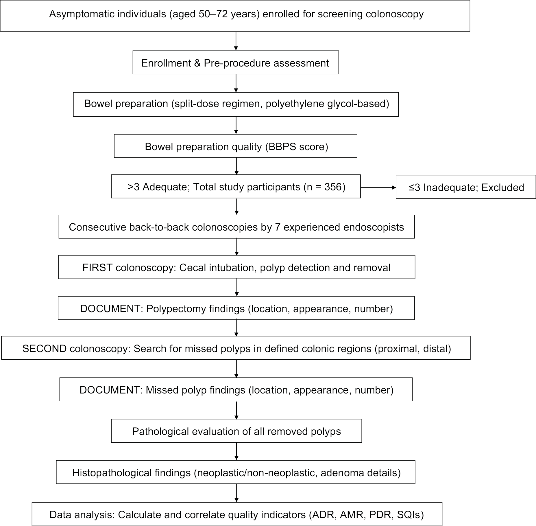
The inclusion criteria were as follows: asymptomatic individuals aged 50–72 years who had undergone screening colonoscopy within the previous 5 years and were enrolled through the outpatient departments of the participating hospitals. The exclusion criteria were as follows: a family history of colorectal cancer (at least 2 first-degree relatives with colorectal cancer, or at least 1 first-degree relative diagnosed with colorectal cancer before the age of 60 years), a personal history of colorectal cancer, any previous colorectal surgery, inadequate bowel preparation, failed cecal intubation, and colorectal cancer detected during the study colonoscopy. Participants with inadequate bowel preparation, defined as a total cumulative Boston Bowel Preparation Scale (BBPS) score ≤3, were excluded from the study.
Tandem colonoscopy
In this study, 356 participants underwent tandem colonoscopies performed by 7 endoscopists at 2 hospitals. The endoscopists were experienced specialists with more than 10 years of experience. All participants followed a bowel-cleansing protocol based on a split-dose bowel preparation regimen, which began the evening before the scheduled colonoscopy. Split-dose bowel preparation consisted of either 2 L of polyethylene glycol with ascorbic acid or 4 L of polyethylene glycol, according to institutional protocols. Colonoscopies were performed 3.5–4.5 h after completion of bowel preparation.
Consecutive colonoscopies were performed by the same endoscopist under identical conditions. Forward-viewing colonoscopes (CF-EZ1500DL/I; Olympus Corp., Tokyo, Japan) were used for all procedures, and the same colonoscope was used for both examinations to minimize instrument-related variability. All participants underwent conscious sedation during the procedures. Colonoscopy was performed with the patient in the left lateral decubitus position, and CO2 insufflation was used throughout the examination. Photographs were obtained upon reaching the ileocecal region, and withdrawal time was recorded using a stopwatch. During withdrawal, endoscopists assessed bowel preparation quality using the Boston Bowel Preparation Scale (BBPS). Inadequate bowel preparation was defined as a total BBPS score ≤3.12 All enrolled participants underwent back-to-back colonoscopies, with the 1st examination immediately followed by the 2nd. During the 1st colonoscopy, the colonoscope was advanced to the cecum, and polyps were identified, counted, and removed during both insertion and withdrawal. Tiny hyperplastic polyps <5 mm in size located in the rectum and sigmoid colon were not removed but were photographed for documentation in accordance with institutional protocol.
During the 2nd colonoscopy, polyps that had not been detected during the 1st colonoscopy were documented according to their location within the colon and their endoscopic appearance. Polyp morphology was classified into 4 categories according to the Japanese classification system: flat, sessile, pedunculated, and laterally spreading tumors. The colon was categorized into 2 anatomical regions: the distal colon (including the descending and sigmoid colon) and the proximal colon (including the cecum, ascending colon, and proximal transverse colon). All removed polyps were subsequently examined by a pathologist. Histopathological findings were broadly classified as neoplastic or non-neoplastic. Neoplastic lesions were further categorized as adenomas, colorectal carcinomas, or advanced adenomas. Advanced adenomas were defined as lesions with a size ≥10 mm, high-grade dysplasia, or villous histology (>25% villous component).16
Colonoscopy quality indicators
The ADR was calculated as the proportion of participants with at least 1 adenoma detected during the 1st colonoscopy relative to the total number of 1st colonoscopies performed. The PDR was calculated as the proportion of participants with at least 1 polyp (including adenomas) detected during the 1st colonoscopy relative to the total number of 1st colonoscopies. Adenomas per colonoscopy (APC) was defined as the total number of adenomas detected during the 1st colonoscopy divided by the total number of 1st colonoscopies performed. Adenoma detection rate plus (ADR-Plus) was calculated as the ratio of the total number of additional adenomas detected after identification of the 1st adenoma during the 1st colonoscopy to the total number of 1st colonoscopies performed. Adenomas per positive participant (APP) was calculated as the ratio of the total number of adenomas detected during the 1st colonoscopy to the total number of participants with at least 1 adenoma detected during the 1st colonoscopy. The AMR was calculated as the ratio of the total number of adenomas missed during the 1st colonoscopy to the total number of adenomas identified during both the 1st and 2nd colonoscopies.
Statistical analyses
A total of 356 participants who underwent back-to-back colonoscopies (356 1st colonoscopies followed by 356 2nd colonoscopies) performed by 7 endoscopists at 2 hospitals were included in the study. Each endoscopist performed colonoscopies in approx. 50 participants, except for 1 endoscopist (A), who examined 56 participants. The minimum sample size was calculated based on the assumptions used in a previous study.16 Spearman correlation coefficients (ρ) regarding baseline quality indicators and AMR ranged widely from –0.560 to 0.680. All evaluated associations with AMR remained statistically non-significant (p > 0.050). The calculated minimum sample size was 50 participants. Descriptive statistics were used to summarize demographic characteristics, including sex, age, smoking status, body mass index (BMI), history of colorectal cancer, and colonoscopy-related variables such as bowel preparation quality and cecal intubation rate. The χ2 test of independence and one-way analysis of variance (ANOVA) were used for categorical variables and normally distributed continuous variables, respectively. The Kolmogorov–Smirnov test was used to assess the normality of continuous variables. Bartlett’s test was used to evaluate the homogeneity of variances for normally distributed continuous variables. All quality indicators were calculated as described previously and compared with AMR using Spearman’s rank correlation coefficient (p < 0.05). The assumption of monotonicity for Spearman’s correlation was not formally tested. All demographic characteristics and colonoscopy-related variables were included in the regression analysis to identify factors associated with missed lesions. All statistical analyses were performed using Descriptive analytics and correlation coefficients were computed using SPSS Statistics v. 27.0 (IBM Corp., Armonk, USA). Forest plots, funnel plots, and Z-score plots were generated using metaanalysisonline.com, v. 2024–2025.
Results
Participant characteristics
A total of 356 participants were enrolled and examined by 7 endoscopists. Each endoscopist examined 50 participants, except for endoscopist A, who examined 56 participants. All enrolled participants underwent back-to-back colonoscopies. The characteristics of the participants examined by the 7 endoscopists are presented in Table 1. A total of 356 colonoscopies were performed during the 1st round and 356 during the 2nd round, resulting in 712 colonoscopies overall. The mean age of the participants was 57.10 ±5.65 years. Of the 356 participants, 182 (51.12%) were male and 174 (48.88%) were female. The cecal intubation rate reported by all 7 endoscopists was 100% for all 712 colonoscopies performed. Participant allocation across endoscopists was determined by institutional availability rather than sex-concordant preferences, with the single female endoscopist evaluating 50 of the 356 total participants. Endoscopist F was the sole female provider in the cohort; of the 50 participants evaluated by this endoscopist, 13 were male (26.000%) and 37 were female (74.000%). A significantly higher proportion of female participants was evaluated by endoscopist F (p = 0.001, χ2 test of independence), reflecting an operational asymmetry in patient distribution. In addition, the prevalence of smoking was significantly lower among participants examined by endoscopist F (1 participant; 2%; p < 0.001, χ2 test of independence). The adequacy of bowel preparation ranged from 98% to 100% across all groups, and the mean withdrawal time was 11.55 ±1.70 min per participant. The mean withdrawal time among the 7 endoscopists ranged from 10.08 ±1.90 min to 13.02 ±2.22 min. Although all endoscopists exceeded the recommended minimum withdrawal time of 6 min, significant variation in withdrawal time was observed among the 7 endoscopists (p < 0.001).
Quality indicators, adenoma miss rate, and adenoma detection rate
The results presented in Table 2 summarize the total number of adenomas detected during colonoscopy. A total of 221 participants (62%; 221/356) were found to have adenomas. These participants had a total of 351 adenomas detected during the 1st colonoscopy. In addition, a total of 132 additional adenomas were identified after the first adenoma detection across the cohort (ADR-Plus). During tandem colonoscopy, a total of 88 adenomas missed during the 1st colonoscopy were identified, whereas 60 participants had at least 1 missed adenoma after the 1st examination. Significant variation in ADR, PDR, and the number of additional adenomas detected after identification of the first adenoma per colonoscopy was observed among the 7 endoscopists (Table 3). The quality indicators reported for each endoscopist differed significantly (Table 3). The ADRs for endoscopists A, B, C, D, E, F, and G were 53.571%, 72.000%, 64.000%, 70.000%, 72.000%, 40.000%, and 64.000%, respectively (p = 0.151). The corresponding PDRs were 52%, 60%, 64%, 58%, 48%, 60%, and 62%, respectively (p = 0.658). The numbers of adenomas per positive participant (APP) were 1.15, 1.26, 1.59, 1.89, 1.05, 1.55, and 1.60 for endoscopists A–G, respectively (p = 0.012). Individual provider performance varied widely, with endoscopist-specific AMRs ranging from a minimum of 12.727% (endoscopist G) to a maximum of 27.778% (endoscopist A), culminating in a collective pooled lesion-level AMR of 20.046% (Table 3). No significant variation in AMR was observed among the 7 endoscopists.
Correlations among the quality indicators
Spearman’s rank correlation analysis revealed that ADR (ρ = 0.290) and PDR (ρ = 0.680) displayed positive correlations with AMR, whereas a minor negative correlation was noted for APC (ρ = −0.070) (Table 4).
Detection of adenoma and association with the AMR among endoscopists
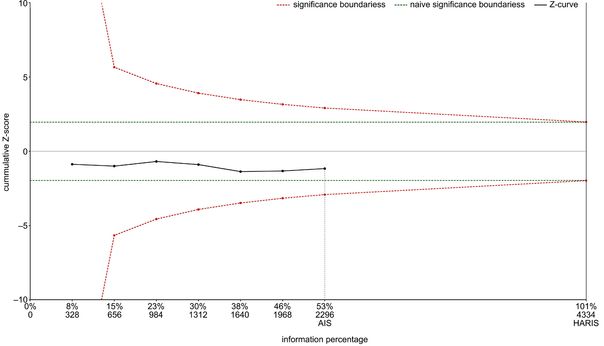
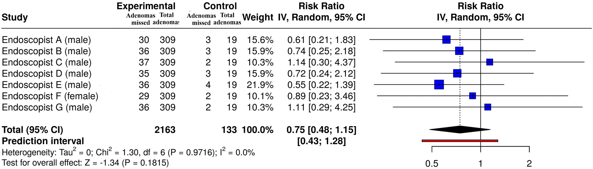
The distribution of adenomas detected during the primary examination was compared with the distribution of missed adenomas across the endoscopist cohorts. The experimental cohort totals (n = 309) and events represent the adjusted baseline detected adenomas, while the control cohort totals (n = 19) and events represent the subgroup of missed adenomas evaluated using the software model (Figure 1). The overall risk ratio (RR) was 0.75 (95% confidence interval (95% CI): 0.48–1.15). The analysis did not reveal significant heterogeneity, indicating that the relationship between adenoma detection and polyp miss rate was consistent across endoscopists in both magnitude and direction. The funnel plot (Figure 2) suggested the presence of potential endoscopist-related bias. Egger’s test supported the presence of funnel plot asymmetry (intercept: 2.92, 95% CI: 1.96–3.87, t = 5.963, p = 0.002). Based on the Z-score plot (Figure 3), the cumulative Z-curve (black solid line) terminated at an Actual Information Size (AIS) of 2,296 (53% of the required information size), failing to cross the significance boundaries (red dashed lines) or reach the Heterogeneity-Adjusted Required Information Size (HARIS) of 4,334. A summary of these statistical analyses is presented in Table 5, Table 6, Table 7. These findings indicate that adenoma detection frequency was not significantly associated with AMR during colonoscopy across endoscopists (I2 = 0%). A study flowchart is presented in Figure 4. Details of the statistical test assumptions for each variable are provided in Table 8.
Discussion
The most effective method for determining AMR is tandem (back-to-back) colonoscopy, in which 2 consecutive colonoscopies are performed on the same patient, on the same day, and under identical conditions. However, this approach may be limited by poor patient compliance due to the discomfort associated with undergoing 2 colonoscopic examinations on the same day. Adenoma detection rate, which is more practical for routine clinical use, indicates only that at least 1 adenoma was detected during colonoscopy, whereas AMR directly quantifies missed adenomas and therefore provides a more accurate assessment of detection performance. Nevertheless, AMR can only be evaluated through tandem colonoscopy, which is typically performed on the same day to identify lesions missed during the 1st examination. Consequently, considerable effort has been devoted to identifying surrogate indicators of AMR. A meta-analysis that included data from approximately 15,000 colonoscopies reported a mean AMR of 26%.15 In a study by Han et al.,16 the mean AMR was 20%. In the present study, the overall AMR was, which is consistent with the findings of these previous studies.
Adenoma detection rate is regarded as the benchmark indicator for adenoma detection. Knowledge of quality indicators may help identify AMR, which could contribute to improving the quality of colonoscopy in colorectal cancer prevention. In a report by Waldmann et al., ADR values <25% and ≥25% were not associated with substantial differences in colorectal cancer occurrence among participants.17 Additionally, Aniwan et al.11 demonstrated that an ADR >40% was associated with AMRs ranging from 13% to 36%. In the present study, the mean ADR across 7 endoscopists performing tandem colonoscopy was 68% (i.e., >40%), whereas the AMR ranged from 1.81% to 3.88%, suggesting that a very high ADR does not necessarily correspond to a low AMR. Previous studies have reported that, although ADR is an important quality indicator, it is insufficient on its own for assessing endoscopist performance.7 Endoscopists with a high ADR may still have a substantial AMR among participants with adenomas. In the present study, a discrepancy between ADR and AMR was observed among endoscopists. Endoscopists C (74%) and G (72%) achieved the highest ADRs among the 7 endoscopists; however, their corresponding AMRs were 2.37% and 3.88%, respectively. In addition, ADR and AMR point estimates are likely to have relatively wide confidence intervals when based on only 50 colonoscopies per endoscopist (Figure 3).18
Although ADR is considered the gold standard for adenoma detection, it has certain limitations because it requires pathological evaluation of all detected polyps. In contrast, PDR has the advantage of not requiring pathological confirmation, is readily available from endoscopy reports, and has been shown to correlate well with ADR.13, 17 In the present study, ADR was strongly correlated with PDR (ρ = 0.93, p = 0.002). However, this correlation does not necessarily reflect improved adenoma detection, as both indicators were calculated independently.
When assessing the associations between surrogate quality indicators and mucosal clearance patterns, Spearman’s rank correlation analysis revealed that ADR (ρ = 0.290, p = 0.520) and PDR (ρ = 0.680, p = 0.420) were positively correlated with AMR (Table 4). Conventionally, a higher ADR or PDR indicates that a greater volume of adenomas or polyps is being detected, which might theoretically be expected to result in a lower miss rate due to more thorough examination. However, our finding of a positive correlation highlights a distinct clinical paradox: higher diagnostic detection thresholds do not mechanically guarantee an absolute reduction in missed lesions.
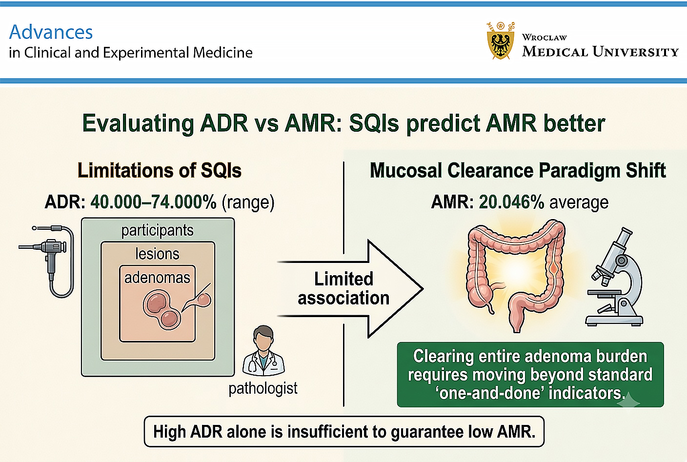
Conversely, when examining advanced metrics, only APC demonstrated a minor negative correlation with AMR (ρ = –0.070, p = 0.880; Table 4), suggesting that maximizing the absolute adenoma yield per procedure may track more closely with reduced miss rates. In contrast, APP (ρ = 0.350, P = 0.480) and ADR-Plus (ρ = 0.280, p = 0.550) both exhibited weak positive correlations with AMR (Table 4). These multi-directional variations among sub-metrics emphasize that clearing the entire adenoma burden requires a paradigm shift beyond standard “one-and-done” indicators, a conceptual framework that aligns closely with the performance variations reported by Aniwan et al.11
In the present study, all 7 endoscopists (6 males and 1 female) were experienced specialists from different institutions. All were qualified and highly experienced in performing colonoscopy. The 7 endoscopists achieved a high mean ADR of 68%, along with a 100% cecal intubation rate. Bowel preparation quality was adequate in all cases (BBPS > 3), and the mean withdrawal time exceeded 6 min, although the AMR ranged up to 3.88%. In addition, polyp detection was not significantly associated with AMR among endoscopists (Figure 1). These findings suggest that a high ADR may still be accompanied by a variable AMR. APC and ADR-Plus are closely related to ADR because all 3 indicators are influenced by the number of adenomas detected during colonoscopy. However, they differ conceptually from ADR. APC incorporates the total number of adenomas detected, whereas ADR-Plus reflects the number of additional adenomas identified after detection of the first adenoma. APP also correlates strongly with ADR; however, it is not entirely determined by ADR because APP is calculated only among participants with adenomas, whereas ADR is based on the entire study population.19 Therefore, indicators such as APC, APP, and ADR-Plus may better reflect the burden of adenoma detection and have been proposed as complementary measures of both ADR and AMR. Overall, quality indicators such as APC, APP, and ADR-Plus may exhibit high values without being associated with a significant reduction in AMR. Therefore, further studies involving populations with a high burden of adenomas are needed to confirm the usefulness of these quality indicators in real-world clinical practice.
Limitations of the study
First, there was an imbalance in the number of male and female participants enrolled in the study. Second, the participant distribution may have introduced heterogeneity, as subjects were recruited from multiple hospitals rather than from a single institution or geographical area. Third, although each of the 7 endoscopists examined approx. 50 participants, one endoscopist (A) examined 56 participants. In addition, the total sample size of 356 participants was substantially smaller than those reported in several previously published studies. Fourth, the study was based on tandem colonoscopies performed on the same day, a design that may have limited patient acceptance and consequently restricted participant recruitment. Fifth, the correlation analyses were conducted at the endoscopist level (n = 7), resulting in limited statistical power and an inability to fully utilize participant-level information. This may have contributed to unstable estimates and inconsistent findings (Figure 3). The small sample size further limited the ability to detect true associations. Finally, in tandem colonoscopy studies, the repeat procedure is often performed by a different endoscopist to maximize the likelihood of detecting missed lesions. In the present study, however, consecutive colonoscopies were performed by the same endoscopist under identical conditions. This represents an important limitation of the study. Future studies should consider using different endoscopists for the repeat examination.
Conclusions
The present study demonstrated that a high ADR alone does not ensure a lower AMR in tandem colonoscopy for colorectal cancer screening. Moreover, in this limited provider-level sample, no statistically significant association was observed between surrogate quality indicators, such as APC, APP, and ADR-Plus, and AMR. Further large-scale studies are needed to identify the optimal quality indicators that can accurately reflect AMR across diverse populations and clinical settings.
Data Availability Statement
The datasets supporting the findings of the current study are openly available in Zenodo at https://doi.org/10.5281/zenodo.22077108.
Consent for publication
Not applicable.
Use of AI and AI-assisted technologies
Not applicable.
.jpg)