Abstract
Background. Nonalcoholic fatty liver disease (NAFLD) is increasingly recognized as a multisystem disorder with potential cardiovascular consequences. Its role in patients with unstable angina (UA), a high-risk manifestation of coronary artery disease (CAD), remains underexplored.
Objectives. To evaluate the impact of NAFLD on long-term cardiovascular outcomes in patients with UA.
Materials and methods. This prospective cohort study enrolled 150 patients diagnosed with UA, who were categorized into NAFLD and non-NAFLD groups based on imaging and clinical criteria. Baseline metabolic and liver function parameters were recorded. Patients were followed for 3 years to document major adverse cardiovascular events (MACE). Kaplan–Meier analysis was used to assess event-free survival, and multivariate logistic regression was performed to identify independent predictors of MACE.
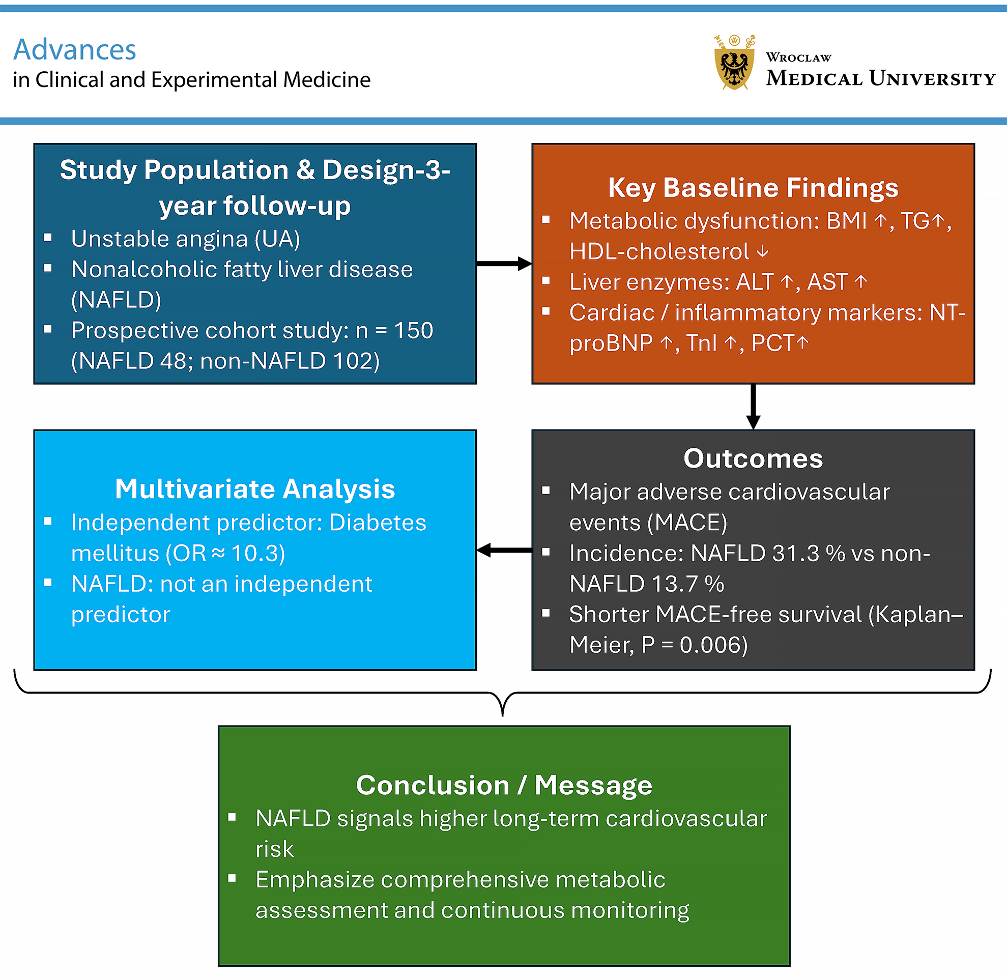
Results. Patients with NAFLD exhibited worse metabolic and liver function profiles than those without NAFLD. During the 3-year follow-up period, the incidence of MACE was significantly higher in the NAFLD group. Kaplan–Meier analysis revealed shorter MACE-free survival time among patients with NAFLD (p < 0.05). However, in multivariate logistic regression analysis, diabetes mellitus, but not NAFLD, remained an independent predictor of MACE after adjustment for potential confounding factors.
Conclusions. Nonalcoholic fatty liver disease is associated with metabolic dysfunction and an increased risk of cardiovascular events in patients with UA. Although it is not an independent predictor of MACE, its presence may indicate a higher long-term cardiovascular risk and should be considered in clinical risk stratification.
Key words: nonalcoholic fatty liver disease, unstable angina, cardiovascular diseases, Kaplan–Meier analysis, logistic regression
Background
Unstable angina (UA) is a manifestation of acute coronary syndrome (ACS) and is often considered a precursor to myocardial infarction, representing a critical cardiovascular emergency. It is characterized by new-onset, worsening, or rest angina without an obvious cause.1 Epidemiological data indicate that UA is one of the most common cardiovascular events among older adults, with its incidence increasing with age. The condition is particularly prevalent in individuals with known coronary artery disease (CAD), who are at higher risk of progressing to myocardial infarction or sudden cardiac death.2, 3 The clinical significance of UA lies not only in its symptomatic presentation but also in its associated complications and unfavorable long-term prognosis.4 Research indicates that individuals with UA face an elevated risk of myocardial infarction or cardiac death within a short period, particularly when timely treatment is not administered.5, 6 Long-term outcomes in these patients are influenced by several factors, including the severity of coronary artery stenosis, coexisting conditions such as diabetes mellitus and hypertension, and the timeliness and effectiveness of medical interventions.7 Therefore, UA is considered a cardiovascular emergency requiring prompt diagnosis and treatment to reduce mortality and improve prognosis.
Nonalcoholic fatty liver disease (NAFLD), a chronic liver condition associated with metabolic syndrome, is characterized by fat accumulation in the liver in the absence of significant alcohol consumption. Its prevalence is increasing globally, particularly among adults with obesity or type 2 diabetes (T2D). Nonalcoholic fatty liver disease encompasses a broad pathological spectrum, ranging from simple steatosis to nonalcoholic steatohepatitis, which may progress to fibrosis or cirrhosis.8, 9, 10 Increasing evidence highlights its strong association with systemic metabolic disorders and cardiovascular diseases. Studies suggest that NAFLD increases the risk of major adverse cardiovascular events (MACE) and is associated with poorer outcomes in patients with pre-existing cardiovascular conditions.11, 12
Mechanistically, NAFLD may contribute to atherosclerosis and cardiovascular injury through insulin resistance, endothelial dysfunction, chronic inflammation, and oxidative stress.13, 14 Clinical studies have further identified NAFLD as an independent risk factor for both malignant arrhythmias in patients with ACS and increased CAD severity, as evidenced by its association with higher Gensini scores and an increased risk of coronary heart disease.15, 16 These findings underscore the systemic cardiovascular impact of NAFLD. However, evidence regarding the prognostic value of NAFLD specifically in patients with UA remains scarce, and prospective studies with long-term follow-up in this population are particularly limited. This highlights the need to clarify whether NAFLD independently influences cardiovascular outcomes in this high-risk subgroup.
Objectives
Despite growing evidence linking NAFLD to cardiovascular diseases, studies specifically examining its impact on the long-term prognosis of patients with UA remain limited. This study aimed to investigate the influence of NAFLD on the 3-year incidence of MACE and cardiovascular outcomes in patients with UA using a prospective cohort design. Findings from this research may provide valuable insights into clinical risk stratification and the long-term management of patients with UA, particularly those with coexisting NAFLD.
Materials and methods
Study population
This was a single-center prospective cohort study that enrolled all eligible patients with UA treated at Xi’an Third Hospital (China) between January 2020 and December 2021. A total of 150 patients aged >18 years were included and divided into 2 groups based on the presence of NAFLD: the non-NAFLD group and the NAFLD group. The diagnostic criteria for UA were as follows17: 1) typical anginal chest pain; 2) ischemic changes on electrocardiography (ECG), including new or transient ST-segment depression ≥0.1 mV or T-wave inversion ≥0.2 mV; and 3) normal cardiac biomarker levels, including cardiac troponin T (TnT), cardiac troponin I (TnI), and creatine kinase-MB isoenzyme (CK-MB).
Exclusion criteria included: 1) other cardiovascular conditions, such as stable angina, myocardial bridging, coronary artery anomalies, cardiomyopathy, valvular or congenital heart disease, or heart failure; 2) a history of percutaneous coronary intervention (PCI) or coronary artery bypass grafting (CABG); 3) recent use (within 1 month) of steroids, anti-inflammatory agents, antibiotics, non-steroidal anti-inflammatory drugs (NSAIDs), or immunosuppressants; 4) chronic liver diseases, including hepatitis B, hepatitis C, autoimmune liver disease, or drug-induced liver injury; and 5) severe infections, renal dysfunction, malignancies, or other significant comorbidities.
Nonalcoholic fatty liver disease was diagnosed based on the following criteria: 1) absence of significant alcohol consumption, defined as an average ethanol intake of <210 g/week for men and <140 g/week for women, maintained consistently over the preceding 6 months; 2) exclusion of other specific liver diseases, such as viral hepatitis; and 3) characteristic findings on abdominal B-mode ultrasonography, including increased hepatic echogenicity compared with the spleen and kidney, uniform enhancement of the near field with posterior attenuation, indistinct intrahepatic vascular architecture, and mild-to-moderate hepatomegaly with rounded borders. All patients provided written informed consent. The present study was conducted in accordance with the Declaration of Helsinki and was approved by the Ethics Committee of Xi’an Third Hospital (approval No. XATH-20200041).
The required sample size was calculated using a cohort study formula, with parameters based on preliminary estimates of NAFLD prevalence among patients with UA. Using a 95% confidence level (Z = 1.96), an estimated NAFLD proportion (p = 0.28), and a margin of error (d = 0.072), the sample size was calculated as follows:
n = Z2 × p × (1 − p)/d2 = 1.962 × 0.28 × (1 − 0.28)/0.0722 ≈ 150.
Thus, a sample size of 150 was considered adequate to detect significant differences in cardiovascular outcomes between the groups.
Data collection
Demographic, epidemiological, and clinical data were collected, including age, sex, body mass index (BMI), smoking status, alcohol consumption history, and comorbidities such as hypertension and diabetes mellitus. Blood tests were performed using an automated biochemical analyzer (Hitachi 7600; Hitachi Corporation, Tokyo, Japan) to assess the following parameters: lipid metabolism markers, including total cholesterol (TC), triglycerides (TG), high-density lipoprotein cholesterol (HDL-C), and low-density lipoprotein cholesterol (LDL-C); liver function markers, including alanine aminotransferase (ALT) and aspartate aminotransferase (AST); cardiac biomarkers, including N-terminal pro-B-type natriuretic peptide (NT-proBNP), CK-MB activity, and TnI; and inflammatory markers, including C-reactive protein (CRP) and procalcitonin (PCT), as previously described.18, 19
Follow-up and observation indicators
All patients were followed for 3 years after discharge. For patients who died during follow-up, the time and cause of death were recorded. MACE were defined as the occurrence of any of the following events during follow-up: sudden cardiac death, heart failure, cardiogenic shock, recurrent myocardial infarction, or arrhythmias associated with hemodynamic instability. MACE-free survival time was documented for each patient.
Statistical analyses
Statistical analyses were performed using IBM SPSS v. 25.0 (IBM Corp., Armonk, USA). Data normality was assessed separately in each group. The Shapiro–Wilk test was used for the NAFLD group (n = 48), whereas the Kolmogorov–Smirnov test was applied to the non-NAFLD group (n = 102), in accordance with standard recommendations based on sample size (Supplementary Table 1).
Continuous variables were expressed as mean ± standard deviation (SD) for normally distributed data or as median (range, interquartile range (IQR)) for non-normally distributed data. Comparisons between groups were conducted using Student’s t-test or the Mann–Whitney U test, as appropriate. For variables that were normally distributed in both groups, equality of variances was assessed using Levene’s test; if variances were unequal, Welch’s t-test was applied instead of the standard Student’s t-test.
Categorical variables were compared using Pearson’s χ2 test of independence, as all cross-tabulations had a minimum expected cell count greater than 5. Survival analysis was performed using the Kaplan–Meier method, and differences in survival distributions were assessed with the log-rank test. Multivariate logistic regression analyses were conducted to identify independent predictors of MACE.
The assumptions of logistic regression were assessed as follows: linearity of continuous variables with the logit of the outcome was tested using the Box–Tidwell procedure; multicollinearity was evaluated using variance inflation factors (VIFs); and potential outliers or influential observations were examined using Cook’s distance and studentized residuals obtained from the SPSS regression diagnostics output. Model fit was assessed using the Hosmer–Lemeshow goodness-of-fit test and Nagelkerke’s R2.
A two-sided p < 0.05 was considered statistically significant. This study involved several hypothesis tests. No formal adjustment for multiple comparisons was performed, as all comparisons were conducted using standard statistical methods (Student’s t-test, Mann–Whitney U test, and χ2 test).
Results
Comparison of demographic characteristics, liver function, and lipid metabolism indicators between the NAFLD and non-NAFLD groups
As shown in Table 1, there were no significant differences between the NAFLD and non-NAFLD groups in terms of age, sex distribution, smoking status, alcohol consumption history, or the prevalence of hypertension and diabetes mellitus. However, the NAFLD group had a significantly higher BMI (p < 0.001), accompanied by an unfavorable lipid profile, including elevated TG levels (p < 0.001) and decreased HDL-C levels (p < 0.001), whereas no significant differences were observed in TC or LDL-C levels. Additionally, liver function markers were significantly impaired in the NAFLD group, as evidenced by higher ALT and AST levels (both p < 0.001). Median MACE-free survival time was also significantly shorter in the NAFLD group than in the non-NAFLD group (p = 0.006). The original results of Levene’s test are presented in Supplementary Table 2.
Comparison of cardiac biomarkers and inflammatory factor levels between the NAFLD and non-NAFLD groups
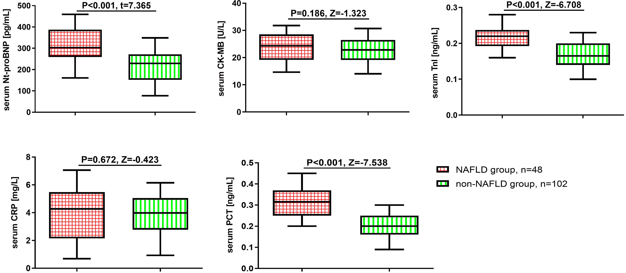
Cardiac biomarker and inflammatory marker levels were further compared between the 2 groups. As shown in Figure 1, the NAFLD group had significantly higher levels of NT-proBNP, TnI, and PCT (all p < 0.001). No significant differences were observed in CK-MB or CRP levels. These findings suggest that patients with NAFLD may have more pronounced myocardial injury and systemic inflammation.
Comparison of MACE incidence between the NAFLD and non-NAFLD groups
To further evaluate long-term outcomes, the incidence of MACE was compared between the 2 groups. During the 3-year follow-up period, a total of 29 patients (19.33%) developed MACE. The NAFLD group exhibited a significantly higher incidence of MACE than the non-NAFLD group (p = 0.003; Table 2). Post hoc power analysis (α = 0.05, two-sided), based on the observed MACE incidence rates (31.3% vs 13.7%) and sample sizes (48 vs 102), yielded a statistical power of 95.6%. Although the NAFLD group showed numerically higher rates of sudden cardiac death, myocardial infarction, and heart failure, none of these differences reached statistical significance. These findings suggest a potential association between NAFLD and an increased overall cardiovascular risk in patients with UA.
Kaplan–Meier analysis of MACE-free survival in the NAFLD and non-NAFLD groups
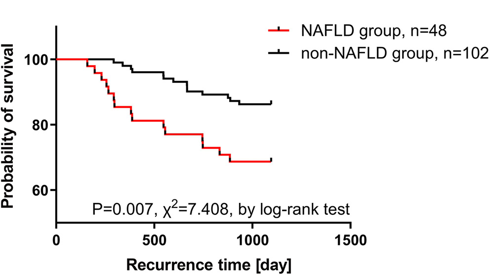
To further assess the prognostic impact of NAFLD, Kaplan–Meier analysis was performed to compare MACE-free survival between the 2 groups. As shown in Figure 2, the NAFLD group had significantly shorter MACE-free survival than the non-NAFLD group (p = 0.006), indicating a higher cumulative incidence of cardiovascular events over time.
Multivariate logistic regression analysis of risk factors for MACE
To identify independent risk factors for MACE, a multivariate binary logistic regression analysis was performed, including all clinically relevant variables. Given their highly skewed distributions and unstable estimates in the initial model, TnI and PCT were transformed into binary variables using the median as the cutoff value to improve interpretability and model stability. The model demonstrated acceptable goodness of fit, as indicated by a non-significant Hosmer–Lemeshow test (p = 0.254) and a Nagelkerke R2 value of 0.357. Multicollinearity was assessed using variance inflation factors (VIFs), which showed that all variables had VIF values <5, except for NAFLD (VIF = 7.046). Given its clinical importance and predefined role as the primary exposure variable, NAFLD was retained in the model despite its elevated VIF (Supplementary Table 3). However, we acknowledge that its inclusion introduces potential multicollinearity and therefore represents a methodological compromise that may have affected its statistical significance. Residual diagnostics revealed no studentized residuals exceeding ±3.0, and all Cook’s distance values were below 1.0, indicating the absence of influential outliers. Leverage values were within acceptable limits, supporting the robustness of the regression model (Supplementary Table 4). Linearity between continuous predictors and the logit of the outcome was assessed using the Box–Tidwell test. All interaction terms were non-significant (p > 0.05), except for AST (p = 0.019), which showed a mild deviation from linearity. However, given the overall model stability and clinical relevance, AST was retained in the final model (Supplementary Table 5). As shown in Table 3, diabetes mellitus was independently associated with an increased risk of MACE (odds ratio (OR) = 10.269, 95% confidence interval (95% CI): 3.148–33.495, p < 0.001). No other variables, including NAFLD, age, lipid parameters, liver enzymes, cardiac biomarkers, or inflammatory markers, reached statistical significance in the multivariate model.
Discussion
Unstable angina is a clinical concern not only because of its acute symptomatic burden but also due to its high instability and potential to progress to myocardial infarction or sudden cardiac death.20 In this prospective cohort study, we found that patients with coexisting NAFLD had a significantly higher incidence of MACE during the 3-year follow-up period compared with those without NAFLD.
MACE occurrence in patients with UA is influenced by various factors. Alongside established cardiovascular risk factors such as hypertension, diabetes mellitus, hyperlipidemia, smoking, and family history, key determinants of prognosis include the severity of coronary artery lesions, cardiac function, inflammatory marker levels, and the timeliness and effectiveness of therapeutic interventions. Studies have identified several predictors of MACE in patients with UA. For instance, 1 study found a higher incidence of MACE in men than in women (4.1% vs 2.6%, p < 0.05), despite similar in-hospital cardiac mortality rates.21 Another study identified advanced age, diabetes mellitus, reduced left ventricular ejection fraction (LVEF), elevated high-sensitivity C-reactive protein (hs-CRP), and high SYNTAX scores as independent predictors of MACE within 2 years after discharge.22 Additionally, unstable plaques and elevated CD4+/CD8+ ratios have been associated with increased plaque instability.23 Emerging serum markers, such as NLRP3 and growth differentiation factor 15 (GDF-15), have also been associated with prognosis in patients with UA.24, 25
In our study, the overall 3-year incidence of MACE was 19.3%, which is comparable to long-term MACE rates reported in population-based cohorts with NAFLD. For instance, an Australian cohort study reported an 18.8% incidence of 5-point MACE over a mean follow-up period of approx. 18 years.26 Given that our study focused on a hospitalized UA population with an inherently higher baseline risk, the observed event rate is clinically plausible.
Furthermore, patients with NAFLD had a significantly higher incidence of MACE during follow-up; however, NAFLD was not identified as an independent predictor after adjustment for confounding factors. This finding suggests that the adverse cardiovascular outcomes observed in patients with UA and NAFLD may be mediated by coexisting metabolic abnormalities, particularly diabetes mellitus, which showed the strongest independent association in our model. In addition, multicollinearity between NAFLD and metabolic parameters such as BMI and triglyceride levels may have attenuated the independent statistical contribution of NAFLD in the multivariate analysis.
Although age did not reach statistical significance in the multivariate model (p = 0.062), the observed trend suggests a potential association that may warrant further investigation in larger cohorts. Among the analyzed variables, diabetes mellitus showed the strongest independent association with MACE, indicating that metabolic dysfunction may be the primary driver of poor prognosis in this subgroup. This finding is consistent with studies in other disease populations, in which cardiovascular risk factors have been shown to independently affect disease phenotype and prognosis, such as in patients with polycythemia vera and essential thrombocythemia.27
Compared with previous studies that identified various inflammatory and structural indicators as predictors of MACE, our findings highlight the need to clarify whether NAFLD acts as a mediator, amplifier, or merely a marker of metabolic risk in predicting cardiovascular outcomes in patients with UA.
Nonalcoholic fatty liver disease is an established independent risk factor for cardiovascular diseases and is linked to metabolic disorders and chronic inflammation. It increases the risk of hypertension, coronary heart disease, cardiomyopathy, and arrhythmias, thereby contributing to higher cardiovascular morbidity and mortality.28 A cohort study involving more than 210,000 participants confirmed NAFLD as a predictor of all-cause mortality and adverse cardiovascular outcomes, highlighting the value of incorporating NAFLD into cardiovascular risk assessment.29 Additionally, NAFLD has been associated with an increased incidence of myocardial infarction, irrespective of other risk factors.30 A meta-analysis further revealed that female patients with NAFLD face significantly higher rates of coronary events and mortality.31
The pathophysiological mechanisms underlying NAFLD are closely related to insulin resistance, which leads to dysfunction of adipokines produced by adipose tissue, particularly adiponectin. Furthermore, increased formation of reactive oxygen species (ROS) promotes the oxidation of free fatty acids, while triglyceride accumulation enhances de novo lipogenesis, resulting in a pro-inflammatory state and increased oxidative stress. These mechanisms may contribute to the formation and progression of atherosclerotic plaques, potentially increasing cardiovascular risk in patients with NAFLD.12
Although NAFLD is widely recognized as an independent cardiovascular risk factor in the general population, its role as an independent predictor of MACE in patients with UA was not confirmed in our cohort after adjustment for confounding factors. Nonetheless, the prospective design of our study and the 3-year follow-up period strengthen the evidence for an association between NAFLD and adverse cardiovascular outcomes in patients with UA, even if NAFLD was not independently predictive in the multivariate analysis.
Limitations of the study
While this study provides valuable insights into the impact of NAFLD on patients with UA, several limitations should be acknowledged. First, this was a single-center study with a relatively limited sample size, which may affect the generalizability of the findings. Although potential confounding factors were considered, influences such as lifestyle and genetic background may not have been fully accounted for. In addition, the 3-year follow-up period may be insufficient to capture the full long-term effects of NAFLD in patients with UA. Furthermore, due to the relatively low number of MACE events and the inclusion of multiple clinically relevant variables, the multivariate model may have been affected by limited statistical power and potential overfitting. Nevertheless, the modeling approach was retained to preserve a comprehensive assessment of cardiovascular risk. In addition, CK-MB was measured as enzymatic activity rather than mass concentration, which may be more susceptible to false-positive results due to skeletal muscle injury or macro-CK interference and should therefore be considered a potential source of diagnostic imprecision. Finally, because multiple hypotheses were tested, there is a potential risk of type I error, and the results should therefore be interpreted with caution.
Conclusions
In patients with UA, coexisting NAFLD was associated with a higher incidence of major adverse cardiovascular events during a 3-year follow-up period, although it was not identified as an independent predictor after adjustment for confounding factors. These findings suggest that NAFLD may serve as a marker of increased cardiovascular risk and highlight the importance of comprehensive metabolic assessment and long-term follow-up in this population.
Supplementary data
The supplementary materials are available at: https://doi.org/10.5281/zenodo.17370159. The package contains the following files:
Supplementary Table 1. Results of normality tests.
Supplementary Table 2. Original Student’s t-test results for Table 1 of the main text, including Levene’s test results.
Supplementary Table 3. VIF results for the logistic regression analysis presented in Table 3.
Supplementary Table 4. Residual and influence diagnostics for the logistic regression model presented in Table 3, including studentized residuals, Cook’s distance, leverage values, and Mahalanobis distance.
Supplementary Table 5. Box–Tidwell test results for assessing the linearity of continuous variables in the logistic regression model (Table 3).
Supplementary Table 6. χ2 test results for sex (Table 1).
Supplementary Table 7. χ2 test results for smoking status (Table 1).
Supplementary Table 8. χ2 test results for alcohol consumption history (Table 1).
Supplementary Table 9. χ2 test results for hypertension (Table 1).
Supplementary Table 10. χ2 test results for diabetes mellitus (Table 1).
Supplementary Table 11. χ2 test results for sudden cardiac death (Table 2).
Supplementary Table 12. χ2 test results for myocardial infarction (Table 2).
Supplementary Table 13. χ2 test results for heart failure (Table 2).
Supplementary Table 14. χ2 test results for MACE (Table 2).
Data Availability Statement
The datasets supporting the findings of the present study are openly available in Zenodo at https://doi.org/10.5281/zenodo.17370189.
Use of AI and AI-assisted technologies
This manuscript underwent language editing using GPT-4o, a large language model developed by OpenAI, to improve clarity and readability. All scientific content, data interpretation, and conclusions were generated by the authors.
Consent for publication of personal information
Not applicable.