Advances in Clinical and Experimental Medicine

Title abbreviation: Adv Clin Exp Med
5-Year IF – 2.0, IF – 1.9, JCI (2024) – 0.43
Scopus CiteScore – 4.3
Q1 in SJR 2024, SJR score – 0.598, H-index: 49 (SJR)
ICV – 161.00; MNiSW – 70 pts
Initial editorial assessment and first decision within 24 h

ISSN 1899–5276 (print), ISSN 2451-2680 (online)
Periodicity – monthly

Download original text (EN)

Advances in Clinical and Experimental Medicine

Ahead of print

doi: 10.17219/acem/211596

Publication type: review

Language: English

License: Creative Commons Attribution 3.0 Unported (CC BY 3.0)

Download citation:

  • BIBTEX (JabRef, Mendeley)
  • RIS (Papers, Reference Manager, RefWorks, Zotero)

Cite as:


Gadour E, Miutescu B, AlQahtani MS, et al. Diagnostic challenges in progressive familial intrahepatic cholestasis type 3 (PFIC3) misdiagnosed as Wilson’s disease: A systematic review [published online as ahead of print on January 21, 2026]. Adv Clin Exp Med. 2026. doi:10.17219/acem/211596

Diagnostic challenges in progressive familial intrahepatic cholestasis type 3 (PFIC3) misdiagnosed as Wilson’s disease: A systematic review

Eyad Gadour1,2,A,B,C,D,E,F, Bogdan Miutescu3,4,A,E,F, Mohammed Saad Alqahtani1,5,A,B,E,F, Deiana Vuletici3,4,B,C,D, Ghassan Elsayed6,B,D,E,F, Ielmina Domilescu7,B,C,D,E, Antonio Facciorusso8,A,B,C,D,E,F

1 Multiorgan Transplant Centre of Excellence, Liver Transplantation Unit, King Fahad Specialist Hospital, Dammam, Saudi Arabia

2 Department of Medicine, Zamzam University College, School of Medicine, Khartoum, Sudan

3 Department of Gastroenterology and Hepatology, University of Medicine and Pharmacy Victor Babes, Timișoara, Romania

4 Advanced Regional Research Centre, University of Medicine and Pharmacy Victor Babes, Timișoara, Romania

5 Department of Surgery, Imam Abdulrahman Bin Faisal University, Dammam, Saudi Arabia

6 Department of Gastroenterology, Mediclinic Middle East Hospital, Abu Dhabi, UAE

7 Doctoral School, Department of Gastroenterology and Hepatology, Faculty of Medicine, Victor Babes, Timișoara, Romania

8 Department of Surgical and Medical Sciences, Section of Gastroenterology, University of Foggia, Italy

Graphical abstract


Graphical abstracts

Highlights


• Progressive familial intrahepatic cholestasis type 3 caused by ABCB4 mutations is frequently misdiagnosed as Wilson’s disease due to overlapping abnormalities in copper metabolism.
• A systematic review of 16 misdiagnosed cases showed that chelation therapy was ineffective, resulting in delayed initiation of appropriate treatment.
• Genetic testing for ABCB4 mutations should be considered in atypical Wilson’s disease presentations, particularly in patients with normal ceruloplasmin levels and absence of Kayser–Fleischer rings.
• Switching to ursodeoxycholic acid therapy led to clinical stabilization, highlighting the importance of early and accurate diagnosis.

Abstract

Progressive familial intrahepatic cholestasis type 3 (PFIC3) is a rare liver disorder caused by biallelic mutations in the ABCB4 gene, leading to multidrug resistance protein 3 (MDR3) deficiency. PFIC3 often presents with clinical and biochemical features that overlap with Wilson’s disease (WD), including hepatic copper accumulation and elevated urinary copper excretion. These similarities contribute to frequent misdiagnosis, resulting in inappropriate chelation therapy and delayed appropriate management. This systematic review examines reported cases of PFIC3 initially misdiagnosed as WD to highlight diagnostic challenges and assess patient outcomes. A comprehensive search across PubMed, ScienceDirect and Google Scholar identified 11 eligible studies involving 16 patients. Most cases were first treated as WD, receiving chelation therapy without clinical improvement. Diagnosis was later revised to PFIC3 following negative ATP7B mutation testing and identification of ABCB4 variants, often via whole-genome sequencing. Upon switching to ursodeoxycholic acid (UDCA), most patients experienced clinical stabilization. The findings underscore the need for heightened awareness of PFIC3 as a differential diagnosis in atypical WD cases, especially when ceruloplasmin is normal and Kayser–Fleischer (KF) rings are absent. Early genetic testing is essential to avoid mismanagement. Further observational studies are warranted to estimate misdiagnosis frequency and guide diagnostic protocols.

Key words: progressive familial intrahepatic cholestasis type 3, Wilson disease, misdiagnosis, genetic testing, ABCB4 gene

Introduction

Progressive familial intrahepatic cholestasis type 3 (PFIC3) represents the most severe phenotype associated with multidrug resistance protein 3 (MDR3) deficiency.1, 2 This condition arises due to biallelic pathogenic variations in the ATP-binding cassette subfamily B member 4 (ABCB4) gene.3 Patients typically exhibit symptoms such as hepatosplenomegaly, jaundice, discolored stools, and a history of pruritus.4 In the majority of cases, the disease advances to portal hypertension, cirrhosis and liver failure within the first 2 decades of life.5 The management of PFIC3 primarily involves the administration of ursodeoxycholic acid (UDCA), which is the current first-line treatment. Ursodeoxycholic acid therapy has demonstrated efficacy in ameliorating symptoms in PFIC3 patients and improving liver function.6 This diagnostic overlap has substantial clinical implications, as inappropriate chelation therapy not only fails to halt disease progression but can also contribute to unnecessary treatment-related adverse effects and delayed liver transplantation.

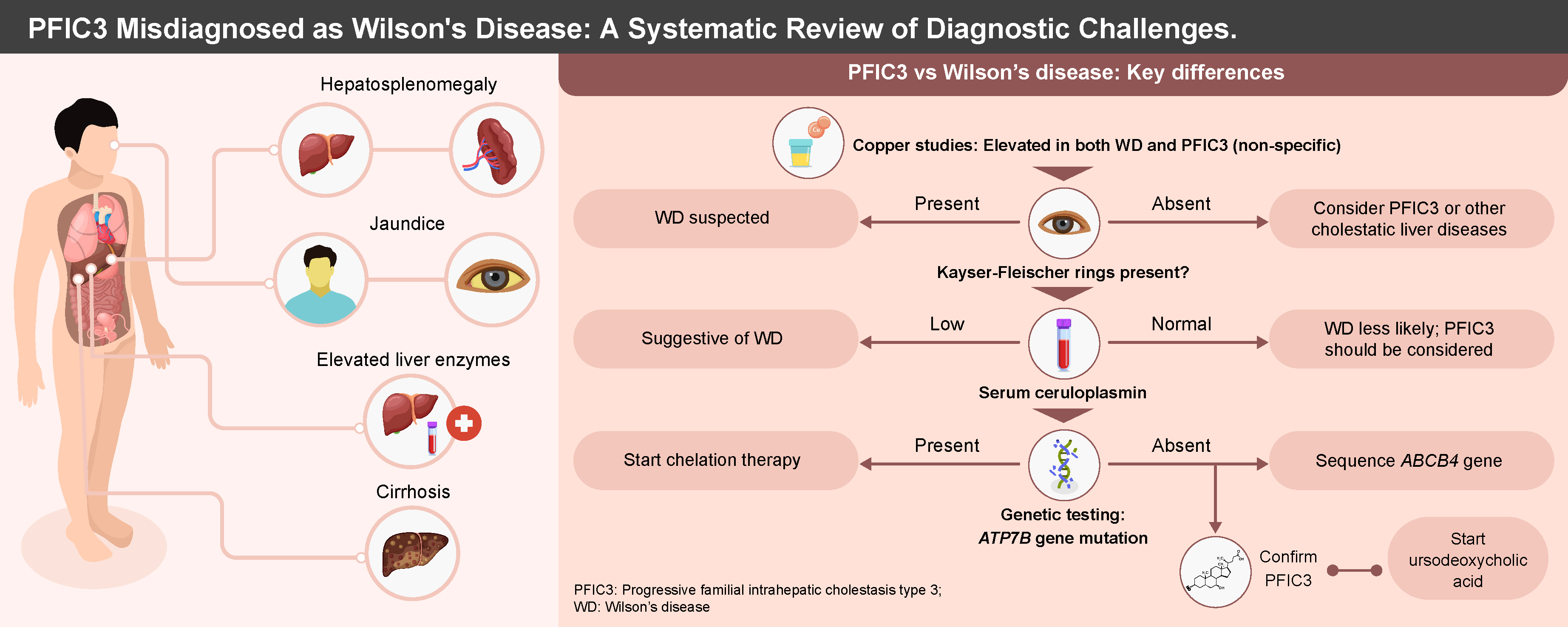
A notable characteristic of PFIC3 is the disruption of copper metabolism secondary to intrahepatic cholestasis, as indicated by elevated hepatic and urinary copper levels. The elevated copper levels in PFIC3 lead to the mimicry of other diseases of impaired copper metabolism, such as Wilson’s disease (WD). Wilson’s disease is a rare autosomal recessive copper metabolism disorder caused by pathogenic mutations in the copper transporter gene ATP7B. According to the Leipzig diagnostic criteria for WD presented in Supplementary Table 2, patients are diagnosed with WD if they have a score ≥2. However, most PFIC3 patients also meet these requirements for WD, leading to some being misdiagnosed with WD.7 This misdiagnosis results in the administration of chelation therapy, to which PFIC3 patients do not respond, ultimately leading to disease progression and deterioration of liver function over time.7 Therefore, we conducted a systematic review to examine current reports of PFIC3 cases mimicking WD, identify diagnostic challenges and assess patient outcomes.

Objectives

The primary objectives of this systematic review are: 1) to examine current reports of PFIC3 cases mimicking WD; 2) to identify diagnostic challenges in differentiating PFIC3 from WD; and 3) to assess patient outcomes in cases of PFIC3 initially misdiagnosed as WD.

Materials and methods

Protocol and registration

The methodology employed in this review conformed to the PRISMA 2020 guidelines (Preferred Reporting Items for Systematic Reviews and Meta-Analyses).8 This study has been registered in PROSPERO with registration No. CRD420251065032.

Literature search

Two independent authors conducted an extensive literature review for pertinent articles up to May 2025. The initial strategy employed a specified search criterion across 3 databases: PubMed, ScienceDirect and Google Scholar. Boolean expressions were utilized to combine keywords for the electronic search (progressive familial intrahepatic cholestasis OR PFIC3 OR ABCB4 disease OR MDR3 deficiency) AND (mimicking OR misdiagnosed). Following the completion of the database search, the reviewers manually examined the references of the identified studies to locate additional relevant studies.

Eligibility criteria

Following the retrieval of studies from the databases, each study was evaluated against a predetermined set of eligibility criteria prior to inclusion in the review. A study was incorporated into the review upon satisfying the following inclusion criteria: studies involving patients with PFIC3 presenting as WD, and studies designed as case reports, case series or observational studies.

The exclusion criteria for the review were applied to omit studies that did not meet the following conditions: 1) studies published in languages other than English; 2) studies that did not include patients with PFIC3; 3) studies that did not involve patients with a misdiagnosis or potential misdiagnosis of PFIC3 as WD; and 4) secondary studies such as narrative reviews, meta-analyses and systematic reviews.

Data extraction

Two reviewers independently performed the data extraction for this review. In instances where consensus was not initially achieved during the data extraction process, the reviewers engaged in discussions to resolve any discrepancies until agreement was reached. In accordance with the PRISMA guidelines, the authors examined all references obtained through various phases prior to data extraction. The initial phases involved screening abstracts to assess the relevance of the articles, leading to the elimination of irrelevant articles. Subsequently, the remaining articles were retrieved and evaluated to determine their eligibility. All studies meeting the eligibility criteria were included in the review, and their data were extracted. The information obtained from each article encompassed the authors, participants’ age and sex, initial presentation, initial laboratory investigations, first diagnosis and management regimen, response to the initial regimen, point of diagnosis change, alteration in management therapy, and patient outcomes.

Quality assessment

We employed the Joanna Briggs Institute (JBI) critical appraisal checklists for case series and case reports to evaluate their methodological quality.9, 10 Subsequently, the results of the methodological quality assessment were presented in tabular form. Although formal metrics for inter-rater reliability were not computed, any differences were addressed through discussion until agreement was reached, adhering to the standards of qualitative synthesis.

Results

Search results

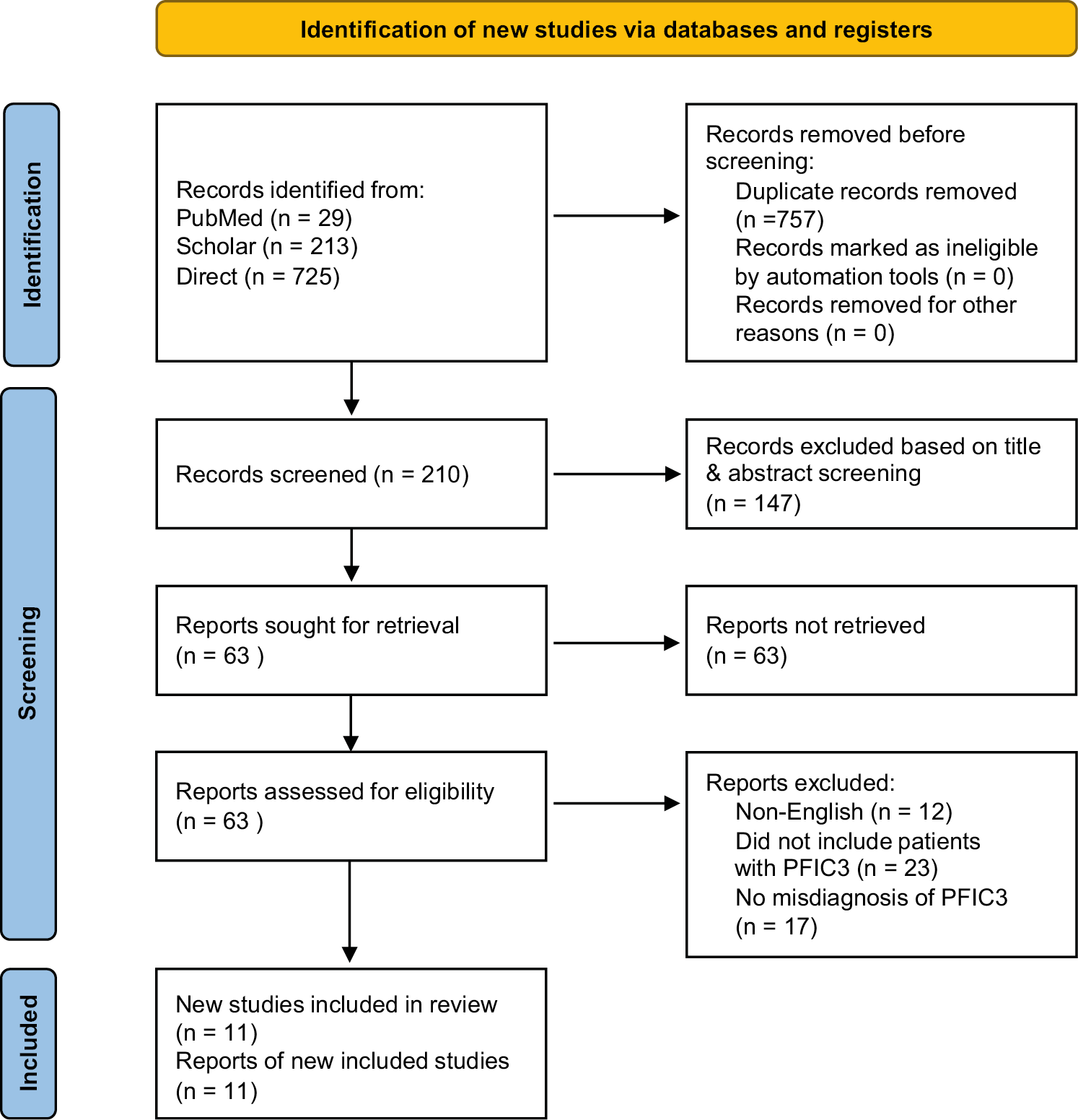
A comprehensive search across the databases yielded 967 references. After identifying and removing 757 duplicates, 210 references remained for relevance assessment. Of these, 147 abstracts were excluded due to irrelevance to the study. Subsequently, 63 articles were retrieved and evaluated against the eligibility criteria for inclusion in the review. Ultimately, 11 studies were included, having met the inclusion criteria. The remaining articles were excluded based on the following criteria: 12 were not published in English, 23 did not involve patients with PFIC3 and 17 did not address a misdiagnosis of PFIC3 as WD. Figure 1 presents the PRISMA diagram, which summarizes the search strategy.

Characteristics of the included studies

This review encompasses 11 studies, of which 9 are case reports11, 12, 13, 14, 15, 16, 17, 18, 19 and 2 are case series.20, 21 These studies present evidence from 16 patients diagnosed with PFIC3, who initially presented as WD and were predominantly diagnosed and treated as such. The cohort consisted of 8 males, 5 females and 1 individual whose sex was not reported. The age of the participants across the studies was normally distributed (Shapiro–Wilk test, W = 0.918, p = 0.206), and is presented as mean ± standard deviation (SD) (19 ±12 years). A summary of the characteristics of all patients is provided in Table 1.11, 12, 13, 14, 15, 16, 17, 18, 19, 20, 21

Methodological quality

Most of the included case reports demonstrated high methodological quality and comprehensively reported all relevant information regarding the treatment and diagnosis of PFIC3 in the included patients. However, a few studies did not adequately document the patients’ medical histories, while others failed to report the interventions administered following the diagnosis of PFIC3. A complete summary of the methodological quality is provided in Supplementary Table 1.

Discussion

This systematic review synthesizes case reports concerning the diagnostic challenges associated with PFIC3. The studies included in this review highlight that both the failure to diagnose PFIC3 and its misdiagnosis as WD result in significant long-term consequences, as chelation therapy proves ineffective, leading to a progressive deterioration of liver function in patients.20, 21 Consequently, it is imperative to identify common diagnostic pitfalls to facilitate the differentiation between these 2 conditions. We also present a summary of the differences identified in the features of PFIC3 and WD, which can aid in differential diagnosis (Supplementary Table 3).

Presentation of PFIC3 patients

The typical age of symptom onset in these patients is within the first 2 decades of life.18 The earliest reported presentation occurs at 2 years of age, with the latest initial presentation occurring during the teenage years.18 Common symptoms among these patients include elevated liver indices, hepatosplenomegaly, ascites, increased liver copper levels, elevated urinary copper excretion, and evidence of cirrhosis upon liver biopsy.16, 17 Unlike in WD, these patients do not exhibit Kayser–Fleischer (KF) rings upon ophthalmic examination.17, 18, 22 Additionally, their ceruloplasmin levels are typically normal.16, 22 The presentation of impaired copper metabolism and accumulation often leads to the consideration of WD as the likely etiology of the observed liver disease in these patients.23 Patients with PFIC3 also do not typically present with neurologic symptoms, unlike WD patients. Pathognomonic signs, especially in the putamen and globus pallidus, are common findings on brain magnetic resonance imaging (MRI) in WD patients.24

A distinguishing feature in the presentation of these patients is the absence of mutations in the ATP7B gene upon complete genome sequencing.18, 21 In most cases, ruling out WD due to the absence of this mutation prompts consideration of PFIC3 as a differential diagnosis, thereby reducing the likelihood of misdiagnosis.16, 20 Furthermore, the lack of ATP7B mutations often leads to further sequencing of the ABCB4 gene, revealing mutations that confirm the diagnosis of PFIC3.20 It is important to note that testing for MDR3 deficiency in biopsy specimens or explant samples may not yield conclusive results.17 For example, Bansal and Rastogi reported negative findings for MDR3 deficiency in tissue samples, only to identify the mutation through whole-genome sequencing.17 Therefore, whole-genome sequencing is recommended in cases of suspected PFIC3. Another distinguishing feature of these 2 conditions is the etiology of copper accumulation. In WD, copper accumulation is primarily due to impairment in copper transport, while in PFIC3, the cause of copper accumulation is cholestasis in most cases. Radiocopper testing, which typically relies on liver metabolism and transport of copper, is therefore a good diagnostic test for differentiating these 2 diseases. However, its use in the WD and PFIC3 diagnostic pitfalls is yet to be demonstrated.25

Management of PFIC3 in the context of misdiagnosis

In all included studies, the initial diagnosis of WD resulted in patients being placed on chelation therapy with either penicillamine, trientine or zinc.16, 19, 20 However, evidence suggests that these patients do not benefit from chelation therapy, and liver function continues to deteriorate during the treatment period.16, 20 In some instances, this deterioration necessitates liver transplantation.11, 12, 17 However, once PFIC3 is diagnosed and management is adjusted to UDCA at an initial dose of 20 mg/kg/day, most patients experience stable liver function, and the liver disease does not progress.19, 20 While liver transplantation remains the definitive treatment for PFIC3, failure to diagnose PFIC3 can still lead to worsening liver function, as reported by Bansal and Rastogi, where a patient’s liver indices became deranged 4 months post-liver transplantation. However, following the management change to UDCA, the patient responded well to treatment.17

Limitations of the study

This systematic review employed a comprehensive search strategy to examine all studies reporting PFIC3 presenting as WD. Nevertheless, the study encountered certain limitations. Primarily, we included only case reports, which suggests that these occurrences are rare. Consequently, while the findings may enhance awareness regarding the potential misdiagnosis of PFIC3, they cannot be generalized to the broader population. Furthermore, the exclusive reliance on case reports resulted in a minimal sample size from which our data were derived. The total sample size of 16 patients across 11 studies is quite small. Additionally, there is limited information provided on how data extraction and quality assessment were conducted – e.g., no formal metrics for inter-rater reliability are reported. The methodological quality assessment using JBI checklists is mentioned but details are lacking on how this was applied. There is also no formal synthesis or meta-analysis of data across studies due to the nature of the included reports. Finally, potential publication bias is not addressed, as case reports of successful diagnoses may be more likely to be published than misdiagnoses that were not eventually corrected.

Conclusions

Evidence synthesized from the included studies indicates that PFIC3 can manifest with symptoms resembling those of WD, potentially leading to misdiagnosis. It is important for clinicians to recognize that patients with PFIC3 do not exhibit KF rings upon slit lamp examination and typically present with normal ceruloplasmin levels. Consequently, whole-genome sequencing (WGS) of the ATP7B and ABCB4 genes should be conducted to distinguish between WD and PFIC3. Furthermore, additional retrospective observational studies are warranted to ascertain the frequency with which PFIC3 is misdiagnosed as WD. This will facilitate the identification of diagnostic challenges and contribute to the development of diagnostic guidelines and protocols. Moreover, prospective studies should be undertaken to evaluate whether dual genetic testing for ATP7B and ABCB4 gene mutations yields beneficial outcomes in the management of patients with either WD or PFIC3.

Supplementary data

The supplementary materials are available at https://doi.org/10.5281/zenodo.17242238. The package contains the following files:

Supplementary Table 1. A JBI checklist summarizing the methodological quality of the included case reports.

Supplementary Table 2. Leipzig criteria for WD diagnosis.

Supplementary Table 3. Differences in the presentation of WD and PFIC3.

Use of AI and AI-assisted technologies

Not applicable.

Tables


Table 1. Characteristics of the included studies

Reference

Sex (age [years])

Presentation, initial diagnostic findings

Initial diagnosis and therapy

Response to initial therapy

Point of change in diagnosis

Change in management

Outcome after final diagnosis

Sticova et al., 202011

male (11)

Growth retardation, marked hepato-splenomegaly, right upper quadrant pain, intermittent pruritus, hepatomegaly, hirsutism, short stature, craniofacial dysmorphia, elevated liver enzymes, increased copper, fibrosis, no ATP7B mutations.

Wilson’s disease was treated with conservative therapy.

Disease progression with further deterioration in laboratory indices necessitated split liver transplantation (LT).

After LT, immune-histological examination of the liver explant revealed the absence of the hepato-canalicular ABCB4/MDR3 expression. PFIC3 was then confirmed through targeted resequencing of ABCB4.

No change since LT had already been done.

Doing well after LT.

Bardak et al., 202512

male (33)

Fatigue, pruritus, jaundice, elevated liver enzymes, bilirubin; liver biopsy shows hepatitis, inflammation, fibrosis, copper accumulation; gallstones detected; increased urinary copper excretion, especially after penicillamine challenge.

WD (penicillamine) was changed to zinc and trientine after 3 years due to tremors.

The liver enzymes, conjugated bilirubin (0.21–2.17 mg/dL), remained elevated. Overall, there was an inadequate response to chelation therapy.

Repeat tests showed similar lab abnormalities; no Kayser–Fleischer rings on slit lamp; MRI revealed atypical basal ganglia changes; no ATP7B mutation; heterozygous ABCB4 mutations confirmed PFIC3.

Awaiting liver transplantation.

Panda et al., 202413

male (12)

Progressive abdominal distention, flank fullness, edema, jaundice; hepatosplenomegaly; stunted growth; elevated liver enzymes, bilirubin; low albumin, pancytopenia; ascites; esophageal varices; no Kayser–Fleischer rings.

WD, which was managed by chelation therapy with D-penicillamine, increased to 20 mg/kg/day.

Liver function worsened, serum albumin decreased, transaminases increased, and an increase in INR.

Genetic analysis of ATP7B mutations came back negative. Further analysis found the ABCB4 gene mutation, confirming the PFIC3 diagnosis.

Chelation therapy stopped.

Ursodeoxycholic acid was started, and supportive management included fat-soluble vitamin supplementation and nutritional support.

Still deteriorating and awaiting liver transplant.

Flanagan et al., 202114

female (6)

Epistaxis, abdominal pain, facial edema, pallor, fever; clubbing, icterus, splenomegaly; severe anemia, thrombocytopenia; elevated liver enzymes, bile acids; low ceruloplasmin; high urinary and hepatic copper; cirrhosis; no KF rings.

WD. Managed by chelation therapy before genetic testing.

Not reported

The cholestatic gene panel indicated the ABCB4 gene variant, resulting in PFIC3 diagnosis.

Chelation therapy stopped. UDCA commenced after trientine was stopped.

Clinically stable, awaiting liver transplant.

Sondhi et al., 202515

female (33)

Fatigue, elevated liver enzymes (2–3 years), painless progressive jaundice; high bilirubin; urinary copper 185 mcg; liver biopsy shows prominent cholestasis.

WD based on a history of jaundice in siblings. WD was managed by chelation therapy with oral D-penicillamine.

No improvement in the symptoms.

After failure of symptom improvement, whole-genome sequencing found the ABCB4 mutation but did not find the ATB7B mutation, suggesting PFIC3 and ruling out WD.

Not reported.

Not reported.

Boga et al., 201516

not reported (15)

Fatigue, esophageal varices, elevated urinary copper (342 µg), normal ceruloplasmin, absent Kayser–Fleischer rings, no ATP7B mutations; liver biopsy shows bridging fibrosis and high hepatic copper (1471 µg/g).

The original diagnosis of WD was managed with zinc, which was later switched to trientine due to the GI side effects of zinc. Nadolol was initiated as a prophylaxis against variceal bleeding.

No improvement after 12 months of trientine therapy. All the liver transaminases were elevated; urine copper excretion was elevated.

Due to poor response and lack of symptoms suggestive of WD (absent KF rings and lack of ATB7B mutation), other differential diagnoses were sought. Sequencing for the ABCB4 gene was performed, and heterozygosity was found in the ABCB4 gene mutations.

Patients were placed on the trial of UDCA, and after PFIC3 confirmatory diagnosis, the therapy was continued.

UDCA therapy reduced transaminases but not to normal; biopsy confirmed cirrhosis from initial presentation; mild persistent liver enzyme elevation; stable synthetic function; esophageal varices treated with banding.

Bansal and Rastogi 201717

male (21)

Weakness, jaundice, ascites, abdominal pain, melena; grade III esophageal varices treated; anemia; high bilirubin; normal renal function; elevated urinary copper (1732.8 μg/24 h); hepatosplenomegaly; no Kayser–Fleischer rings.

Initial diagnosis of acute liver failure with a living donor transplant. WD was given as the etiology of cirrhosis observed on microscopic examination of the liver explant.

The patient was stable after LT, but liver enzymes became deranged after 4 months.

Explant tissues and biopsy slides were tested for the MDR3 gene, all of which came back negative. A gene analysis was not possible, and thus, PFIC3 was diagnosed based on immunohistochemical findings.

Patients were placed on UDCA therapy.

All patients responded well to treatment.

Shneider et al., 201118

female (2)

Abdominal distention, failure to thrive; elevated liver enzymes and copper; normal albumin, mild thrombocytopenia; high serum bile acids; liver biopsy shows bridging fibrosis; MRCP excludes sclerosing cholangitis.

Patient placed on UDCA.

Responded well to UDCA and is doing well with improvement in both liver and growth parameters.

Anheim et al., 201019

male (35)

Bilateral keratoconus surgery; splenomegaly, cholelithiasis; elevated ALT, GGT (10x ULN); high bilirubin; thrombocytopenia; normal ceruloplasmin, serum, and urine copper; liver fibrosis with elevated copper; no ATP7B mutations.

A diagnosis of WD was evoked, and zinc was initiated.

Mild response

The patient presented with an epileptic seizure with delirium. Further evaluation led to a diagnosis of acanthocytosis. Liver function further worsened, and liver transplantation was performed, and a diagnosis of PFIC3 was made and confirmed by mutations found in the ABCB4 genes.

UDCA was commenced alongside the acanthocytosis management regimen.

The patient is responding well to treatment.

Ramraj et al., 201220

female (11)

male (6)

Abdominal distention, hepatosplenomegaly, moderate ascites; liver biopsy shows elevated copper (860 µg/g); increased urinary copper; normal ceruloplasmin.

Scleral icterus, hepatosplenomegaly; elevated AST, ALT, ALP, GGT; conjugated bilirubin 0.6 mg/dL; urine copper 66 µg/d; ceruloplasmin 44.6 mg/dL; liver biopsy shows bridging fibrosis, inflammation, and high copper (863 µg/g).

WD, which was treated with trientine as chelation therapy.

Provisional diagnosis of WD, and the patient started on chelation therapy.

There was initial mild improvement in liver diseases.

Not reported since the patient has not been on chelation therapy for long.

Patient referred for liver transplantation 2 years later.

On evaluation, liver indices were elevated, and copper was also elevated. A liver biopsy showed inflammation and cirrhosis. ATP7B gene mutations were negative, so other etiologies were sought. After full gene sequencing, ABCB4 gene mutations were discovered, and thus, PFIC3 was diagnosed.

Whole gene sequencing found no ATB7B mutations but found an ABCB4 gene mutation, and thus, PFIC3 was given as the final diagnosis.

Chelation therapy with trientine was discontinued, and UDCA was initiated at 20 mg/kg/d.

Chelation therapy was discontinued, and UDCA was commenced at 20 mg/kg/d.

During a follow-up of 4 years, liver indices remained normal.

The liver indices were stable during the follow-up of 18 months.

Nagral et al., 202421

female (23)

6-year-old with jaundice, ascites; urinary copper 162 mcg/day; liver copper 1,692 mcg/g; positive penicillamine challenge; Leipzig score 4.

male (27)

15-year-old with anger, poor school performance; ceruloplasmin 13.9 mg/dl; urinary copper 367.5 mcg/day; liver copper 90.4 mcg/g; Leipzig score 5.

Wilson’s disease was managed with penicillamine.

No improvement was observed.

After reviewing the family history, whole gene sequencing was performed, and ABCB4 mutations were found.

Not reported

Not reported.

male (31)

24-year-old with abnormal LFTs; absent Kayser–Fleischer rings; urinary copper 192 mcg/day; liver biopsy shows elevated copper (166 mcg/g).

Wilson’s disease was managed.

Presented after 7 years with worsening LFTs accompanied by itchiness.

The diagnosis was reviewed, whole-exome sequencing was performed, ABCB4 gene mutations were found, and PFIC3 was diagnosed.

Not reported

Not reported.

ALT – alanine aminotransferase; AST – aspartate aminotransferase; CB – conjugated bilirubin; GGT – gamma-glutamyl transferase; INR – International Normalized Ratio; KF – Kayser–Fleischer; LT – liver transplantation; PFIC3 – progressive familial intrahepatic cholestasis type 3; TB – total bilirubin; UDCA – ursodeoxycholic acid; USG – ultrasonography; MRI – magnetic resonance imaging; ALP – alkaline phosphatase; Hb – hemoglobin; WBC – white blood cell count; GI – gastrointestinal; SAP – serum alkaline phosphatase; LFT – liver function test; MRCP – magnetic resonance cholangiopancreatography.

Figures


Fig. 1. Preferred Reporting Items for Systematic Reviews and Meta-Analyses (PRISMA) flow diagram

References (25)

  1. Srivastava A. Progressive familial intrahepatic cholestasis. J Clin Exp Hepatol. 2014;4(1):25–36. doi:10.1016/j.jceh.2013.10.005
  2. Agarwal S, Lal BB, Rawat D, Rastogi A, Bharathy KGS, Alam S. Progressive familial intrahepatic cholestasis (PFIC) in Indian children: Clinical spectrum and outcome. J Clin Exp Hepatol. 2016;6(3):203–208. doi:10.1016/j.jceh.2016.05.003
  3. Gonzales E, Gardin A, Almes M, et al. Outcomes of 38 patients with PFIC3: Impact of genotype and of response to ursodeoxycholic acid therapy. JHEP Rep. 2023;5(10):100844. doi:10.1016/j.jhepr.2023.100844
  4. Rosmorduc O, Poupon R, Hermelin B. MDR3 gene defect in adults with symptomatic intrahepatic and gallbladder cholesterol cholelithiasis. Gastroenterology. 2001;120(6):1459–1467. doi:10.1053/gast.2001.23947
  5. Jacquemin E, Bernard O, Hadchouel M, et al. The wide spectrum of multidrug resistance 3 deficiency: From neonatal cholestasis to cirrhosis of adulthood. Gastroenterology. 2001;120(6):1448–1458. doi:10.1053/gast.2001.23984
  6. Chen R, Yang FX, Tan YF, et al. Clinical and genetic characterization of pediatric patients with progressive familial intrahepatic cholestasis type 3 (PFIC3): Identification of 14 novel ABCB4 variants and review of the literatures. Orphanet J Rare Dis. 2022;17(1):445. doi:10.1186/s13023-022-02597-y
  7. European Association for the Study of the Liver. EASL Clinical Practice Guidelines: Wilson’s disease. J Hepatol. 2012;56(3):671–685. doi:10.1016/j.jhep.2011.11.007
  8. Page MJ, McKenzie JE, Bossuyt PM, et al. The PRISMA 2020 statement: An updated guideline for reporting systematic reviews. BMJ. 2021;372:n71. doi:10.1136/bmj.n71
  9. Barker TH, Stone JC, Sears K, et al. Revising the JBI quantitative critical appraisal tools to improve their applicability: An overview of methods and the development process. JBI Evid Synth. 2023;21(3):478–493. doi:10.11124/JBIES-22-00125
  10. Gadour E. Lesson learnt from 60 years of liver transplantation: Advancements, challenges, and future directions. World J Transplant. 2025:18;15(1):93253. doi:10.5500/wjt.v15.i1.93253
  11. Sticova E, Neroldova M, Kotalova R, Subhanova I, Jirsa M. ABCB4 disease mimicking morbus Wilson: A potential diagnostic pitfall. Biomed Pap Med Fac Univ Palacky Olomouc Czech Repub. 2020;164(1):121–125. doi:10.5507/bp.2019.054
  12. Bardak AE, Kalaycı T, Çavuş B, Çifçibaşı Örmeci A, Demir K. From chelation to transplantation: Lessons from a progressive familial intrahepatic cholestasis type 3 case initially managed as Wilson’s disease. Gastroenterol Rep (Oxf). 2025;13:goaf017. doi:10.1093/gastro/goaf017
  13. Panda K, Pradhan S, Dash M, Pati GK. Progressive familial intrahepatic cholestasis 3 camouflaging as Wilson disease in a 12-year-old: A diagnostic odyssey. Gastroenterol Hepatol Bed Bench. 2024;17(3):320–324. doi:10.22037/ghfbb.v17i3.2999
  14. Flanagan M, Little R, Siddiqui I, Jones N, Ng V. A215 MDR3 deficiency mimicking Wilson disease. J Can Assoc Gastroenterol. 2021;4(Suppl 1):247–248. doi:10.1093/jcag/gwab002.213
  15. Sondhi P, Singh J, Kaur R, Kathait A, Brar AS. The neurological and hepatic nexus: Analyzing PFIC3 presentation as Wilson disease. Molecular genetic diagnosis and treatment response (P8-5.022). Neurology. 2025;104(7 Suppl 1):3239. doi:10.1212/WNL.0000000000210882
  16. Boga S, Jain D, Schilsky ML. Presentation of progressive familial intrahepatic cholestasis type 3 mimicking Wilson disease: Molecular genetic diagnosis and response to treatment. Pediatr Gastroenterol Hepatol Nutr. 2015;18(3):202. doi:10.5223/pghn.2015.18.3.202
  17. Bansal N, Rastogi M. An itchy experience: PFIC 3 masquerading as Wilson’s disease. Learning from mistakes. Oncol Gastroenterol Hepatol Rep. 2017;6(2):67–71. https://pdfs.semanticscholar.org/dab0/307f092b889864a2ab1bb71f8440a789abc7.pdf.
  18. Shneider BL. ABCB4 disease presenting with cirrhosis and copper overload-potential confusion with Wilson disease. J Clin Exp Hepatol. 2011;1(2):115–117. doi:10.1016/S0973-6883(11)60131-X
  19. Anheim M, Chamouard P, Rudolf G, et al. Unexpected combination of inherited chorea-acanthocytosis with MDR3 (ABCB4) defect mimicking Wilson’s disease. Clin Genet. 2010;78(3):294–295. doi:10.1111/j.1399-0004.2010.01386.x
  20. Ramraj R, Finegold MJ, Karpen SJ. Progressive familial intrahepatic cholestasis type 3: Overlapping presentation with Wilson disease. Clin Pediatr (Phila). 2012;51(7):689–691. doi:10.1177/0009922812451076
  21. Nagral N, Poyekar S, Shah R, Wadia P, Nagral A. PFIC-3 mimicking Wilson disease: A case series. J Clin Exp Hepatol. 2024;14:102033. doi:10.1016/j.jceh.2024.102033
  22. Schilsky ML, Roberts EA, Bronstein JM, et al. A multidisciplinary approach to the diagnosis and management of Wilson disease: Executive summary of the 2022 Practice Guidance on Wilson disease from the American Association for the Study of Liver Diseases. Hepatology. 2023;77(4):1428–1455. doi:10.1002/hep.32805
  23. Wungjiranirun M, Sharzehi K. Wilson’s disease. Semin Neurol. 2023;43(4):626–633. doi:10.1055/s-0043-1771465
  24. Singh P, Ahluwalia A, Saggar K, Grewal C. Wilson’s disease: MRI features. J Pediatr Neurosci. 2011;6(1):27. doi:10.4103/1817-1745.84402
  25. Członkowska A, Rodo M, Wierzchowska-Ciok A, Smolinski L, Litwin T. Accuracy of the radioactive copper incorporation test in the diagnosis of Wilson disease. Liver Int. 2018;38(10):1860–1866. doi:10.1111/liv.13715