


Abstract
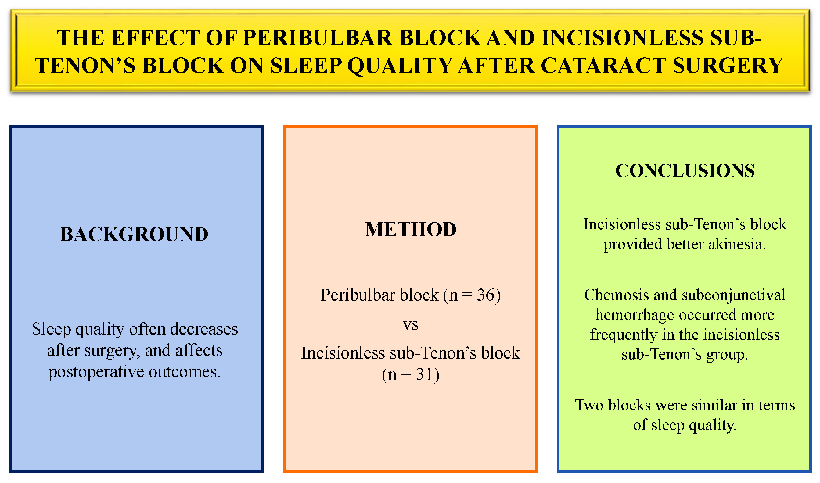
Background. Sleep quality often decreases in patients after surgery and may affect postoperative outcomes.
Objectives. The aim of this study was to compare the effects of peribulbar block and incisionless sub-Tenon’s block on postoperative sleep quality.
Materials and methods. Patients who underwent cataract surgery under peribulbar block (n = 36) or incisionless sub-Tenon’s block (n = 31) were included in the study. The 2 blocks were compared with each other in terms of eyelid and globe movements, corneal sensation, complications, pain, and postoperative sleep quality (evaluated using the Richard–Campbell Sleep Questionnaire (RCSQ) score).
Results. Sixty-seven patients with a mean age of 67 years were included in the study. At the 15th minute after the block (p = 0.066) and at the end of surgery (p = 0.098), akinesia was better in the incisionless sub-Tenon’s group, with p-values close to the level of statistical significance. Chemosis was found to be significantly lower in the peribulbar block group than in the incisionless sub-Tenon’s block group (p = 0.033) 3 h after surgery. All minor complications, including chemosis, subconjunctival petechiae, and subconjunctival hemorrhage, were observed less frequently in the peribulbar block group, although the differences were not statistically significant (p > 0.05). Pain scores were comparable between the groups (p > 0.05). The total RCSQ score (p = 0.396) and overall sleep perception (p = 0.355) were also comparable between the groups.
Conclusions. The incisionless sub-Tenon’s block was superior to the peribulbar block in terms of providing akinesia and reducing the need for maintenance anesthetic medications such as sedatives or analgesics. Although chemosis and subconjunctival hemorrhage occurred more frequently in the incisionless sub-Tenon’s group, all events were transient and had no serious clinical significance. The 2 regional techniques were similar in terms of postoperative sleep quality and patient satisfaction.
Key words: cataract surgery, peribulbar block, incisionless sub-Tenon’s block, postoperative sleep quality
Background
Cataract surgery is among the most commonly performed operations worldwide. It is generally an outpatient procedure, and early return to routine life is an important goal of this surgery.1 Sleep quality often decreases in patients after surgery, which may affect postoperative outcomes, including conditions such as postoperative delirium, long-term chronic pain, and potentially serious cardiovascular events.2 In this regard, postoperative sleep quality is highly important. Improving sleep quality accelerates the recovery process and shortens the time required to return to daily life.
Sleep quality after cataract surgery can be affected by several factors, such as sex, anxiety, comorbidities, pain, anesthetic drugs, and perioperative complications. Postoperative pain is one of the leading factors affecting sleep quality and is directly associated with anesthesia.2, 3 Cataract surgery can be performed under local, regional, or general anesthesia. In recent years, regional block techniques, such as peribulbar and sub-Tenon’s blocks, have gained popularity because they provide better analgesia and eyeball immobilization than topical anesthesia. In addition, cataract surgery is more frequently performed in older patients with a higher anesthesia risk due to the presence of comorbidities. Therefore, preventing potential problems associated with general anesthesia has made regional block techniques more preferable.1, 2, 3, 4
The peribulbar block is one of the most commonly used anesthesia methods for ocular procedures because of its low complication rate and ease of application. In contrast, the sub-Tenon’s block has recently been increasingly preferred because it provides better akinesia and analgesia and causes less damage to the vasculature and optic nerve.5, 6, 7 Many studies in the literature have compared peribulbar and sub-Tenon’s blocks, most of which focused on intraoperative eye movements, postoperative analgesia, and complications.7, 8 However, there are no studies comparing these techniques in terms of postoperative sleep quality.
Objectives
In this study, 2 regional anesthesia techniques, peribulbar block and incisionless sub-Tenon’s block, were compared in terms of postoperative sleep quality in patients undergoing cataract surgery.
Materials and methods
Ethics approval
After obtaining approval from the Clinical Research Ethics Committee (approval No. 25.07.2023/6), the study was conducted at the Department of Anesthesiology and Reanimation at Eskisehir Osmangazi University (Turkey) between August 2023 and March 2024, in accordance with Good Clinical Practice guidelines and the principles of the Declaration of Helsinki.
Patient selection
Sixty-seven adult patients who underwent cataract surgery using one of 2 regional anesthesia techniques – peribulbar block (n = 36) or incisionless sub-Tenon’s block (n = 31) – were included in the study. All patients were diagnosed with elective cataract by the same surgical team. Inclusion criteria included age ≥18 years, an American Society of Anesthesiologists (ASA) physical status of 1–3, and elective surgery. Patients with intellectual disability, dementia, or psychiatric disorders; those receiving medical therapy affecting the central nervous system, such as benzodiazepines or antipsychotics; and those who developed an allergic reaction to local anesthetics were excluded from the study. All patients were informed in detail about the stages of the study, and written informed consent was obtained from each participant. Patient age, sex, ASA physical status, body mass index (BMI), surgical procedure, and type of regional block were recorded.
Anesthetic management
The patients were taken to the regional block room and monitored using electrocardiography (ECG), pulse oximetry, and noninvasive blood pressure measurement. Intravenous (IV) access was established using a 20-G cannula placed in a hand vein, and a saline infusion was initiated to maintain vascular access. Premedication included dexmedetomidine (5–10 µg), midazolam (0.5–2 mg), and fentanyl (25–50 µg). Subsequently, proparacaine (1 drop) and 1% povidone–iodine (1 drop) were instilled into the eye. All regional blocks were performed by the 1st author (Y.K.). Thereafter, the patients were transferred to the operating room. Before surgery, IV paracetamol (Partemol 1 g/100 mL; VEM, Ankara, Turkey) was administered to all patients. During the intraoperative period, additional sedoanalgesic medications, including dexmedetomidine (Dekstomid 200 µg/2 mL; Polifarma, Tekirdag, Turkey), fentanyl (Fentanyl-PF 100 µg/2 mL; Polifarma) or midazolam (Zolamid 5 mg/5 mL; VEM), were administered as needed. Additional analgesics were given when the patient reported pain (visual analogue scale (VAS) ≥4) or restlessness (Ramsay sedation score = 1).
Hemodynamic parameters, including heart rate (HR), mean arterial pressure (MAP), and peripheral oxygen saturation (SpO2), were recorded at 5 time points: before sedation (baseline), 15 min after the regional block, at the end of surgery, 3 h after surgery, and 1 day after surgery.
Block techniques
In the peribulbar block, after the skin was cleaned with alcohol, the technique was performed by inserting a 25-gauge needle through the skin (25 mm for patients with an axial eyeball length of 20–26 mm and for those with an axial length <20 mm) at the junction of the medial 2/3 and lateral 1/3 of the orbital rim. If contact with bone was encountered, the needle was redirected slightly upward. The tip of the needle was maintained in the extraconal peribulbar space. With the patient’s eye in a neutral position, a local anesthetic mixture of 0.5% bupivacaine and 2% lidocaine was administered into the extraconal area, with a volume of 4–7 mL adjusted according to the axial length of the eyeball, through the main inferotemporal quadrant.
In the incisionless sub-Tenon’s block, a proparacaine eye drop was instilled into the conjunctiva, followed by a drop of aqueous povidone–iodine into the conjunctival sac. A wired eyelid speculum was inserted to separate the eyelids. Thereafter, the conjunctiva and Tenon’s capsule were grasped and punctured with forceps 5–8 mm away from the limbus in the inferonasal quadrant, while the patient was looking upward and outward. A disposable plastic cannula (22-gauge) was then advanced along the curvature of the eyeball. After negative aspiration, a local anesthetic mixture of 0.5% bupivacaine and 2% lidocaine, at a volume of 4–7 mL, was administered into the sub-Tenon’s space through the cannula.
Outcome measures
The motor effects of the regional blocks included eyelid and globe movements, whereas the sensory effects included corneal sensitivity. Eyelid and globe movements were recorded in 4 primary gaze positions (superonasal, superotemporal, inferonasal, and inferotemporal) at 5 time points (before sedation, 15 min after the regional block, at the end of surgery, 3 h after surgery, and on the first postoperative day) using a 3-point scale (1 – movement, 2 – mild/moderate movement, 3 – akinesia). Corneal sensitivity was assessed based on patients’ reported sensation in response to 1% povidone–iodine eye drops at the same time points, using a 3-point scale (0 – no burning, 1 – mild sensation, 2 – strong sensation).
Minor complications included chemosis, subconjunctival petechiae, and hemorrhage and were recorded at 5 time points (before sedation, 15 min after the regional block, at the end of surgery, 3 h after surgery, and 1 day after surgery). These complications were assessed in 4 quadrants of the eye (superonasal, superotemporal, inferonasal, and inferotemporal) using a 2-point scale ranging from 0 (absent) to 1 (present in any quadrant).
Pain was evaluated using a VAS score ranging from 0 (no pain) to 10 (the worst imaginable pain) before sedation, 3 h after the operation, and on the 1st postoperative day. Sleep quality was evaluated using the validated version of the Richard–Campbell Sleep Questionnaire (RCSQ) on the first postoperative day.9 This questionnaire consists of 6 items that assess depth of sleep, time to fall asleep, frequency of awakenings, time spent awake after awakening, sleep quality, and environmental noise level. Each item is scored on a scale from 0 to 100. The average of the first 5 items was calculated as the total score and represented overall sleep perception.
Overall sleep perception was classified into 4 categories: 0–25 points – very poor sleep perception; 26–50 points – poor sleep perception; 51–75 points – moderate/good sleep perception; and 76–100 points – very good sleep perception. The 6th item evaluates the noise level of the sleeping environment and is not included in the total score. The score obtained from the 6th item was analyzed to determine whether the sleeping environments of the 2 groups were comparable.
Statistical analyses
Because the study population was not fully known (as there are no similar studies in the literature comparing the effects of peribulbar and incisionless sub-Tenon’s blocks on postoperative sleep quality), the power and sample size of the present study were calculated based on the study by Duran and Öztürk.10 A total of 48 patients were required at a power level of 80%. The effect size of the study was 0.35 (medium). Statistical analyses were performed using IBM SPSS Statistics for Windows v. 20.0 (IBM Corp., Armonk, USA). Descriptive statistics are presented as mean and standard deviation (SD) for continuous variables and as frequency and percentage for categorical variables. Data distribution was evaluated using the Shapiro–Wilk test. The Mann–Whitney U test was used to analyze differences in nonparametric continuous variables between groups, and the χ2 test of independence was used for categorical variables (Table 1). Changes in HR, MAP, and SpO2 (Table 2), as well as changes in eyelid movement, globe movement (akinesia), and corneal sensation (Table 3) over time in the peribulbar block and incisionless sub-Tenon’s block groups, were evaluated separately using two-way analysis of variance (ANOVA) with repeated measures. Post hoc analyses were performed using Bonferroni correction for both within-group and between-group comparisons (Table 4, Table 5, Table 6, Table 7). The results of checking the assumptions for repeated-measures ANOVA (eyelid movement, globe movement, corneal sensation, chemosis, and subconjunctival hemorrhage) are presented as supplementary material (Supplementary Table 1). The subconjunctival petechiae variable could not be analyzed because of the low number of observed cases. The results of assumption testing are presented as supplementary material (Supplementary Table 2). For the analysis of repeated binary outcomes (presence of chemosis or hemorrhage at 4 time points), a generalized linear mixed model was used. The model included time, block type, and their interaction as fixed effects, and patient ID (PROTOCOL) as a random effect.
The model was specified as:
model < lmer(move_score ~ time × block +
(1 | PROTOCOL), data = long_df).
P-values were calculated using the Satterthwaite approximation for degrees of freedom. A p-value of less than 5% was considered statistically significant.
Results
Sixty-seven adult patients were included in the study (mean age: 67 years; range: 25–90 years). There were 36 men (53.7%) and 31 women (46.3%). Patients were classified into 2 groups according to the anesthesia technique used: peribulbar block (n = 36) and incisionless sub-Tenon’s block (n = 31). The 2 groups had similar baseline patient characteristics (Table 1).
Heart rate, MAP, and SpO2 values measured at all time points (before sedation, 15 min after the block, at the end of surgery, 3 h after surgery, and on the 1st postoperative day) were similar between the peribulbar block group and the incisionless sub-Tenon’s block group (Table 2).
Before sedation, eyelid and globe movements were completely normal in all patients. In addition, no corneal sensation was observed in either group. The incisionless sub-Tenon’s block provided better akinesia than the peribulbar block, although the difference was not statistically significant (p > 0.05) (Table 3). Post hoc within-group analyses were performed using the Bonferroni correction (Table 4, Table 5).
Before sedation, no patient had chemosis, subconjunctival petechiae, or hemorrhage. Chemosis was significantly lower in the peribulbar block group than in the incisionless sub-Tenon’s block group (p = 0.033) at 3 h after surgery. Although minor complications were less frequent in the peribulbar block group, the differences were not statistically significant (p > 0.05). (Table 8). Post hoc within-group analyses were performed using the Bonferroni correction (Table 6, Table 7).
In both groups, none of the patients reported pain before sedation. Visual analogue scale scores measured at 3 h after the block and on the 1st postoperative day were comparable between the groups (p > 0.05). The percentages of patients in the peribulbar block group and the incisionless sub-Tenon’s block group were also comparable with regard to the 6th item of the RCSQ (79.2 vs 75.48; p = 0.843). The mean total RCSQ score was 381.2 ±56.9 (range: 280–490) in the peribulbar block group, whereas patients in the incisionless sub-Tenon’s block group had a mean total RCSQ score of 365.8 ±67.7 (range: 115–500) (p = 0.396). Overall sleep perception was comparable between the groups (p = 0.355) (Figure 1). Twenty patients (55.5%) in the peribulbar block group and 15 patients (48.3%) in the incisionless sub-Tenon’s block group reported “very good sleep perception.” Finally, patient satisfaction was assessed on the 1st postoperative day. Satisfaction levels were comparable between the 2 groups (p = 0.417) (Table 9).
Discussion
In this study, 2 increasingly used regional anesthesia techniques in cataract surgery – peribulbar block and incisionless sub-Tenon’s block – were compared in several aspects, including their ability to provide essential surgical conditions such as eyelid immobilization and akinesia, their associated perioperative complications, and their potential effects on postoperative sleep quality.
Akinesia and eyelid immobility are essential for the safety and success of cataract surgery. In the present study, compared with the peribulbar block, the incisionless sub-Tenon’s block was found to be superior in providing akinesia, with p-values close to the level of statistical significance. There are conflicting results in the literature regarding this issue. In a study by Antony et al., the proportion of patients with complete akinesia was found to be significantly greater in the peribulbar block group than in the sub-Tenon’s block group. However, the authors reported that the onset time of akinesia was significantly longer in the peribulbar block group than in the sub-Tenon’s block group.7 We did not measure the onset time of akinesia in our study; however, our findings were consistent with the results of that study. In another study, patients in the sub-Tenon’s block group had better akinesia than those in the peribulbar block group.11 On the other hand, Iganga et al. reported that the 2 regional blocks provided comparable levels of adequate akinesia during cataract surgery.12 We frequently perform these 2 regional blocks for cataract surgery and many other ocular procedures in our clinic. Based on both the results of our study and our clinical experience, we believe that the sub-Tenon’s block provides better akinesia than the peribulbar block.
There is a clear interaction between postoperative pain status and sleep quality.13 The relationship between pain and sleep disorders, such as short sleep duration or insomnia, has been well established. However, this relationship is bidirectional; while pain disrupts sleep quality, sleep disorders can also exacerbate pain.3, 14 In our study, postoperative pain scores were similar between the peribulbar block and incisionless sub-Tenon’s block groups, consistent with previous studies.12, 15, 16
No major complications were observed in the present study. Therefore, minor complications, including chemosis, subconjunctival petechiae, and hemorrhage, were evaluated. According to the results, chemosis and subconjunctival hemorrhage were observed more frequently in the sub-Tenon’s block group than in the peribulbar block group. Similar findings have been reported in the majority of previous studies.12, 15, 16 In one of the first studies on sub-Tenon’s blocks, it was reported that such minor complications were common with this technique but did not cause intraoperative complications.17 Based on our clinical experience, we agree with this observation, as all of these complications resolved within a few days after surgery and no serious clinical problems developed in any patient.
The primary aim of the present study was to compare the effects of a peribulbar block and an incisionless sub-Tenon’s block on postoperative sleep quality. Postoperative sleep disorders are important factors that can negatively affect the recovery process and increase patient morbidity and mortality. Approximately half of patients experience sleep problems during the first days after surgery. The anesthesia technique has also been suggested to be one of the factors affecting postoperative sleep quality.18, 19 Anesthetic drugs can reduce sleep quality by disrupting the normal sleep–wake cycle or affecting the natural rhythm of melatonin, one of the key neurotransmitters involved in the sleep process.14, 20, 21, 22 Studies have shown that general anesthesia impairs sleep patterns, and that regional anesthesia is advantageous over general anesthesia with regard to the development of postoperative sleep disorders.21, 23 Despite its importance for surgical success and postoperative recovery, the effect of anesthesia methods on sleep quality has not received sufficient attention. Based on this, the present study investigated the effects of the 2 most commonly used regional block techniques on postoperative sleep quality in patients undergoing cataract surgery, one of the most frequently performed procedures in routine clinical practice.
Statistically, there was no difference in sleep quality between the 2 regional techniques. Sleep is a physiological process that can be influenced by many individual and environmental factors.20 Therefore, in studies evaluating sleep quality, creating homogeneous patient groups and ensuring environmental conditions that are as similar as possible allow for more reliable statistical outcomes.14 In our study, the 2 anesthesia groups were similar in terms of baseline patient and surgical characteristics. Noise levels in the sleeping environments were also comparable between the groups.
It should be noted that the need for maintenance anesthetic medications (sedatives and/or analgesics) was greater in the peribulbar block group than in the incisionless sub-Tenon’s block group. Considering the effect of postoperative pain levels on sleep quality, the lower requirement for analgesic medication in the sub-Tenon’s block group may make this anesthesia technique more favorable, even though statistically both regional blocks had similar effects on postoperative sleep quality. In the present study, all patients were premedicated with dexmedetomidine in addition to midazolam and fentanyl. We routinely use dexmedetomidine in ophthalmic surgeries because of its effectiveness in providing sedoanalgesia and reducing intraocular pressure. Palte et al. also demonstrated in their studies that early administration of a dexmedetomidine bolus (8–20 µg) before midazolam (1–2 mg) and/or low-dose fentanyl (0.5 µg/kg) created ideal conditions for the administration of peribulbar, retrobulbar, or sub-Tenon’s blocks. They reported that the use of dexmedetomidine reduced the need for propofol and provided prolonged sedation.24
Limitations of the study
Several limitations of this study should be acknowledged. First, it was conducted at a single center, which may limit the generalizability of the results. Second, sleep quality was assessed subjectively rather than objectively, using methods such as polysomnography. Finally, the relatively small sample size may be considered another limitation of this work. However, owing to its prospective design and the fact that it is the first study to compare 2 regional block techniques in terms of postoperative sleep quality, this study remains scientifically valuable.
Conclusions
Incisionless sub-Tenon’s block was superior to the peribulbar block in terms of providing akinesia and reducing the need for maintenance anesthetic medications such as sedatives or analgesics. Although minor complications, including chemosis and subconjunctival hemorrhage, occurred more frequently in the incisionless sub-Tenon’s group, all were transient and had no serious clinical significance. The 2 regional techniques were similar with regard to postoperative sleep quality and patient satisfaction.
Supplementary data
The supplementary materials are available at https://doi.org/10.5281/zenodo.13826194. The package contains the following files:
Supplementary Table 1. Test statistics 1.
Supplementary Table 2. Test statistics 2.
Supplementary Table 3. Test statistics 3.
Supplementary Table 4. Test statistics 4.
Supplementary Table 5. Test statistics 5.
Supplementary Table 6. Test statistics 6.
Supplementary Table 7. The results of checking the ANOVA assumptions for repeated measures.
Supplementary Table 8. Test assumption checking.
Data Availability Statement
The datasets supporting the findings of the current study are openly available in Zenodo repository at https://doi.org/10.5281/zenodo.16358814.
Consent for publication
Not applicable.
Use of AI and AI-assisted technologies
Not applicable.
.jpg)