Advances in Clinical and Experimental Medicine

Title abbreviation: Adv Clin Exp Med
5-Year IF – 2.0, IF – 1.9, JCI (2024) – 0.43
Scopus CiteScore – 4.3
Q1 in SJR 2024, SJR score – 0.598, H-index: 49 (SJR)
ICV – 161.00; MNiSW – 70 pts
Initial editorial assessment and first decision within 24 h

ISSN 1899–5276 (print), ISSN 2451-2680 (online)
Periodicity – monthly

Download original text (EN)

Advances in Clinical and Experimental Medicine

Ahead of print

doi: 10.17219/acem/204078

Publication type: original article

Language: English

License: Creative Commons Attribution 3.0 Unported (CC BY 3.0)

Download citation:

  • BIBTEX (JabRef, Mendeley)
  • RIS (Papers, Reference Manager, RefWorks, Zotero)

Cite as:


Paczwa K, Szeretucha M, Romanowska-Próchnicka K, et al. The correlation between nailfold capillaroscopic findings and adaptive optics imaging of retinal microvasculature in patients with systemic sclerosis [published online as ahead of print on September 17, 2025]. Adv Clin Exp Med. 2026. doi:10.17219/acem/204078

The correlation between nailfold capillaroscopic findings and adaptive optics imaging of retinal microvasculature in patients with systemic sclerosis

Katarzyna Paczwa1,A,B,C,D, Magdalena Szeretucha1,B,C,D, Katarzyna Romanowska-Próchnicka2,B,C, Sylwia Ornowska3,B, Marzena Olesińska3,E, Radosław Różycki1,E, Joanna Gołębiewska1,E,F

1 Department of Ophthalmology, Military Institute of Aviation Medicine, Warsaw, Poland

2 Department of Biopysics, Physiology and Pathophysiology, Medical University of Warsaw, Poland

3 Department and Polyclinic of Systemic Connective Tissue Diseases, National Institute of Geriatrics, Rheumatology and Rehabilitation, Warsaw, Poland

Graphical abstract


Graphical abstracts

Highlights


• Adaptive optics unveils retinal microvascular changes in systemic sclerosis (SSc): First study to apply high-resolution AO imaging in scleroderma patients.
• Concordance between nailfold capillaroscopy and adaptive optics metrics: Combined modalities enhance accuracy in detecting SSc microangiopathy.
• Thinner retinal vessel walls in SSc vs controls: Quantitative AO analysis shows significant retinal wall thinning.
• Adaptive optics as a novel diagnostic tool for SSc microvascular injury: Promises noninvasive, early detection and monitoring of microvascular changes in scleroderma.

Abstract

Background. Vascular injury is a central and early feature of systemic sclerosis (SSc) pathogenesis. Although nailfold capillaroscopy (NC) effectively visualizes characteristic peripheral arteriolar and capillary changes, the retinal microcirculation provides a noninvasive, high-resolution view into subtler vascular dysfunction. Consequently, retinal vascular imaging may offer an ideal modality for monitoring microvascular injury and detecting early manifestations of SSc.

Objectives. To compare retinal microvascular parameters between SSc patients and healthy controls using adaptive optics (AO) imaging, and to evaluate the correlation between adaptive optics-derived retinal measurements and NC findings in SSc.

Materials and methods. The study included 31 patients with SSc and 41 healthy controls. The AO images of the retinal arteries were obtained in both groups and the measurements were compared. Nailfold capillaroscopy was also performed in the SSc cohort, and its findings were directly compared with the AO imaging results.

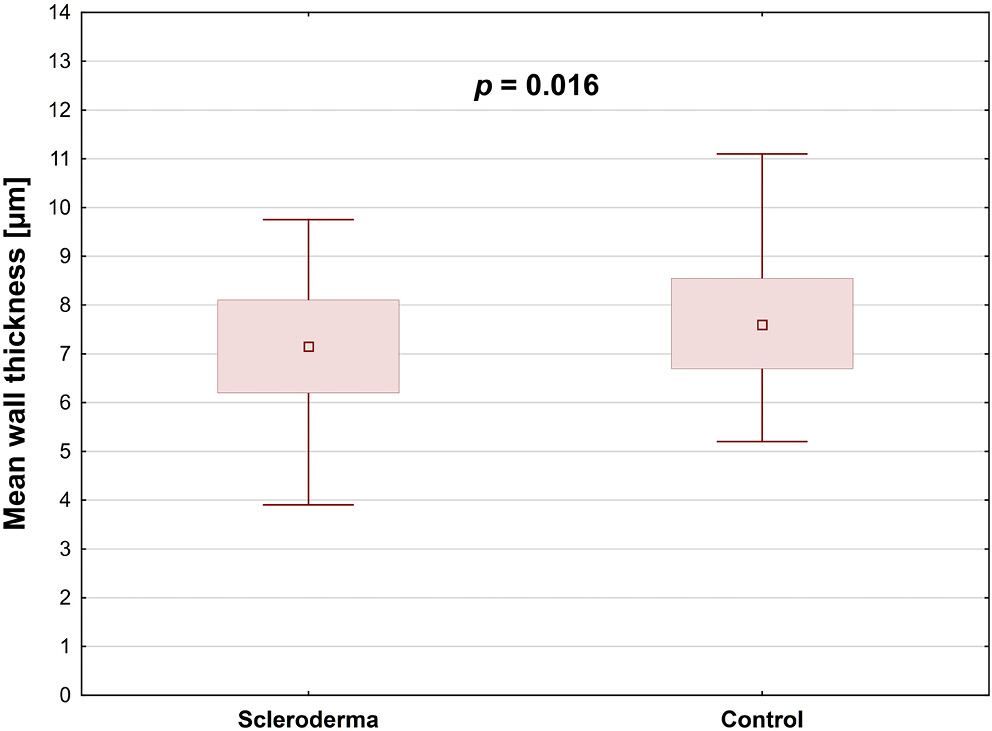
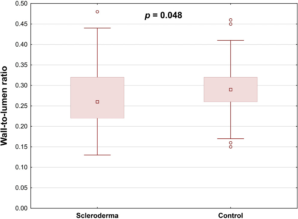
Results. Retinal arterial wall thickness was significantly lower in SSc patients than in healthy controls (p = 0.016), and the wall-to-lumen ratio was similarly reduced in the SSc group (p = 0.048). Within the SSc cohort, hypertensive patients exhibited a significantly greater wall cross-sectional area compared to those without hypertension (p = 0.026).

Conclusions. Adaptive optics retinal imaging demonstrated a significant reduction in mean arterial wall thickness in SSc patients compared with healthy controls. However, no correlation was identified between the AO findings and the NC parameters or the disease stage. Our analysis revealed that alterations in retinal vascular parameters were confined to SSc patients with comorbid hypertension or those receiving sildenafil therapy. To fully establish the clinical utility of adaptive optics imaging in SSc, and to elucidate its relationship with NC findings, larger, multicenter studies with more diverse patient cohorts are warranted.

Key words: systemic sclerosis, scleroderma, adaptive optics, nailfold capillaroscopy, retinal microvasculature

Background

Systemic sclerosis (SSc) is a connective tissue disorder caused by autoimmunity. It is characterized by obliterative vasculopathy, widespread fibrosis and a dysregulated immune response.1, 2 Vascular involvement represents an initial and pivotal factor in the pathogenesis of the disease, as well as in the development of multiple organ dysfunction. The vasculopathy in SSc includes thrombosis, vasospasm and vessel lumen obliteration leading to microangiopathy.2 Microcirculatory changes observed in the capillaries are the hallmark of the disease.

Nailfold capillaroscopy (NC), as a noninvasive imaging technique, is routinely used to assess peripheral capillary abnormalities in scleroderma patients and is included in the diagnostic criteria for SSc.3, 4 Therefore, this tool plays a pivotal role in both diagnosing and monitoring the progression of the disease.

A range of ocular manifestations have been observed in patients with scleroderma, including those affecting the posterior segment of the eye.5, 6, 7, 8, 9, 10 Recent research has concentrated on retinal and choroidal alterations observed in these individuals. Advanced imaging methods, such as optical coherence tomography angiography (OCTA), have facilitated the detection of structural changes in the retinal microvasculature linked to systemic diseases.

Adaptive optics (AO) imaging represents a noninvasive tool for the visualization of retinal structures, including photoreceptors, blood vessels and nerve fibers, in vivo at the microscopic level. The camera employs infrared illumination with a wavelength of 850 nm.11 The AO technology is employed for the evaluation of retinal vessels in healthy eyes and arterial hypertension, as well as in ocular pathologies such as diabetic retinopathy, glaucoma, retinal vessel occlusion and retinal vasculitis.12, 13, 14, 15, 16, 17, 18, 19, 20, 21, 22

Objectives

The study aimed to assess and compare the retinal microvasculature parameters in scleroderma patients and healthy controls using AO imaging. Furthermore, we aimed to evaluate the relationship between NC findings, disease characteristics and AO results within the SSc group.

Materials and methods

The study was conducted at the Military Institute of Aviation Medicine (Warsaw, Poland) between March 2022 and May 2023. The study received approval from the Ethics Committee Reviewing Biometric Research at the Military Institute of Aviation Medicine (approval No. 1/2022) and followed the tenets of the Declaration of Helsinki.

The purpose and procedures of the study were thoroughly explained to the participants, and written consent was subsequently obtained from individuals. The study sample comprised patients with SSc from the Department and Polyclinic of Systemic Connective Tissue Diseases at the National Institute of Geriatrics, Rheumatology and Rehabilitation in Warsaw. Healthy participants were enrolled during routine appointments at the Department of Ophthalmology in the Military Institute of Aviation Medicine.

Participants

A total of 31 patients (61 eyes) diagnosed with SSc and 41 healthy controls (81 eyes) were included in this and our previous study.23 No significant differences were observed between the scleroderma patients and the control group regarding age, gender distribution or axial length (Table 1, Table 2). Within the study cohort, 71.4% of patients were diagnosed with diffuse cutaneous SSc, while 28.6% were diagnosed with limited cutaneous SSc. Nailfold capillaroscopy revealed an early scleroderma pattern in 5 patients, an active pattern in 12 and a late pattern in 9. Within the SSc cohort, 2 patients had pulmonary arterial hypertension (PAH), 8 developed digital ulcers and 8 were hypertensive. A total of 13 participants were treated with sildenafil, while 12 were treated with amlodipine. Interstitial lung disease was identified in 11 patients, of whom 64% were classified as having nonspecific interstitial pneumonia (NSIP) and 36% as having usual interstitial pneumonia (UIP). Elevated pro-B-type natriuretic peptide (proBNP) levels were observed in 16.1% of the studied cases (Table 3).

Methods

A comprehensive ophthalmic examination was performed on all participants. This included assessments of best-corrected visual acuity (BCVA), intraocular pressure, refraction, anterior segment examination, fundoscopy with pupillary dilatation, and axial length (AL) measurement. Exclusion criteria were refractive errors beyond −3.0 or +3.0 diopters and any pathological ocular condition, including glaucoma or retinal and choroidal diseases. Moreover, eyes with low-quality AO images were also excluded. The BCVA was measured monocularly using LogMAR charts (Lighthouse International, New York, USA) at 5 m, while AL was assessed using the IOL Master 500 (Carl Zeiss Meditec AG, Jena, Germany).

All participants with scleroderma underwent a clinical evaluation and NC. The clinical data collected from each SSc patient comprised the following: The type of SSc (either limited or diffuse cutaneous scleroderma), the presence of interstitial lung disease (classified as either NSIP or UIP), hypertension, elevated proBNP, and the treatment method (administrations of sildenafil or amlodipine). The NC was conducted using the CapillaryScope 200 Pro (Dino-Lite, Taipei, Taiwan), following the manufacturer’s guidelines. To ensure consistency, all images were captured by the same expert, maintaining uniformity in lighting, focus and positioning.

The study aimed to categorize scleroderma microangiopathy using the Cutolo classification and to assess multiple capillaroscopic parameters – vessel density, hemorrhages, capillary morphology, giant capillaries, and avascular areas. The classification was based on identifying specific capillary abnormalities linked to different disease stages. In each NC image, vessel density was classified as normal (≥7 capillaries), reduced (4–6 capillaries) or severely reduced (≤3 capillaries). This classification offered valuable insights into the microvascular changes linked to the progression of scleroderma. For each participant, we recorded capillaroscopic features – including hemorrhages, tortuous (branched) vessels, giant capillaries, and avascular zones – offering crucial insight into the microvascular abnormalities characteristic of SSc.24 The Dino-Lite capillaroscope underwent routine quality control checks, during which calibration images were acquired to confirm accurate color reproduction and resolution.

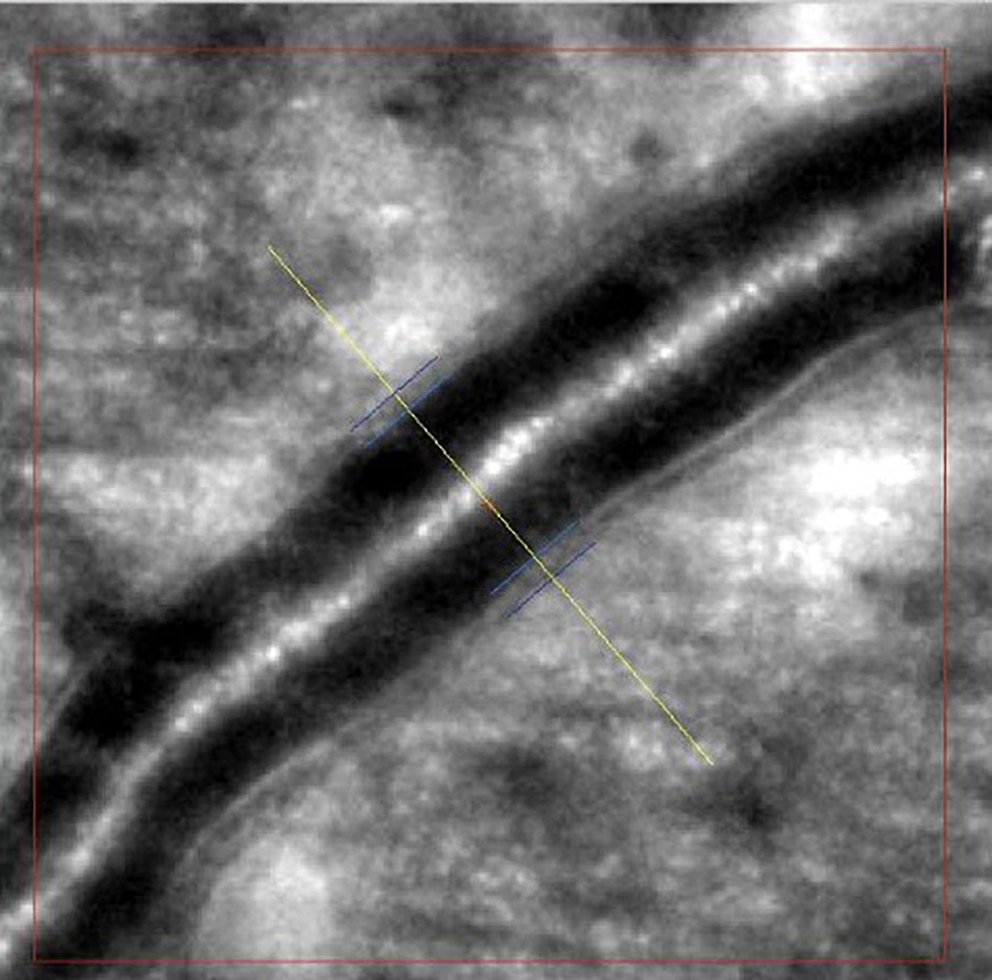
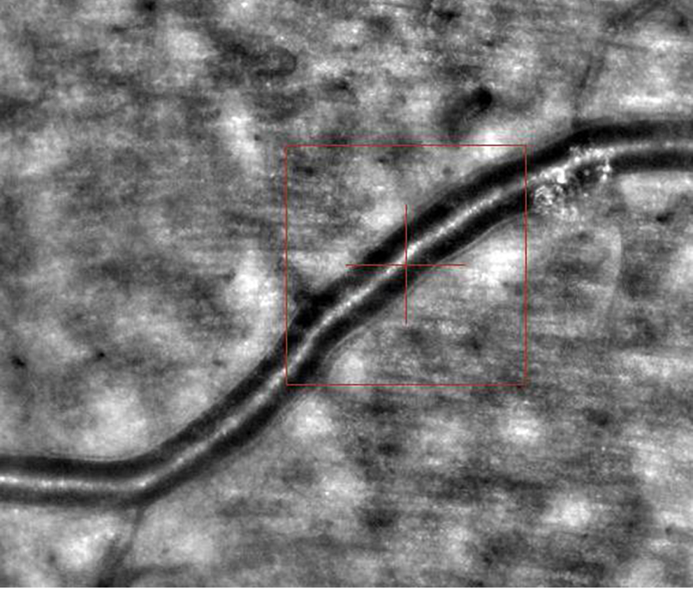
Images of retinal arterioles using an AO retinal camera (rtx1™; Imagine Eyes, Orsay, France) were captured in both patient groups. Before imaging, participants received 1 drop of 1% tropicamide for pupil dilation. Retinal artery images were analyzed with AOdetectArtery software (rtx1™; Imagine Eyes), focusing on the superior quadrant arterioles with a lumen diameter of at least 40 μm and no bifurcations. Two measurements were taken, and the highest-quality scan was selected for analysis.

The main vascular parameters obtained with AO were total vessel diameter (TD), lumen diameter (LD) and wall thickness (WT). The software also calculates 2 additional vascular metrics: The wall-to-lumen ratio (WLR), defined as wall thickness (WT) divided by lumen diameter (LD), and the wall cross-sectional area (WCSA), computed as π × [(TD/2)2 – (LD/2)2], which represents the area occupied by the vessel wall (Figure 1, Figure 2).

Statistical analyses

Qualitative variables were shown as integers with percentages. Quantitative parameters were presented according to their weighted arithmetic mean, median, standard deviation (SD), and min–max values. Relationships among the examined qualitative variables were illustrated using contingency tables. Relationships among categorical variables were assessed using Pearson’s χ2 test when all expected cell counts were ≥5, and Fisher’s two-tailed exact test when any expected count fell below 5. The Shapiro–Wilk W test was used to assess the normality of distribution and Levene’s test was fitted to check the homogeneity of variances. Differences between independent groups for normally distributed numerical variables with homogeneous variances were assessed by one-way analysis of variance (ANOVA) without replication. Variables failing to meet normality or homogeneity of variance assumptions were analyzed with nonparametric tests – namely, the Mann–Whitney U test for dichotomous independent variables and the Kruskal–Wallis H test for variables with more than 2 categories. A level of p < 0.050 was deemed statistically significant. All the statistical procedures were performed using STATISTICA v. 13.3 (TIBCO Software Inc., Palo Alto, USA).

Results

The study included 31 SSc patients (61 eyes) and 41 healthy controls (81 eyes). No significant differences between the 2 groups regarding retinal arteries TD, LD, second wall thickness, and WCSA were found (Table 4). Mean retinal arterial wall thickness was significantly lower in SSc patients than in healthy controls (p = 0.016), and the wall-to-lumen ratio was also notably decreased in the SSc group (p = 0.048) (Table 4; Figure 3, Figure 4).

Retinal vessel parameters obtained through AO, including TD, LD, wall thicknesses, WLR, and WCSA, were analyzed in relation to NC findings and clinical features of SSc. No statistically significant correlation was observed between retinal vessel parameters and capillaroscopic patterns, nor between these parameters and the type of SSc (Table 5, Table 6, Table 7). The WCSA was significantly higher in hypertensive SSc patients compared to those without hypertension (p = 0.026; Table 7).

Discussion

Vascular damage is a hallmark of SSc and plays a pivotal role in its pathogenesis. It has been established that microvascular changes primarily affect peripheral arterioles and capillaries. Generalized vasculopathy has been demonstrated to contribute significantly to perfusion disorders in both the retina and the choroid. Therefore, the retinal microvasculature offers an ideal window for monitoring disease progression and detecting early pathological changes. Previous studies have shown that retinal findings, such as hard exudates, vascular tortuosity and arterial narrowing, as well as microhemorrhages, have been discovered in patients with SSc during fundus examination.25, 26, 27 The presence of small retinal capillary abnormalities can be detected through a range of diagnostic techniques, including OCTA and fluorescein angiography (FA). The morphological changes in the retinal caliber, shape, or lumen diameter are valuable for assessing the state of microcirculatory perfusion. Therefore, we hypothesized that adaptive optics retinal imaging, as a novel noninvasive method, could aid in the detection of abnormalities in the structure of retinal vessels.

In this study, AO was used for retinal artery assessment in both groups. To the best of our knowledge, our study is the first to use AO imaging in SSc. A comparison of the 2 groups revealed no statistically significant differences in terms of LD, TD and WLR. According to previous studies, WLR and WCSA are optimal parameters for evaluating vascular remodeling in cases of diabetic retinopathy and arterial hypertension.11, 28, 29, 30, 31, 32 Żmijewska et al. analyzed arterial measurements using AO in 36 patients with nonproliferative diabetic retinopathy (DR). Their results demonstrated thicker arterial walls, increased WLR and WCSA in DR group than in controls.29 Matuszewski et al.33 and Cristescu et al.34 also demonstrated comparable outcomes. However, our results showed that the WLR was significantly decreased among SSc patients compared to the healthy group (p = 0.048).

Lombardo et al.35 reported that, in eyes with nonproliferative DR, the parafoveal capillary lumen diameter was significantly reduced compared to healthy controls. Similarly, Zaleska-Żmijewska et al.18 demonstrated that prediabetic patients exhibit distinct alterations in both capillary lumen diameter and wall-to-lumen ratio relative to control subjects.

An elevated WLR reflects both thickening of the vessel wall and concomitant narrowing of the lumen. Piantoni et al.36 used AO in patients with active rheumatoid arthritis treated with abatacept to evaluate cardiovascular risk factors. Their results indicated a significant reduction in the WLR of the arterioles after abatacept treatment. However, our results showed that the mean arterial wall was thinner in the scleroderma group than in the controls (p = 0.016). It is possible that the observed results may be attributed to the vasodilator treatment employed in the management of SSc.37

Pache et al.37 demonstrated that in 10 healthy volunteers, a single dose of sildenafil induced significant dilation of both retinal arterioles and venules 30 minutes after administration (p < 0.001). Moreover, Polak at al.38 reported increased retinal blood flow in healthy controls after sildenafil administration. Sildenafil can enhance the vasodilatory effect of nitric oxide (NO), the primary regulator of vascular smooth muscle tone. In addition, we compared the measurements of the retinal arteries obtained in AO with the results of the NC. However, we did not find a statistically significant correlation with the type of scleroderma, finger ulcers and scleroderma patterns, number of vessels, avascular areas, hemorrhages, or branched vessels visualized in NC. In our previous study, we found that scleroderma microangiopathy patterns correlated significantly with both superficial and deep foveal avascular zone (FAZ) areas, as well as with deep retinal vessel density measured by OCTA.23 Several studies have explored the relationship between peripheral capillary density measured with nailfold capillaroscopy and both choriocapillaris vessel density39 and overall retinal perfusion.40 However, these findings have been inconsistent. It is therefore possible that high-resolution imaging of the retinal microvasculature may yield stronger correlations with NC abnormalities.

These findings corroborate earlier reports that hypertension is associated with increased wall cross-sectional area, reflecting arteriolar remodeling.11 Furthermore, in our study, we evaluated the impact of vasodilator medications employed for the management of complications associated with scleroderma vasculopathy. Contrary to expectations, the findings of this study indicated a decreased LD in patients treated with sildenafil (p = 0.017). It is worth noting that most patients in our NC cohort exhibited a late scleroderma pattern – reflecting severe vasculopathy and advanced disease – so the observed arterial lumen narrowing likely reflects the chronicity and severity of SSc.

Limitations

It is important to acknowledge the limitations of the research, including the relatively small number of participants and the cross-sectional nature of the study.

Conclusions

Our study analysis of the AO retinal imaging data set revealed a reduced mean arterial wall thickness in patients with SSc compared to healthy controls. However, no correlation was identified between the AO findings and the NC parameters or the disease stage. The retinal vessel parameters revealed significant differences only in hypertensive or sildenafil-treated patients. Further studies on a larger cohort of patients are necessary to draw reliable conclusions about the usefulness of AO in the diagnosis of SSc and its possible correlation with capillaroscopic findings.

Data Availability Statement

All data generated or analyzed during this study are included in this article.

Consent for publication

Not applicable.

Use of AI and AI-assisted technologies

Not applicable.

Tables


Table 1. Baseline characteristics of the study cohort

Analyzed trait*

Statistic

Number of participants, n (%)

72 (100.00)

Number of eyes, n (%)

142 (100.00)

Gender

female

49 (60.06)

male

23 (31.94)

Age [years], Me (Q1–Q3)

46 (36–55)

Shapiro–Wilk W test for normality

W = 0.933

p < 0.001

*Statistic parameters used: n – absolute number; % – percentage; Me – median; Q – quartiles.
Table 2. Baseline characteristics of the study cohort by occurrence of scleroderma

Analyzed trait

Scleroderma

p-value

present

absent

Number of participants, n (%)

31 (43.06)

41 (56.94)

Number of eyes, n (%)

61 (42.96)

81 (57.04)

Gender, n (%)

female

22 (70.97)

27 (65.85)

χ2 = 0.209

df = 1

p = 0.647

male

9 (29.03)

14 (34.15)

Age [years], Me (Q1–Q3)

Shapiro–Wilk test

48 (40–54)

W = 0.935

p = 0.003

41 (35–55)

W = 0.0867

p < 0.001

Levene’s test

F (1, 140) = 1.137

p = 0.288

Mann–Whitney test

U = 2070.000

Zcorr. = 1.651

p = 0.099

AL [mm], Me (Q1–Q3)

Shapiro–Wilk test

23.53 (22.88–24.20)

W = 0.968

p = 0.114

23.78 (23.13–24.35)

W = 0.970

p = 0.129

Levene’s test

F (1, 121) = 0.475

p = 0.492

ANOVA

F (1, 121) = 0.138

p = 0.711

*Statistic parameters used: n – absolute number; % – percentage; Me – median; Q – quartiles; ANOVA – analysis of variance; df – degrees of freedom; AL – axial length.
Table 3. Baseline characteristics of the scleroderma group (n = 31)

Trait

n

M (SD)

%

Me (Q1–Q3)

Gender

female

22

71.0

male

9

29.0

Scleroderma

localized

8

28.6

systemic

20

71.4

capillaroscopy

27

96.4

Scleroderma pattern

none

2

7.1

early

5

17.9

active

12

42.9

late

9

32.1

anti-PM/SCL

1

3.6

PC TH fibrillarin

3

10.7

anti-Scl-70-antibodies

17

54.8

Centromere

8

25.8

Number of vessels

normal

2

7.1

reduced

20

71.5

very reduced

6

21.4

Avascular are

16

57.1

Giant capillaries

14

50.0

Hemorrhages

11

39.3

Branched vessels

10

35.7

MES

Shapiro–Wilk test

2.2 (1.1)

W = 0.848

2.5 (1.1–3.1)

p < 0.001

Interstitial lung disease

12

42.9

PAH

2

7.1

NSIP

7

22.6

UIP

4

14.3

MMF

13

41.9

MTX

7

25.0

mRSS

Shapiro–Wilk test

8.1 (7.9)

W = 0.771

2.0 (2.0–12.0)

p < 0.001

Finger ulcers

8

28.6

RVSP > 35 mm Hg

3

1.1

Elevated proBNP

5

18.5

Arterial hypertension

8

25.8

Administration of sildenafil

13

41.9

Administration of amlodipine

12

38.7

Missing data were pair-wise deleted. For discrete variables: n – number; % – percentage. For numerical traits: M – mean; SD – standard deviation; Me – median; Q – quartiles; anti-PM/SCL – anti-polymyositis/scleroderma antibodies; PAH – pulmonary arterial hypertension; NSIP – non-specific interstitial pneumonia; UIP – usual interstitial pneumonia, MMF – mycophenolat mofetil; MTX – methotrexate, mRSS – modified Rodnan Skin Score; RVSP – right ventricle systolic pressure; MES – the microangiopathy evolution score.
Table 4. Descriptive statistics for the adaptive optics by occurrence of scleroderma (n = 142 eyes)

Adaptive optics

Study group

Statistical parameter

Statistical tests

M

SD

Me

Q1–Q3

Total diameter

[µm]

scleroderma

67.25

11.37

67.15

58.00–75.50

normality test

W = 0.979, p = 0.411

W = 0.966, p = 0.033

Levene’s test

F (1, 135) = 1.083

p = 0.300

Mann–Whitney test

U = 2130.000

Zcorr. = −0.699

p = 0.484

control

69.03

12.88

69.30

58.00–78.40

Wall 1 thickness

[µm]

scleroderma

6.53

1.44

6.60

5.60–7.40

normality test

W = 0.985, p = 0.677

W = 0.325, p < 0.001

Levene’s test

F (1, 135) = 1.337

p = 0.250

Mann–Whitney test

U = 1323.000

Zcorr. = −4.216

p < 0.001

control

8.20

4.78

7.50

6.60–8.90

Wall 2 thickness

[µm]

scleroderma

7.46

1.72

7.45

6.10–8.50

normality test

W = 0.976, p = 0.317

W = 0.950, p = 0.004

Levene’s test

F (1, 135) = 0.686

p = 0.409

Mann–Whitney test

U = 2284.500

Zcorr. = −0.026

p = 0.979

control

7.52

1.61

7.20

6.40–8.10

Mean wall thickness

[µm]

scleroderma

6.99

1.34

7.15

6.20–8.10

normality test

W = 0.983, p = 0.589

W = 0.512, p < 0.001

Levene’s test

F (1, 135) = 0.285

p = 0.594

Mann–Whitney test

U = 1740.000

Zcorr. = −2.398

p = 0.016

control

7.86

2.52

7.60

6.70–8.55

Lumen diameter

[µm]

scleroderma

53.25

10.49

52.65

44.80–61.10

normality test

W = 0.983, p = 0.585

W = 0.962, p = 0.018

Levene’s test

F (1, 135) = 2.123

p = 0.147

Mann–Whitney test

U = 2279.000

Zcorr. = 0.050

p = 0.960

control

53.64

14.03

53.30

44.20–62.40

Wall-to-lumen ratio

scleroderma

0.2710

0.0681

0.2600

0.2200–0.3200

normality test

W = 0.970, p = 0.159

W = 0.946, p = 0.002

Levene’s test

F (1, 135) = 0.024

p = 0.878

Mann–Whitney test

U = 1837.000

Zcorr. = −1.976

p = 0.048

control

0.2940

0.0724

0.2600

0.2200–0.3300

Wall cross-sectional area [µm2]

scleroderma

1338.3

410.0

1386

983–1594

normality test

W = 0.952, p = 0.024

W = 0.960, p = 0.015

Levene’s test

F (1, 135) = 0.006

p = 0.941

Mann–Whitney test

U = 1843.000

Zcorr. = 0.051

p = 0.051

control

1479.3

431.3

1498

1129–1724

M – mean; SD – standard deviation; Me – median; Q – quartiles. Values in bold are statistically significant.
Table 5. Descriptive statistics for the lumen diameter [µm] in the study participants with scleroderma by selected capillaroscopic assessments (n = 61 eyes)

Investigated trait

Statistical parameter

Statistics

M (SD)

Me (Q1–Q3)

Scleroderma

localized

55.81 (13.23)

58.20 (41.83–61.18)

normality test

W = 0.980, p = 0.721

W = 0.929, p = 0.261

Levene’s test

F (1, 50) = 4.027, p = 0.051

ANOVA

F (1, 50) = 2.244

p = 0.140

systemic

51.15 (8.65)

50.00 (44.73–57.20)

Scleroderma pattern

early

48.80 (9.92)

52.40 (41.33–55.83)

normality test

W = 0.964, p = 0.841

W = 0.970, p = 0.692

W = 0.959, p = 0.612

Levene’s test.

F (3, 48) = 1.884, p = 0.145

ANOVA

F (3, 48) = 1.718

p = 0.176

active

55.94 (11.75)

57.20 (45.85–64.53)

late

49.78 (7.85)

49.00 (43.63–55.97)

Number of vessels

normal

52.23 (11.12)

58.20 (42.53–58.95)

normality test

W = 0.784, p = 0.077

W = 0.982, p = 0.811

W = 0.932, p = 0.403

Levene’s test

F (2, 49) = 0.365,

p = 0.696

ANOVA

F (2, 49) = 1.718

p = 0.498

reduced

51.51 (9.43)

50.00 (44.57–59.77)

very reduced

55.59 (12.71)

54.50 (44.07–62.04)

Avascular areas

yes

52.83 (11.23)

51.35 (44.52–61.70)

normality test

W = 0.968, p = 0.667

W = 0.980, p = 0.820

Levene’s test

F (1, 50) = 0.797, p = 0.376

ANOVA

F (1, 50) = 0.072

p = 0.789

no

52.04 (9.03)

52.50 (43.95–58.83)

Giant capillaries

yes

52.66 (12.52)

49.95 (42.23–61.24)

normality test

W = 0.970, p = 0.613

W = 0.968, p = 0.576

Levene’s test

F (1, 50) = 6.718, p = 0.012

ANOVA

F (3, 48) = 0.014

p = 0.907

no

52.33 (7.64)

52.90 (44.78–58.28)

Hemorrhages

yes

49.38 (12.18)

46.65 (40.92–55.28)

normality test

W = 0.978, p = 0.726

W = 0.906, p = 0.054

Levene’s test

F (1, 50) = 1.244, p = 0.270

ANOVA

F (1, 50) = 3.127

p = 0.083

no

54.45 (8.51)

54.55 (48.30–61.49)

Branched vessels

yes

50.38 (9.78)

49.10 (44.56–57.58)

normality test

W = 0.959, p = 0.232

W = 0.975, p = 0.878

Levene’s test

F (1, 50) = 0.116, p = 0.735

ANOVA

F (1, 50) = 1.175

p = 0.283

no

53.62 (10.49)

53.35 (43.68–59.27)

Finger ulcers

yes

54.05 (12.26)

52.70 (46.08–60.95)

normality test

W = 0.951, p = 0.114

W = 0.954, p = 0.586

Levene’s test

F (1, 49) = 0.168, p = 0.683

ANOVA

F (1, 50) = 0.399

p = 0.530

no

52.04 (9.54)

52.50 (43.21–59.10)

Arterial hypertension

yes

56.30 (13.02)

55.15 (49.00–65.50)

normality test

W = 0.975, p = 0.566

W = 0.961, p = 0.731

Levene’s test

F (1, 49) = 1.248, p = 0.269

ANOVA

F (1, 49) = 2.871

p = 0.097

no

52.29 (9.52)

52.50 (44.70–59.10)

Sildenafil

yes

48.92 (8.21)

49.00 (44.10–53.60)

normality test

W = 0.972, p = 0.676

W = 0.985, p = 0.957

Levene’s test

F (1, 49) = 2.625, p = 0.112

ANOVA

F (1, 49) = 6.062

p = 0.017

no

56.54 (10.94)

58.20 (48.50–64.20)

Amlodipine

yes

54.44 (10.87)

54.80 (47.80–59.10)

normality test

W = 0.967, p = 0.491

W = 0.955, p = 0.392

Levene’s test

F (1, 49) = 0.024, p = 0.877

ANOVA

F (1, 49) = 1.554

p = 0.218

no

52.53 (10.34)

49.95 (44.70–61.45)

M – mean; SD – standard deviation; Me – median; Q – quartiles; ANOVA – analysis of variance. Values in bold are statistically significant.
Table 6. Descriptive statistics for the wall-to-lumen ration in the study participants with scleroderma by selected capillaroscopic assessments (n = 61 eyes)

Investigated trait

Statistical parameter

Statistics

M (SD)

Me (Q1–Q3)

Scleroderma

localized

0.2593 (0.0794)

0.2500 (0.2300–0.2850)

normality test

W = 0.983, p = 0.835

W = 0.876, p = 0.041

Levene’s test

F (1, 50) = 0.009, p = 0.925

Mann–Whitney test

U = 212.000

Zcorr. = 1.315

p = 0.188

systemic

0.2816 (0.0641)

0.2800 (0.2267–0.3233)

Scleroderma pattern

early

0.2989 (0.0924)

0.3000 (0.2500–0.3500)

normality test

W = 0.964, p = 0.837

W = 0.917, p = 0.057

W = 0.886, p = 0.040

Levene’s test

F (3, 48) = 0.574, p = 0.635

Kruskal–Wallis test

H (3, n = 52) = 3.453

p = 0.327

active

0.2626 (0.0726)

0.2500 (0.2117–0.2900)

late

0.2753 (0.0516)

0.2700 (0.2267–0.3300)

Number of vessels

normal

0.2367 (0.0971)

0.2600 (0.1517–0.3100)

normality test

W = 0.957, p = 0.600

W = 0.968, p = 0.368

W = 0.963, p = 0.820

Levene’s test

F (2, 49) = 1.151, p = 0.229

ANOVA

F (2, 49) = 2.972

p = 0.061

reduced

0.2895 (0.0696)

0.2800 (0.2500–0.3300)

very reduced

0.2408 (0.0448)

0.2350 (0.2142–0.2658)

Avascular areas

yes

0.2753 (0.0707)

0.2700 (0.2292–0.3300)

normality test

W = 0.911, p = 0.049

W = 0.975, p = 0.678

Levene’s test

F (1, 50) = 0.445, p = 0.508

ANOVA

F (1, 50) = 0.0003

p = 0.986

no

0.2750 (0.0678)

0.2600 (0.2475–0.3200)

Giant capillaries

yes

0.2838 (0.0784)

0.2600 (0.2292–0.3208)

normality test

W = 0.953, p = 0.269

W = 0.933, p = 0.090

Levene’s test

F (1, 50) = 0.447, p = 0.235

ANOVA

F (1, 50) = 0.819

p = 0.370

no

0.2665 (0.0580)

0.2750 (0.2292–0.3200)

Hemorrhages

yes

0.2945 (0.0833)

0.2750 (0.2383–0.3300)

normality test

W = 0.966, p = 0.393

W = 0.943, p = 0.271

Levene’s test

F (1, 50) = 3.232, p = 0.078

ANOVA

F (1, 50) = 2.639

p = 0.111

no

0.2631 (0.0561)

0.2650 (0.2242–0.3158)

Branched vessels

yes

0.2861 (0.0750)

0.2750 (0.2200–0.3317)

normality test

W = 0.944, p = 0.080

W = 0.939, p = 0.277

Levene’s test

F (1, 50) = 0.818, p = 0.370

ANOVA

F (1, 50) = 0.688

p = 0.411

no

0.2694 (0.0658)

0.2600 (0.2300–0.3200)

Finger ulcers

yes

0.2728 (0.0684)

0.2600 (0.2242–0.3200)

normality test

W = 0.968, p = 0.373

W = 0.939, p = 0.374

Levene’s test

F (1, 49) = 0.279, p = 0.599

ANOVA

F (1, 49) = 0.001

p = 0.914

no

0.2720 (0.0652)

0.2700 (0.2300–0.3067)

Arterial hypertension

yes

0.2760 (0.0626)

0.2600 (0.2300–0.3300)

normality test

W = 0.960, p = 0.200

W = 0.978, p = 0.958

Levene’s test

F (1, 49) = 0.081, p = 0.777

ANOVA

F (1, 49) = 0.0002

p = 0.989

no

0.2700 (0.0704)

0.2650 (0.2200–0.3150)

Sildenafil

yes

0.2910 (0.0648)

0.2800 (0.2400–0.3300)

normality test

W = 0.944, p = 0.162

W = 0.949, p = 0.239

Levene’s test

F (1, 49) = 0.011, p = 0.918

ANOVA

F (1, 49) = 2.449

p = 0.124

no

0.2560 (0.0677)

0.2600 (0.2200–0.2900)

Amlodipine

yes

0.2560 (0.0643)

0.2550 (0.2200–0.3000)

normality test

W = 0.938, p = 0.089

W = 0.980, p = 0.914

Levene’s test

F (1, 49) = 0.143, p = 0.707

ANOVA

F (1, 49) = 3.051

p = 0.087

no

0.2800 (0.0697)

0.2700 (0.2250–0.3200)

M – mean; SD – standard deviation; Me – median; Q – quartiles; ANOVA – analysis of variance.
Table 7. Descriptive statistics for the wall cross-sectional area [µm2] in the study participants with scleroderma by selected capillaroscopic assessments (n = 61 eyes)

Investigated trait

Statistical parameter

Statistics

M (SD)

Me (Q1–Q3)

Scleroderma

localized

1399.9 (540.5)

1438 (874–1619)

normality test

W = 0.907, p = 0.122

W = 0.959, p = 0.187

Levene’s test

F (1, 50) = 3.233, p = 0.078

ANOVA

F (1, 50) = 0.802

p = 0.375

systemic

1291.3 (323.1)

1379 (1032–1534)

Scleroderma pattern

early

1235.8 (382.5)

1389 (840–1601)

normality test

W = 0.787, p = 0.015

W = 0.951, p = 0.315

W = 0.946, p = 0.391

Levene’s test

F (3, 48) = 1.493, p = 0.228

Kruskal–Wallis test

H (3, n = 52) = 3.104

p = 0.376

active

1420.4 (458.6)

1428 (1079–1703)

late

1210.5 (314.5)

1244 (897–1434)

Number of vessels

normal

1078.3 (413.4)

912 (796–1444)

normality test

W = 0.879, p = 0.322

W = 0.964, p = 0.265

W = 0.828, p = 0.020

Levene’s test.

F (2, 49) = 0.124, p = 0.883

Kruskal–Wallis test

H (2, n = 52) = 1.857

p = 0.395

reduced

1350.1 (362.8)

1389 (1067–1608)

very reduced

1298.8 (493.5)

1325 (922–1446)

Avascular areas

yes

1322.3 (392.2)

1381 (1048–1496)

normality test

W = 0.919, p = 0.025

W = 0.915, p = 0.060

Levene’s test

F (1, 50) = 0.535, p = 0.468

Mann–Whitney test

U = 310.000

Zcorr. = −0.361

p = 0.718

no

1323.0 (409.1)

1433 (866–1605)

Giant capillaries

yes

1278.2 (461.3)

1405 (892–1620)

normality test

W = 0.931, p = 0.083

W = 0.952, p = 0.262

Levene’s test

F (1, 50) = 1.790, p = 0.187

ANOVA

F (1, 50) = 0.657

p = 0.421

no

1323.0 (319.5)

1356 (981–1532)

Hemorrhages

yes

1249.4 (440.7)

1195 (864–1467)

normality test

W = 0.857, p = 0.007

W = 0.968, p = 0.437

Levene’s test

F (1, 50) = 0.443, p = 0.509

Mann–Whitney test

U = 248.000

Zcorr. = 1.345

p = 0.179

no

1358.3 (364.1)

1397 (1059–1600)

Branched vessels

yes

1260.5 (324.7)

1353 (899–1530)

normality test

W = 0.945, p = 0.353

W = 0.935, p = 0.045

Levene’s test.

F (1, 50) = 0.925, p = 0.341

Mann–Whitney test

U = 267.000

Zcorr. = −0.741

p = 0.459

no

1355.5 (429.1)

1397 (981–1605)

Finger ulcers

yes

1291.3 (376.8)

1353 (896–1576)

normality test

W = 0.884, p = 0.054

W = 0.934, p = 0.033

Levene’s test.

F (1, 49) = 0.154, p = 0.696

Mann–Whitney test

U = 263.000

Zcorr. = −0.134

p = 0.893

no

1377.9 (450.1)

1389 (1079–1489)

Arterial hypertension

yes

1517.5 (476.9)

1466 (1259–1717)

normality test

W = 0.929, p = 0.295

W = 0.940, p = 0.047

Levene’s test

F (1, 49) = 0.296, p = 0.589

ANOVA

F (1, 49) = 5.252

p = 0.026

no

1281.2 (374.6)

1324 (905–1547)

Sildenafil

yes

1224.4 (300.2)

1244 (987–1428)

normality test

W = 0.963, p = 0.478

W = 0.931, p = 0.080

Levene’s test

F (1, 49) = 2.494, p = 0.121

ANOVA

F (1, 49) = 2.821

p = 0.099

no

1424.6 (462.6)

1466 (983–1717)

Amlodipine

yes

1316.4 (422.9)

1356 (912–1541)

normality test

W = 0.895, p = 0.023

W = 0.958, p = 0.291

Levene’s test

F (1, 49) = 0.015, p = 0.903

Mann–Whitney test

U = 308.000

Zcorr. = −0.200

p = 0.842

no

1351.6 (407.4)

1392 (985–1612)

M – mean; SD – standard deviation; Me – median; Q – quartiles; ANOVA – analysis of variance. Values in bold are statistically significant.

Figures


Fig. 1. Adaptive optics (AO) image of the retinal artery in a patient with scleroderma
Fig. 2. Adaptive optics (AO) image of the retinal artery in a patient with scleroderma
Fig. 3. Graphical presentation of the measures of location (median, quartiles) and dispersion (min–max values) of the mean wall thickness [µm] in the study participants’ eyes by occurrence of scleroderma
Fig. 4. Graphical presentation of the measures of location (median, quartiles) and dispersion (min–max values) of the wall-to-lumen ratio in the study participants’ eyes by occurrence of scleroderma

References (40)

  1. Nikpour M, Stevens WM, Herrick AL, Proudman SM. Epidemiology of systemic sclerosis. Best Pract Res Clin Rheumatol. 2010;24(6):857–869. doi:10.1016/j.berh.2010.10.007
  2. Postlethwaite R, Pattanaik D, Brown M. Vascular involvement in systemic sclerosis (scleroderma). J Inflamm Res. 2011;4:105. doi:10.2147/JIR.S18145
  3. Pawlik K, Bohdziewicz A, Maciejewska M, et al. Evaluation of cutaneous microcirculation in systemic sclerosis: An update. Dermatol Rev. 2023;110(4):499–517. doi:10.5114/dr.2023.131385
  4. Lambova SN, Müller-Ladner U. Nailfold capillaroscopy in systemic sclerosis: State of the art. The evolving knowledge about capillaroscopic abnormalities in systemic sclerosis. J Scleroderma Relat Dis. 2019;4(3):200–211. doi:10.1177/2397198319833486
  5. de AF Gomes B, Santhiago MR, Magalhães P, Kara-Junior N, De Azevedo MNL, Moraes HV. Ocular findings in patients with systemic sclerosis. Clinics (Sao Paulo). 2011;66(3):379–385. doi:10.1590/S1807-59322011000300003
  6. de AF Gomes B, Santhiago MR, De Azevedo MNL, Moraes HV. Evaluation of dry eye signs and symptoms in patients with systemic sclerosis. Graefes Arch Clin Exp Ophthalmol. 2012;250(7):1051–1056. doi:10.1007/s00417-012-1938-3
  7. Sahin Atik S, Koc F, Akin Sari S, Sefi Yurdakul N, Ozmen M, Akar S. Anterior segment parameters and eyelids in systemic sclerosis. Int Ophthalmol. 2016;36(4):577–583. doi:10.1007/s10792-015-0165-4
  8. Kozikowska M, Luboń W, Kucharz E, Mrukwa-Kominek E. Ocular manifestations in patients with systemic sclerosis. Reumatologia. 2020;58(6):401–406. doi:10.5114/reum.2020.102004
  9. Tailor R, Gupta A, Herrick A, Kwartz J. Ocular manifestations of scleroderma. Surv Ophthalmol. 2009;54(2):292–304. doi:10.1016/j.survophthal.2008.12.007
  10. Szucs G, Szekanecz Z, Aszalos Z, et al. A wide spectrum of ocular manifestations signify patients with systemic sclerosis. Ocul Immunol Inflamm. 2021;29(1):81–89. doi:10.1080/09273948.2019.1657467
  11. Szewczuk A, Zaleska-Żmijewska A, Dziedziak J, Szaflik JP. Clinical application of adaptive optics imaging in diagnosis, management, and monitoring of ophthalmological diseases: A narrative review. Med Sci Monit. 2023;29:e941926. doi:10.12659/MSM.941926
  12. Yao X, Ke M, Ho Y, et al. Comparison of retinal vessel diameter measurements from swept-source OCT angiography and adaptive optics ophthalmoscope. Br J Ophthalmol. 2021;105(3):426–431. doi:10.1136/bjophthalmol-2020-316111
  13. Bakker E, Dikland FA, Van Bakel R, et al. Adaptive optics ophthalmoscopy: A systematic review of vascular biomarkers. Surv Ophthalmol. 2022;67(2):369–387. doi:10.1016/j.survophthal.2021.05.012
  14. Rosenbaum D, Mattina A, Koch E, et al. Effects of age, blood pressure and antihypertensive treatments on retinal arterioles remodeling assessed by adaptive optics. J Hypertens. 2016;34(6):1115–1122. doi:10.1097/HJH.0000000000000894
  15. Dziedziak J, Zaleska-Żmijewska A, Szaflik JP, Cudnoch-Jędrzejewska A. Impact of arterial hypertension on the eye: A review of the pathogenesis, diagnostic methods, and treatment of hypertensive retinopathy. Med Sci Monit. 2022;28:e935135. doi:10.12659/MSM.935135
  16. Baltă F, Cristescu IE, Mirescu AE, Baltă G, Zemba M, Tofolean IT. Investigation of retinal microcirculation in diabetic patients using adaptive optics ophthalmoscopy and optical coherence angiography. J Diabetes Res. 2022;2022:1516668. doi:10.1155/2022/1516668
  17. Gallo A, Dietenbeck T, Giron A, Paques M, Kachenoura N, Girerd X. Noninvasive evaluation of retinal vascular remodeling and hypertrophy in humans: Intricate effect of ageing, blood pressure and glycaemia. Clin Res Cardiol. 2021;110(7):959–970. doi:10.1007/s00392-020-01680-3
  18. Zaleska-Żmijewska A, Piątkiewicz P, Śmigielska B, et al. Retinal photoreceptors and microvascular changes in prediabetes measured with adaptive optics (rtx1TM): A case-control study. J Diabetes Res. 2017;2017:4174292. doi:10.1155/2017/4174292
  19. Szewczuk A, Wawrzyniak ZM, Szaflik JP, Zaleska-Żmijewska A. Is primary open-angle glaucoma a vascular disease? Assessment of the relationship between retinal arteriolar morphology and glaucoma severity using adaptive optics. J Clin Med. 2024;13(2):478. doi:10.3390/jcm13020478
  20. Hugo J, Chavane F, Beylerian M, Callet M, Denis D, Matonti F. Morphologic analysis of peripapillary retinal arteriole using adaptive optics in primary open-angle glaucoma. J Glaucoma. 2020;29(4):271–275. doi:10.1097/IJG.0000000000001452
  21. Errera MH, Laguarrigue M, Rossant F, et al. High-resolution imaging of retinal vasculitis by flood illumination adaptive optics ophthalmoscopy: A follow-up study. Ocul Immunol Inflamm. 2020;28(8):1171–1180. doi:10.1080/09273948.2019.1646773
  22. Venkatesh R, Mutalik D, Reddy NG, Akkali MC, Yadav NK, Chhablani J. Retinal vessel wall imaging using fluorescein angiography and adaptive optics imaging in acute branch retinal artery occlusion. Eur J Ophthalmol. 2023;33(4):NP85–NP90. doi:10.1177/11206721221113202
  23. Paczwa K, Rerych M, Romanowska-Próchnicka K, Olesińska M, Różycki R, Gołębiewska J. Retinal microvasculature in systemic sclerosis patients and the correlation between nailfold capillaroscopic findings and optical coherence angiography results. J Clin Med. 2024;13(7):2025. doi:10.3390/jcm13072025
  24. Cutolo M, Pizzorni C, Secchi ME, Sulli A. Capillaroscopy. Best Pract Res Clin Rheumatol. 2008;22(6):1093–1108. doi:10.1016/j.berh.2008.09.001
  25. Waszczykowska A, Goś R, Waszczykowska E, Dziankowska-Bartkowiak B, Jurowski P. Prevalence of ocular manifestations in systemic sclerosis patients. Arch Med Sci. 2013;6:1107–1113. doi:10.5114/aoms.2013.39217
  26. Ushiyama O, Ushiyama K, Yamada T, et al. Retinal findings in systemic sclerosis: A comparison with nailfold capillaroscopic patterns. Ann Rheum Dis. 2003;62(3):204–207. doi:10.1136/ard.62.3.204
  27. Shenavandeh S, Afarid M, Hasanaghaei T, Ali Nazarinia M. Prevalence of retinal changes in patients with systemic sclerosis: The association between retinal vascular changes and nailfold capillaroscopic findings. Reumatologia. 2021;59(1):27–34. doi:10.5114/reum.2021.103436
  28. Mehta R, Akkali M, Jayadev C, Anuj A, Yadav N. Morphometric analysis of retinal arterioles in control and hypertensive population using adaptive optics imaging. Indian J Ophthalmol. 2019;67(10):1673. doi:10.4103/ijo.IJO_253_19
  29. Zaleska-Żmijewska A, Wawrzyniak ZM, Dąbrowska A, Szaflik JP. Adaptive optics (rtx1) high-resolution imaging of photoreceptors and retinal arteries in patients with diabetic retinopathy. J Diabetes Res. 2019;2019:9548324. doi:10.1155/2019/9548324
  30. Meixner E, Michelson G. Measurement of retinal wall-to-lumen ratio by adaptive optics retinal camera: A clinical research. Graefes Arch Clin Exp Ophthalmol. 2015;253(11):1985–1995. doi:10.1007/s00417-015-3115-y
  31. Rizzoni D, Agabiti Rosei C, De Ciuceis C, Semeraro F, Rizzoni M, Docchio F. New methods to study the microcirculation. Am J Hypertens. 2018;31(3):265–273. doi:10.1093/ajh/hpx211
  32. Gallo A, Mattina A, Rosenbaum D, Koch E, Paques M, Girerd X. Retinal arteriolar remodeling evaluated with adaptive optics camera: Relationship with blood pressure levels. Ann Cardiol Angeiol (Paris). 2016;65(3):203–207. doi:10.1016/j.ancard.2016.04.021
  33. Matuszewski W, Gontarz-Nowak K, Harazny JM, Bandurska-Stankiewicz E. Evaluation of morphological changes in retinal vessels in type 1 diabetes mellitus patients with the use of adaptive optics. Biomedicines. 2022;10(8):1926. doi:10.3390/biomedicines10081926
  34. Cristescu IE, Zagrean L, Balta F, Branisteanu DC. Retinal microcirculation investigation in type I and II diabetic patients without retinopathy using an adaptive optics retinal camera. Acta Endocrinol (Buchar). 2019;15(4):417–422. doi:10.4183/aeb.2019.417
  35. Lombardo M, Parravano M, Serrao S, Ducoli P, Stirpe M, Lombardo G. Analysis of retinal capillaries in patients with type 1 diabetes and nonproliferative diabetic retinopathy using adaptive optics imaging. Retina. 2013;33(8):1630–1639. doi:10.1097/IAE.0b013e3182899326
  36. Piantoni S, Regola F, Angeli F, et al. Retinal microvascular alterations in patients with active rheumatoid arthritis without cardiovascular risk factors: The potential effects of T cell co-stimulation blockade. Front Med (Lausanne). 2024;11:1247024. doi:10.3389/fmed.2024.1247024
  37. Pache M, Meyer P, Prünte C, Orgül S, Nuttli I, Flammer J. Sildenafil induces retinal vasodilatation in healthy subjects. Br J Ophthalmol. 2002;86(2):156–158. doi:10.1136/bjo.86.2.156
  38. Polak K, Wimpissinger B, Berisha F, Georgopoulos M, Schmetterer L. Effects of sildenafil on retinal blood flow and flicker-induced retinal vasodilatation in healthy subjects. Invest Ophthalmol Vis Sci. 2003;44(11):4872. doi:10.1167/iovs.03-0177
  39. Mihailovic N, Lahme L, Braasch S, et al. Altered ocular microvasculature in patients with systemic sclerosis and very early disease of systemic sclerosis using optical coherence tomography angiography. Sci Rep. 2022;12(1):10990. doi:10.1038/s41598-022-14377-6
  40. Cutolo CA, Cere A, Toma P, et al. Peripheral and ocular microvascular alterations in systemic sclerosis: Observations from capillaroscopic assessments, perfusion peripheral analysis, and optical coherence tomography angiography. Rheumatol Int. 2023;44(1):107–118. doi:10.1007/s00296-023-05495-z