Advances in Clinical and Experimental Medicine

Title abbreviation: Adv Clin Exp Med
5-Year IF – 2.0, IF – 1.9, JCI (2024) – 0.43
Scopus CiteScore – 4.3
Q1 in SJR 2024, SJR score – 0.598, H-index: 49 (SJR)
ICV – 161.00; MNiSW – 70 pts
Initial editorial assessment and first decision within 24 h

ISSN 1899–5276 (print), ISSN 2451-2680 (online)
Periodicity – monthly

Download original text (EN)

Advances in Clinical and Experimental Medicine

Ahead of print

doi: 10.17219/acem/203397

Publication type: meta-analysis

Language: English

License: brak

Download citation:

  • BIBTEX (JabRef, Mendeley)
  • RIS (Papers, Reference Manager, RefWorks, Zotero)

Cite as:


Yang J, Chen L, Zhao L, et al. Noninvasive ventilation for COPD management: A systematic review and meta-analysis [published online as ahead of print on January 5, 2026]. Adv Clin Exp Med. 2026. doi:10.17219/acem/203397

Noninvasive ventilation for COPD management: A systematic review & meta-analysis

Jinyu Yang1,A, Lin Chen1,B, Lihong Zhao1,C, Chengyi Liu1,C, Xiujuan Gu1,B, Wanjun Qi2,E,F, Lei Wang2,D

1 Department of Respiratory and Critical Care Medicine, The Third Hospital of Mianyang (Sichuan Mental Health Center), China

2 Department of Nursing, The Third Hospital of Mianyang (Sichuan Mental Health Center), China

Graphical abstract


Graphical abstracts

Highlights


• Noninvasive ventilation (NIV) in chronic obstructive pulmonary disease (COPD) alleviates dyspnea, optimizes arterial blood gases, and cuts exacerbation rates without requiring intubation.
• Acute-exacerbation benefits of NIV include reduced mortality, fewer complications and shorter hospital stays – while benefits in stable COPD (no flare-ups in 4 weeks) show mixed evidence.
• Pooled randomized controlled trial (RCT) meta-analysis across COPD stages rigorously evaluated NIV’s efficacy from mild to severe disease, offering a unified evidence base.
• Meta-analysis outcomes revealed significant improvements in arterial gas exchange, enhanced survival and lower frequencies of exacerbations and intubations among COPD patients.

Abstract

Background. Noninvasive ventilation (NIV) is an important treatment modality in the management of chronic obstructive pulmonary disease (COPD) by reducing respiratory distress, improving gas exchange and reducing exacerbations without the need for intubation and invasive airways.

Objectives. To synthesize data from randomized controlled trials (RCTs) and perform a meta-analysis to understand the beneficial effects of NIV across different COPD stages.

Materials and methods. A systematic literature review was performed using MED­LINE (PubMed) and Cochrane Register of Controlled Trials (CENTRAL) al databases for RCTs that involved the administration of NIV vs usual treatment (oxygen supplementation, pharmacological agents, nasal cannulation) in patients with stable COPD, acute exacerbations of COPD (AECOPD), and post-exacerbation COPD (PECOPD). Mortality, exacerbation and intubation rates, and arterial blood gases (PaCO2 and PaO2 levels) were assessed in both groups. RevMan software was used to assess the risk of bias and calculate the pooled odds ratio (OR), mean differences (MDs) and subgroup analyses with a random-effects model.

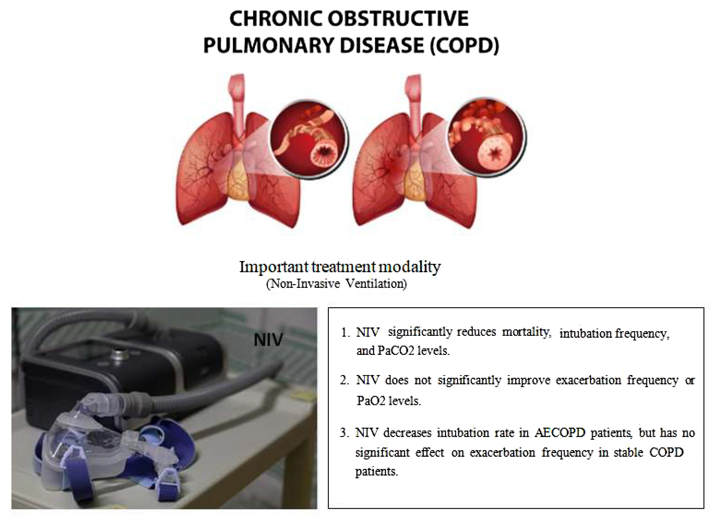
Results. A total of 51 RCTs were included in the meta-analysis with information from 3,775 patients. Meta-analysis of the data showed that there was a significant decrease in mortality outcomes (p < 0.001), intubation frequency (p < 0.001) and PaCO2 levels (p < 0.001) but no significant improvement in exacerbation frequency (p = 0.12) and PaO2 levels (p = 0.69). Subgroup analyses demonstrated no significant difference between COPD stage on mortality outcomes (p = 0.32), PaCO2 level (p = 0.12) and PaO2 level (p = 0.64). There was a significant decrease in intubation rate in AECOPD patients receiving NIV and a statistically nonsignificant difference in exacerbation frequency in stable COPD patients using NIV.

Conclusions. The findings of this meta-analysis indicate a substantial overall enhancement in the frequency of exacerbations and intubations, mortality outcomes, and arterial gas levels among patients in various stages of COPD. Consequently, it is imperative to identify patients with COPD that are most likely to benefit from the use of NIV.

Key words: chronic obstructive pulmonary disease, exacerbation, noninvasive ventilation, arterial blood gases, BiPAP

Introduction

Chronic obstructive pulmonary disease (COPD), encompassing emphysema and chronic bronchitis, is a common, progressive disorder characterized by irreversible airflow limitation resulting from damage to both the airways and the lung parenchyma.1, 2 Globally, COPD remains a leading cause of morbidity and mortality, responsible for an estimated 3.1 million deaths in 2021, with the heaviest burden observed in low- and middle-income countries.3 Beyond its mortality toll, COPD significantly impairs daily functioning and quality of life, and drives substantial healthcare utilization through recurrent exacerbations that often necessitate hospitalization and intensified pharmacotherapy.4

Key risk factors for COPD encompass cigarette smoking; exposure to ambient air pollution; a history of childhood asthma; and α1-antitrypsin deficiency, a rare genetic disorder.4 These insults provoke pathological remodeling of the lung parenchyma, including destruction of alveolar walls, that impairs gas exchange, precipitating hypoxemia and hypercapnia, and in severe cases leading to acute hypercapnic respiratory failure (AHRF).5, 6 Resultant hypoxemia and systemic inflammation manifest as respiratory symptoms (dyspnea, fatigue, wheezing, cough, and chest tightness) and drive extrapulmonary complications, notably pulmonary hypertension and right heart failure, as well as adverse effects on endocrine, gastrointestinal, neuromuscular, and musculoskeletal systems.7, 8

Stable COPD refers to a state where symptoms are manageable and not worsening. Acute exacerbation of COPD (AECOPD) is a sudden worsening of COPD symptoms. Post-exacerbation COPD (PECOPD) describes the recovery phase after an acute exacerbation. The diagnosis of COPD is based on symptom assessment, imaging tests, pulmonary function tests (spirometry), and physical examinations. The Global Initiative for Chronic Obstructive Lung Disease (GOLD) has developed diagnostic criteria, including a post-bronchodilator forced expiratory volume (FEV1)/forced vital capacity (FVC) ratio <0.7. The GOLD criteria also classify the severity of airflow limitations into various stages and are used with patient-reported outcomes and exacerbation history to guide COPD management decisions.2 Treatment plans for COPD aim to improve quality of life, alleviate symptoms and prevent disease progression.

Pharmacological management of acute COPD exacerbations typically includes systemic corticosteroids, inhaled short-acting bronchodilators, and antibiotics when there is clinical or microbiological suspicion of bacterial infection.9, 10 Adjunctive ventilatory support, preferentially noninvasive ventilation (NIV) in cases of hypercapnic respiratory failure, can avert the need for invasive mechanical ventilation, which is reserved for NIV failure or contraindications. However, systemic corticosteroids, while accelerating recovery and reducing relapse rates, carry risks of hyperglycemia, fluid retention and steroid-induced myopathy, and repeated high-dose bronchodilator use may precipitate tachycardia, tremor and tolerance. Pulmonary rehabilitation, although pivotal for restoring functional capacity and reducing future exacerbations, often struggles with poor adherence, transport barriers and limited program availability. Invasive mechanical ventilation requires endotracheal intubation or tracheostomy and increases the risk of ventilator-associated pneumonia, barotrauma and prolonged weaning difficulties in COPD patients.

Noninvasive ventilation is an alternative to invasive ventilation techniques in which ventilator support (pressure-supported airflow) is provided through a noninvasive interface such as a nasal, oronasal or full-face mask to ventilator muscles. It is a comfortable alternative to intubation and avoiding immobility, and is used for managing conditions like acute COPD exacerbations and cardiogenic pulmonary edema-related respiratory failure. It reduces complications like ventilator-associated pneumonia and sinusitis by eliminating the need for sedation and endotracheal intubation, thereby minimizing hospital and intensive care unit (ICU) stays. Bi-level positive airway pressure (BiPAP), continuous positive airway pressure (CPAP) and negative pressure ventilation (NPV) are the most common types of NIV. BiPAP delivers 2 pressure levels for improved ventilation and airway stability, while CPAP provides constant pressure, typically used for milder respiratory issues and sleep apnea. The American Thoracic Society and European Respiratory Journal guidelines recommend the use of BiPAP for acute-on-chronic respiratory acidosis secondary to COPD exacerbations. Studies have shown that NIV has reduced mortality outcomes in patients with acute exacerbations and decreased complications and length of hospital stay.11, 12, 13

Previous meta-analyses have shown mixed results regarding the benefits of NIV in stable COPD patients (generally defined as no exacerbation in last 4 weeks). In general, long-term or domiciliary NIV use resulted in a decrease in mortality and improved quality of life, whereas outcomes such as hospital admissions and gas exchange were variable.10, 14 Effects of NIV in AECOPD patients were associated with lower deaths, intubation rates, and hospital stays in a meta-analysis by Osadnik et al.9 In a meta-analysis comparing NIV with usual care in PECOPD patients, the exacerbation frequency was decreased when NIV was employed, with no significant differences in mortality rates or arterial gases.15 Thus, the beneficial effects of NIV in patients in different COPD stages are heterogeneous in terms of outcomes which can limit its applicability.

Objectives

This study aims to systematically synthesize and critically analyze the available literature on NIV across all GOLD stages of COPD, quantifying its effects on mortality, hospital length of stay, exacerbation frequency, arterial blood gas parameters, and health-related quality of life in both acute exacerbations and stable disease, while comparing different NIV modalities, initiation timings and ventilator settings by patient phenotype, and ultimately developing an evidence-based clinical framework to guide optimal NIV selection, timing and management in acute and chronic COPD.

Materials and methods

Study selection or inclusion/exclusion criteria

We included randomized controlled trials (RCTs) that compared any type of NIV device (BiPAP, nocturnal) or administration device (full face, oronasal or nasal mask) with usual therapy such as oxygen supplementation, long-term oxygen therapy (LTOT), pharmacological treatment (antibiotics, bronchodilators, steroids, theophylline, mucolytic agents, etc.), or sham NIV for our analysis. We included adult patients (≥18 years) with various phases of COPD, including stable COPD, PECOPD and AECOPD in our analysis. Patients diagnosed with COPD as per the Global Initiative for Chronic Obstructive Lung Disease (GOLD) system that uses the FEV1/FVC ratio <0.7 were included. Exclusion criteria included non-randomized studies and prospective and retrospective study designs.

Information sources

We conducted a systematic literature search of MEDLINE (PubMed) and Cochrane Register of Controlled Trials (CENTRAL) in November 2024, encompassing the period of 1990–2024.

Search strategy

We conducted comprehensive searches of PubMed, Embase and the Cochrane Library from inception through May 2025 using both free-text keywords – “non-invasive ventilation,” “NIV,” “non-invasive positive pressure ventilation,” “BiPAP,” “VPAP,” “chronic obstructive pulmonary disease,” “pulmonary disease,” and “pulmonary emphysema” – and their corresponding Medical Subject Headings (MeSH) terms. Titles and abstracts of all retrieved records were screened for relevance, and full texts of potentially eligible studies were reviewed in detail. To ensure completeness, we also examined the reference lists of included articles for additional reports (Table 1).

Data extraction process

Data extraction was performed using a standardized, pre-piloted form to capture key study characteristics and outcomes: study identifiers (authors, publication year), design (e.g. randomized trial, cohort study), intervention and comparator details, duration of follow-up, COPD phase (stable vs exacerbation), participant demographics (mean age), exacerbation frequency and severity, and arterial partial pressure of carbon dioxide (PaCO2) levels.

Data items

We analyzed the following outcomes – mortality, exacerbation frequency, endotracheal intubation rates, and arterial blood gas parameters (PaCO2 and PaO2) – comparing patients receiving NIV with control groups. Article screening and data extraction were performed by a single reviewer using the predefined extraction form to ensure consistency and completeness of the collected data.

Risk of bias assessment

We used the Cochrane Collaboration’s risk of bias tool to assess the methodological quality of the included studies.16 This tool includes the following criteria: randomization, allocation concealment, blinding and completeness of follow-up. The risk of bias for each item was graded as high, low or unclear risk.

Quantitative data synthesis

We performed the meta-analysis and statistical calculations were performed using Review Manager (RevMan, v. 5; The Nordic Cochrane Center, Cochrane Collaboration, Copenhagen, Denmark). Binary outcomes such as mortality, exacerbation, and intubation rates were reported as odds ratios (OR) with corresponding 95% confidence intervals (95% CIs). Meta-analyses for binary outcomes were done using a random-effects model (Mantel–Haenszel method). Continuous outcomes such as PaCO2 and PaO2 levels were reported as mean differences (MDs) with associated 95% CIs using the random-effects model (inverse variance method). Heterogeneity in the included studies was evaluated using I2 statistic, with small heterogeneity for I2 values of <25%, moderate heterogeneity for I2values of 25% to 50% and high heterogeneity for I2 values >50%.17 Forest plots were constructed and p < 0.05 was statistically significant. Subgroup analyses were also performed according to stage of COPD (stable COPD, PECOPD and AECOPD) and type of control treatment or comparator (pharmacological treatment + oxygen, LTOT, high-flow nasal cannula (HFNC), or only pharmacological treatment). Publication bias was assessed using Egger’s test and a funnel plot, where the log OR for each study was plotted against its standard error (SE) for the mortality outcome. The vertical line indicates the pooled OR representing the overall summary effect size.

Results

Identification of studies

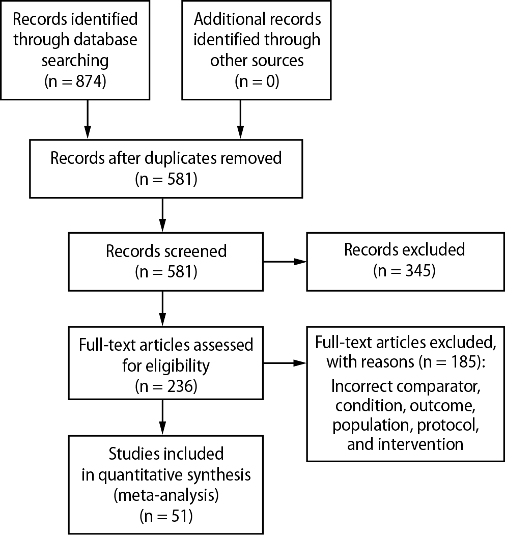
A total of 874 records were identified through database searching. After removing 345 duplicates and irrelevant records, 581 titles and abstracts were screened. Of these, 236 RCTs were assessed for eligibility. However, 185 RCTs were excluded due to reasons such as inappropriate comparator, intervention, condition, or population, missing required outcomes, or duplicate data. The selection process is illustrated in Figure 1. Table 2 shows the results of search strategy and Hits for COPD and NIV literature review.

Study characteristics

A total of 51 RCTs, comprising 3,775 participants, met the inclusion criteria. These included patients with stable COPD (n = 1,187), PECOPD (n = 1,314) and acute exacerbation of AECOPD (n = 1,274). The RCTs compared nocturnal or domiciliary NIV to other COPD treatments, such as LTOT, oxygen supplementation, pharmacologic therapies, HFNC, standard nasal cannula, or sham interventions. Participants were male and female across different COPD stages, with varying baseline PaCO2 levels, presence of hypercapnia, history of recent exacerbations, and differing durations of NIV administration and follow-up. Most studies used BiPAP systems for NIV delivery, administered via nasal, full-face or oronasal masks. Detailed information on interventions and control groups is provided in Table 3.18, 19, 20, 21, 22, 23, 24, 25, 26, 27, 28, 29, 30, 31, 32, 33, 34, 35, 36, 37, 38, 39, 40, 41, 42, 43, 44, 45, 46, 47, 48, 49, 50, 51, 52, 53, 54, 55, 56, 57, 58, 59, 60, 61, 62, 63, 64, 65, 66, 67, 68, 69

Characteristics of participants

The included studies involved patients with stable COPD (19 studies), PECOPD (14 studies) and AECOPD (18 studies). Across all studies, the mean age of participants was over 60 years. In most studies, baseline PaCO2 levels exceeded 6 kPa, and the majority of patients presented with hypercapnia (Table 4).18, 19, 20, 21, 22, 23, 24, 25, 26, 27, 28, 29, 30, 31, 32, 33, 34, 35, 36, 37, 38, 39, 40, 41, 42, 43, 44, 45, 46, 47, 48, 49, 50, 51, 52, 53, 54, 55, 56, 57, 58, 59, 60, 61, 62, 63, 64, 65, 66, 67, 68, 69

Bias assessment

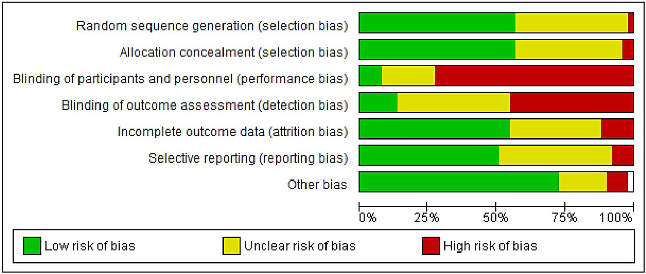
The results of the risk of bias evaluation are presented in Figure 2. Overall, the studies demonstrated a high risk of detection and performance bias. This was primarily due to the inherent differences between NIV devices and their comparators, including interface types (e.g., nasal or oronasal masks), and the practical inability to blind participants and personnel to the intervention. These limitations may have influenced subjective and patient-reported outcomes. However, the funnel plot showed relative symmetry (Figure 3), and Egger’s test returned a p-value of 0.324, exceeding the conventional significance threshold of 0.05, suggesting a low risk of publication bias.

Meta-analysis results

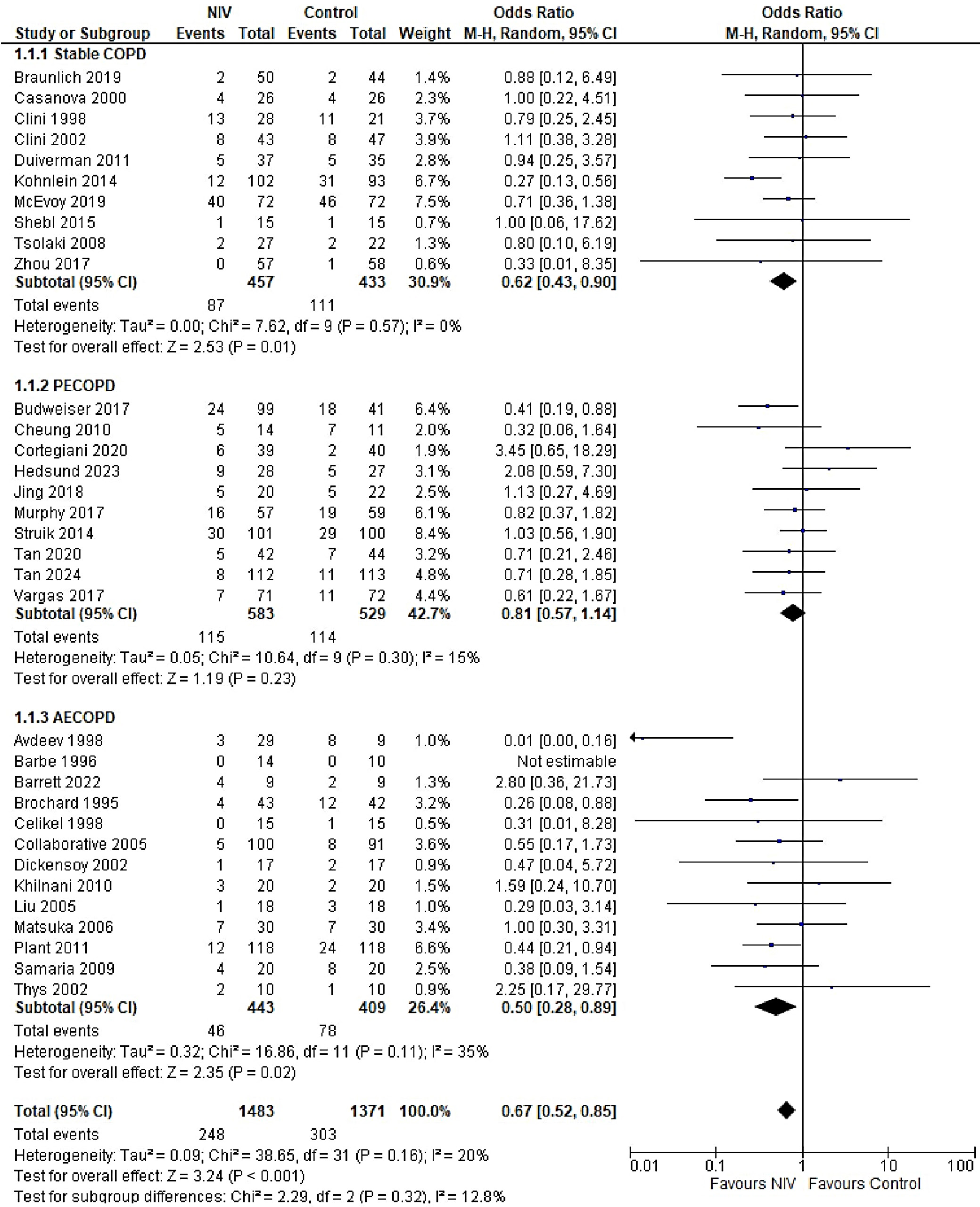
The overall risk of mortality was significantly lower in the NIV group compared to the control group (OR = 0.67, 95% CI: 0.52–0.85, p < 0.001), with low heterogeneity observed across studies (I2 = 20%). However, when stratified by COPD stage, the difference in mortality was not statistically significant (p = 0.32). Mortality outcomes were reported across all COPD subgroups, each exhibiting low-to-moderate heterogeneity (I2 range: 0–35%). The highest heterogeneity was observed in the AECOPD subgroup (I2 = 35%), possibly due to variations in follow-up duration (Figure 4). The funnel plot for mortality outcomes demonstrated approximate symmetry, suggesting a low risk of publication bias, which was supported by Egger’s test results: stable COPD (p = 0.136), PECOPD (p = 0.211) and AECOPD (p = 0.141) (Supplementary Fig. 1).

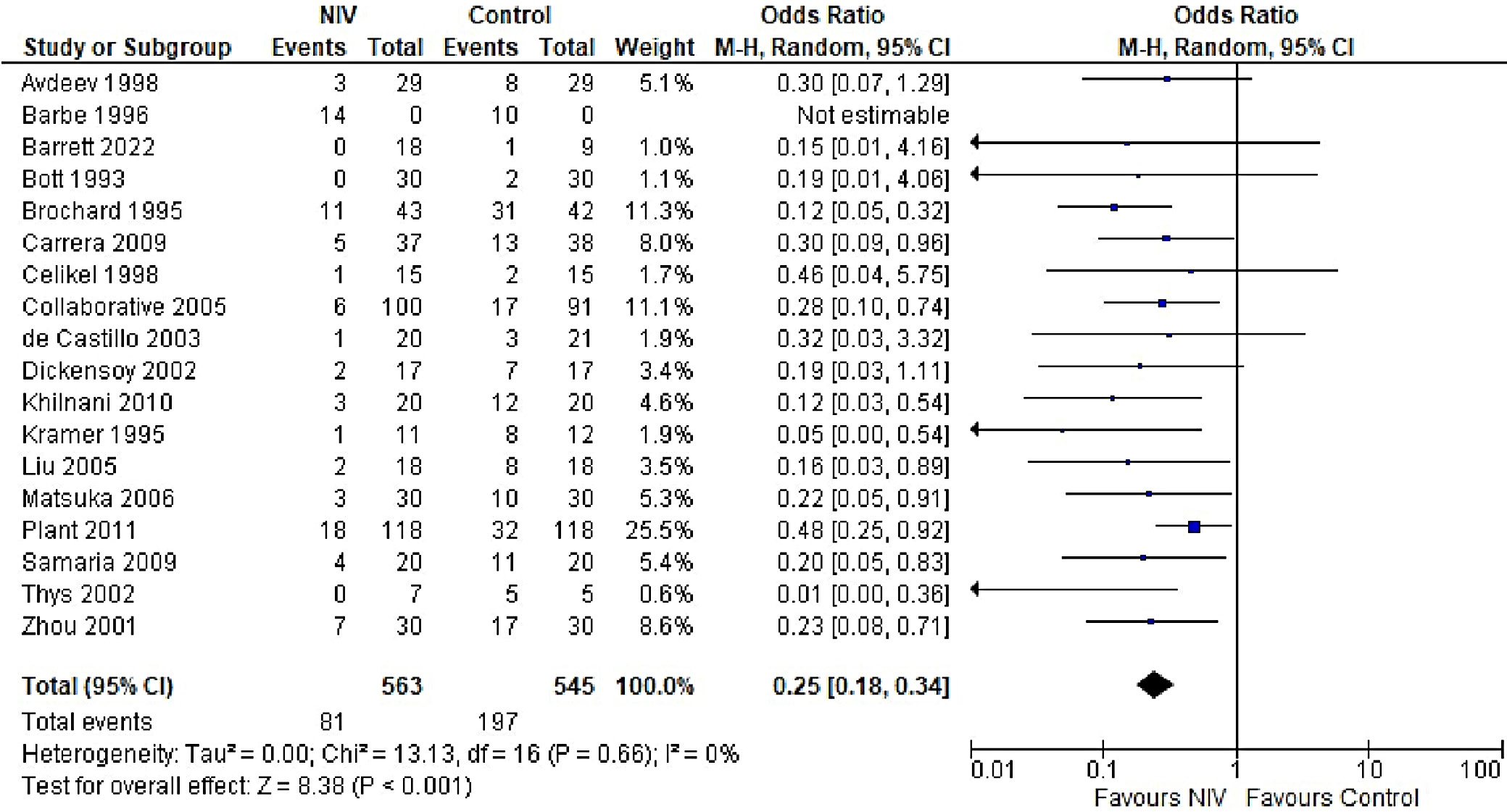
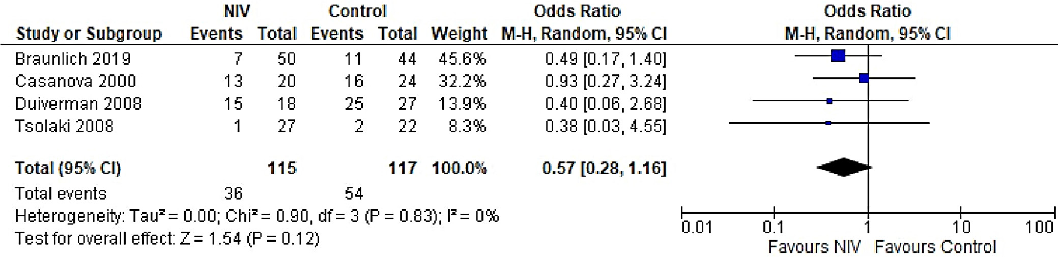
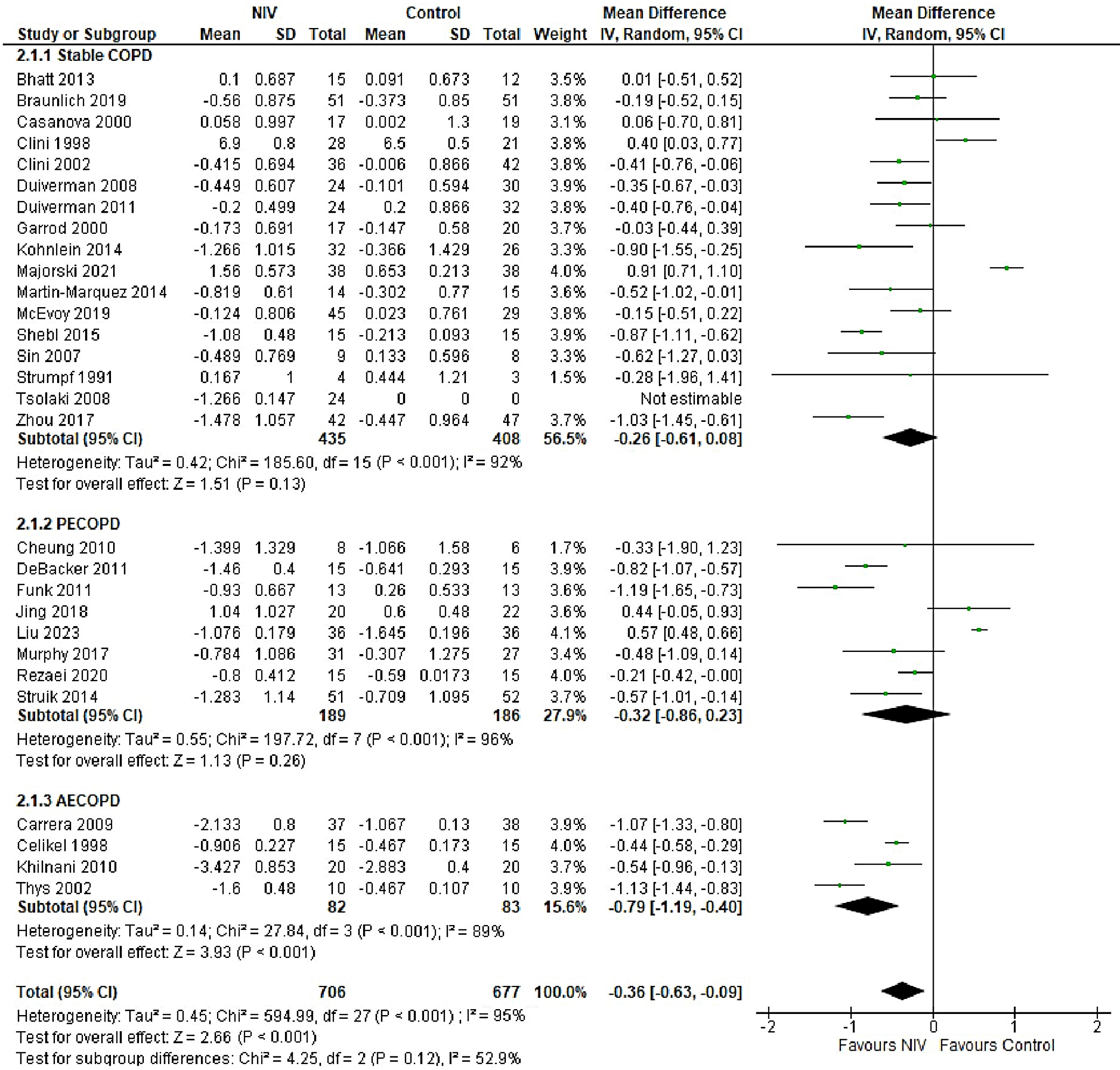
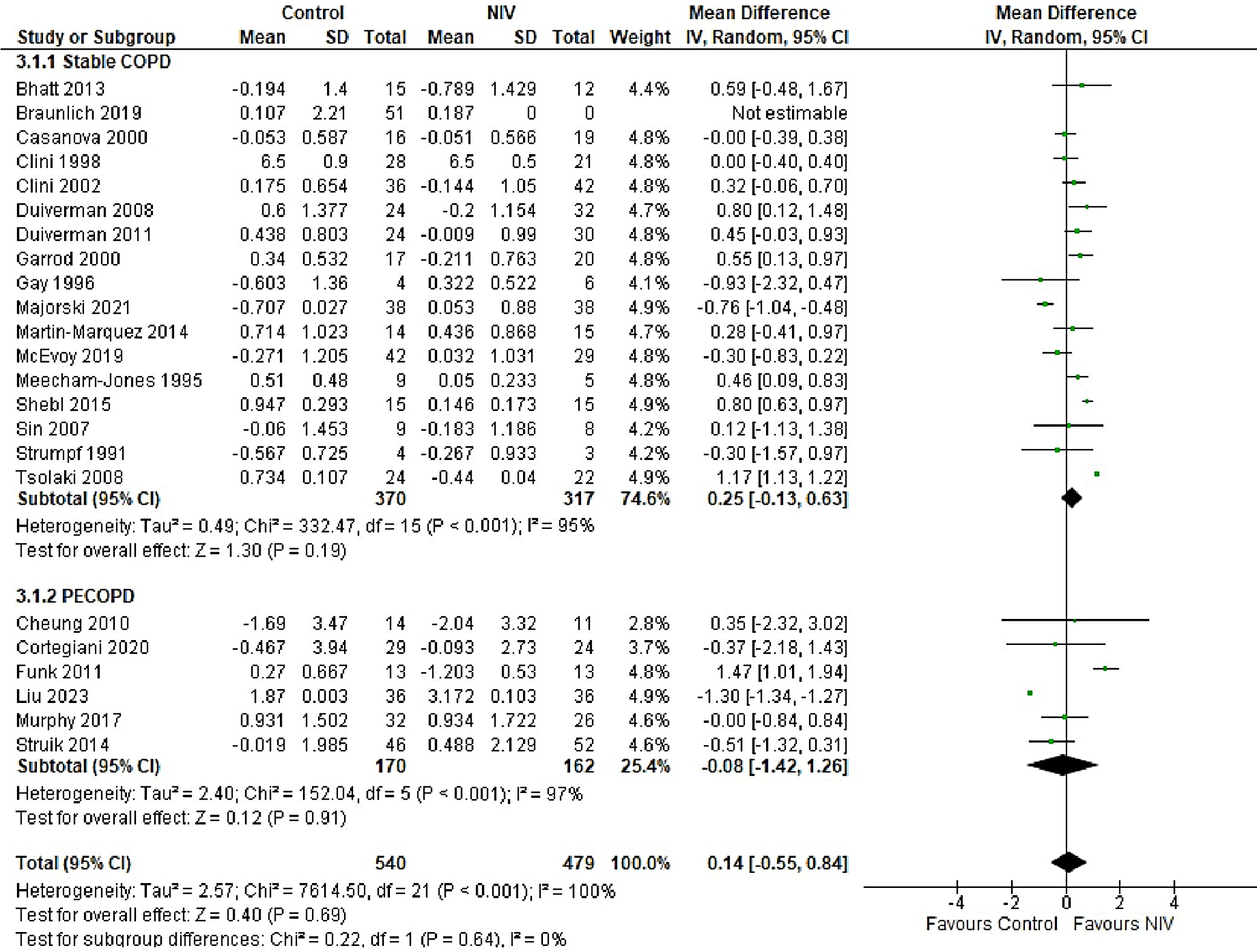
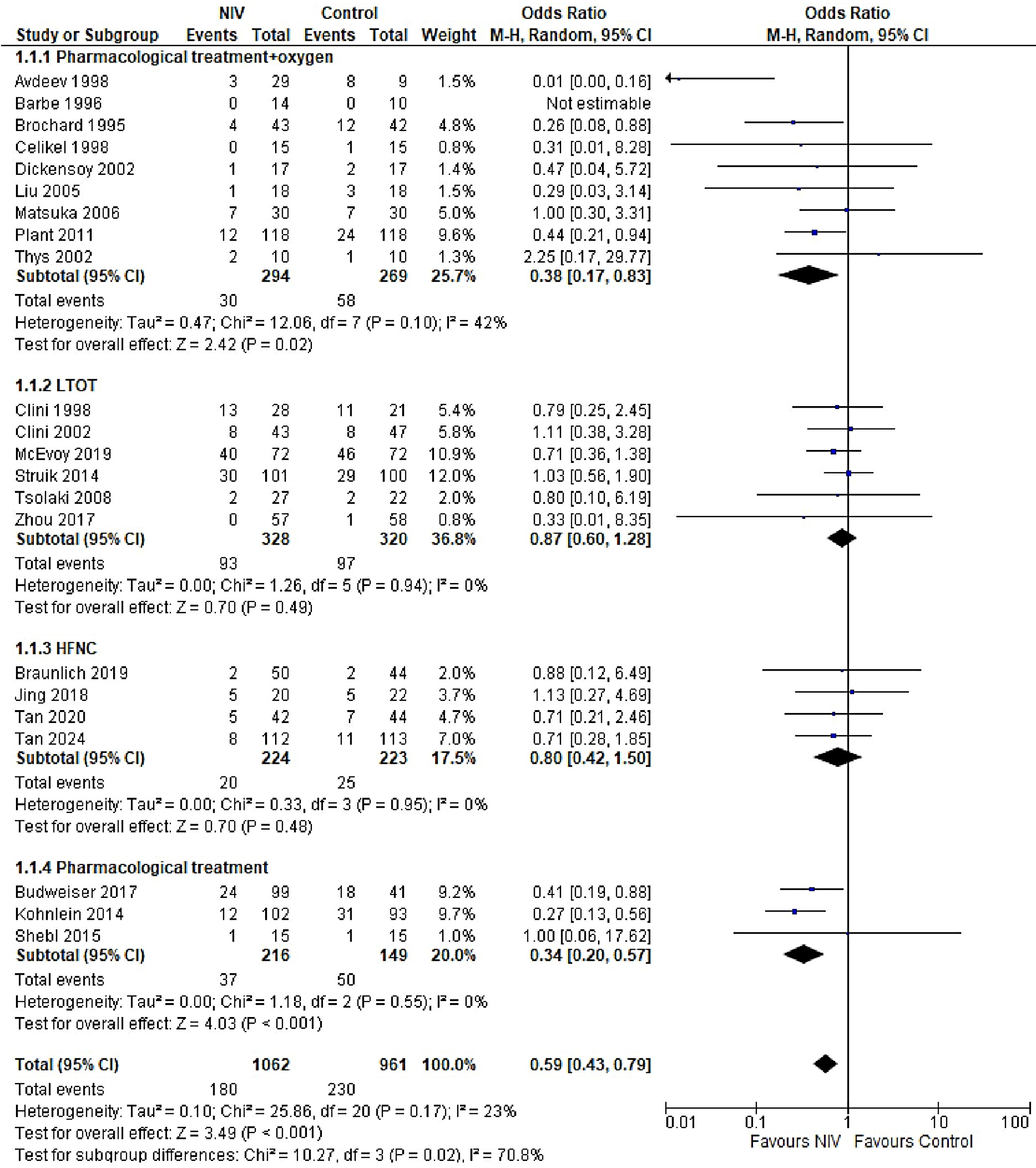
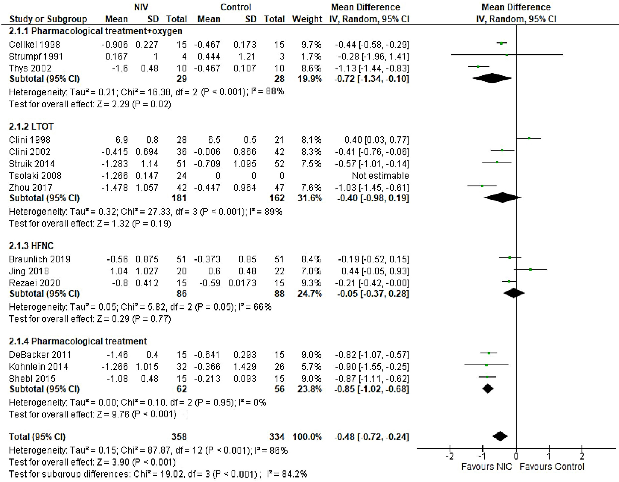
In patients with AECOPD, NIV significantly reduced the risk of intubation compared to the control group (OR = 0.25, 95% CI: 0.18–0.34, p < 0.001). This analysis, based on 18 trials, showed no heterogeneity (I2 = 0%), suggesting consistency in patient characteristics and study conditions (Figure 5). The funnel plot appeared symmetrical, and Egger’s test confirmed a low risk of publication bias (p = 0.173) (Supplementary Fig. 2). In patients with stable COPD, the exacerbation rate did not differ significantly between the NIV and control groups (OR = 0.57, 95% CI: 0.28–1.16, p = 0.83), based on data from 4 studies. The low heterogeneity (I2 = 0%) likely reflects similar patient profiles and consistent definitions of stable COPD (i.e., no exacerbations within the last 4 weeks). One trial27 (Casanova et al.) showed a nonsignificant result, possibly due to a slightly different definition of stability (no exacerbations in the past 3 months). The type of control intervention did not influence the direction of the results (Figure 6). The funnel plot suggested a low risk of publication bias (Supplementary Fig. 3). Continuous outcomes, such as PaCO2 and PaO2 levels, were analyzed across COPD subgroups, reported as changes in arterial blood gases from baseline to follow-up. Noninvasive ventilation was associated with a significantly greater reduction in PaCO2 levels compared to the control group (MD = –0.36, 95% CI: –0.63 to –0.09; p < 0.001), although high heterogeneity was observed (I2 = 95%). When stratified by COPD subgroup, trials involving Acute exacerbation chronic obstructive pulmonary disease (AECOPD) patients demonstrated a significant reduction in PaCO2 with NIV (MD = –0.79, 95% CI: –1.19 to –0.40; p < 0.001; I2 = 89%), while trials in stable COPD and PECOPD populations did not show statistically significant benefits. The high heterogeneity likely reflects variations in patient characteristics, control interventions and follow-up durations (ranging from 3 to 12 months) (Figure 7). The funnel plot appeared symmetrical, suggesting a low risk of publication bias, which was supported by Egger’s test for the stable COPD subgroup (p = 0.119) (Supplementary Fig. 4). No significant difference in PaO2 levels was observed between the NIV and control groups in trials involving stable COPD and PECOPD patients (MD = 0.14, 95% CI: –0.55 to 0.84; p = 0.69) (Figure 8). High heterogeneity (I2 = 100%) was likely attributable to differences in patient characteristics, ventilator settings, comparator treatments, and follow-up durations. The subgroup analysis did not reveal a statistically significant difference (p = 0.64). The funnel plot appeared symmetrical (Supplementary Fig. 5), indicating a low risk of publication bias, which was confirmed by Egger’s test for the stable COPD subgroup (p = 0.159). Subgroup analyses based on the type of control treatment revealed a significant reduction in mortality when the comparator was pharmacological therapy combined with oxygen supplementation (OR = 0.38, 95% CI: 0.17−0.83; p = 0.02; I2 = 42%) or pharmacological therapy alone (OR = 0.34, 95% CI: 0.20−0.57; p < 0.001; I2 = 0%) (Figure 9). However, no significant reduction in mortality was observed when NIV was compared with LTOT or HFNC. The funnel plot for control-treatment subgroups (Supplementary Fig. 6) displayed high symmetry, suggesting a low likelihood of publication bias. Similar effects were observed for PaCO2 levels (Figure 10), with a significant reduction associated with NIV compared to pharmacological treatment combined with oxygen (MD = −0.72, 95% CI: −1.34 to −0.10, p = 0.02, I2 = 88%) and pharmacological treatment alone (MD = −0.85, 95% CI: −1.02 to −0.68, p < 0.001, I2 = 0%). The funnel plot (Supplementary Fig. 7) displayed a high degree of symmetry across various control treatments, indicating a low risk of publication bias.

Discussion

This paper provides an updated synthesis of the evidence for using NIV instead of usual care to manage different stages of COPD. Important outcomes such as mortality, exacerbations, intubation, and arterial gas levels (e.g. PaCO2 and PaO2 were assessed to determine the long- and short-term effects of NIV on patients at different stages of COPD. The findings of this study emphasize the importance of NIV in reducing mortality and morbidity, as well as the incidence of adverse events such as exacerbations and intubations, in COPD patients. Although several trials and meta-analyses have demonstrated the beneficial effects of NIV on survival, hospital admissions and length of stay, as well as improving quality of life, there are challenges associated with NIV devices that limit their applicability. These include mask leaks, difficulty wearing the device, mask discomfort, and severe hypoxia. Therefore, it is necessary to understand which patient baseline characteristics are most likely to benefit from NIV, the ideal length of treatment and continuous monitoring protocols, and training on the appropriate use of devices and ventilator settings.

The risk of bias assessment using the Cochrane tool revealed high performance and detection biases. Blinding of personnel and participants was not possible, as NIV devices and interfaces differ from usual care. Such treatments as pharmacological interventions and sham treatments may not always be feasible. This introduces an inherent bias when subjective outcomes such as quality of life and symptom assessment are measured. Therefore, this study only included objective measurements such as mortality, intubation and exacerbation rates, and arterial blood gases, which are not subject to bias and provide more reliable results regarding the efficacy of NIV. Our study showed that NIV use across different COPD stages decreases mortality. However, there was no significant difference in mortality outcomes between PECOPD patients receiving NIV or usual care.

The effect of NIV on COPD varies between patients with stable COPD and those with PECOPD. Some meta-analyses have reported nonsignificant differences in mortality outcomes among patients with stable COPD and PECOPD. However, other studies, including our analysis, have demonstrated a mortality benefit in patients with stable COPD.14, 70 This benefit may be attributed to persistent hypercapnia commonly observed in stable COPD patients, in contrast to PECOPD patients, where hypercapnia may be transient and often accompanied by additional complications. In patients with AECOPD, NIV was found to significantly reduce mortality rates, which could be linked to a reduced need for intubation – thus minimizing the risk of prolonged hospital and ICU stays and associated infections. The exacerbation rate among patients with stable COPD was significantly lower with NIV compared to usual care. Although this analysis included only a limited number of studies (n = 4), it provides preliminary evidence that long-term or chronic use of NIV may help reduce exacerbation frequency in COPD patients. A greater reduction in PaCO2 levels in stable COPD patients likely contributes to improved alveolar ventilation and respiratory muscle function, particularly in cases of chronic hypercapnia, which may predispose patients to fewer exacerbations. Consistent with previous meta-analyses and the GOLD report, our findings demonstrated a significant improvement in PaCO2 levels in all patients receiving NIV compared to usual care. However, no significant improvement was observed in PaO2 levels between the 2 groups. The beneficial effect of NIV in lowering PaCO2 is attributed to enhanced alveolar ventilation, increased tidal volume and reduced respiratory muscle fatigue as a result of the positive airway pressure delivered by NIV devices, which facilitates more effective CO2 elimination.2, 11, 71 However, destruction of alveolar units and underlying lung pathology can impair oxygen diffusion into the bloodstream, thereby limiting the impact of NIV on arterial oxygenation and often necessitating supplemental oxygen. Moreover, because several studies included oxygen supplementation or LTOT in the standard treatment arms, the differences in PaO2 levels between NIV and comparator groups were often attenuated, rendering the effect of NIV on oxygenation statistically nonsignificant. Subgroup analyses revealed that NIV significantly reduced both mortality and PaCO2 levels when compared to pharmacological or standard oxygen therapy. In contrast, differences were nonsignificant when compared to LTOT and HFNC therapy. These findings may be attributed to the superior ventilatory support provided by NIV, which enhances alveolar ventilation and facilitates CO2 clearance – mechanisms not fully addressed by oxygen or pharmacologic therapy alone. Notably, HFNC delivers heated and humidified oxygen while also generating low-level positive airway pressure, promoting CO2 washout, which may explain its comparable or potentially superior efficacy to NIV in certain clinical scenarios.

Limitations

Despite our study proving a comprehensive and current review and quantitative evidence on the use of NIV in COPD patients in different stages, it has certain limitations that require consideration when generalizing these results. The high heterogeneity present in the results of this analysis, particularly for arterial blood gas levels, PaCO2 and PaO2 is related to heterogenous patient populations and baseline demographics. Differences in baseline PaCO2 levels, comorbidities, and severity of COPD can affect the magnitude of effect of NIV, thereby affecting the efficacy of NIV. Other factors, such as ventilator pressure settings and duration of application, concomitant treatments, comparators such as supplemental oxygen treatment, variable lengths of follow-up between different studies, also result in heterogeneity. However, analyzing studies with homogenous populations regarding patient characteristics results in a limited number of studies included in each subgroup, underpowering the study. Differences in the definitions of outcomes, such as exacerbation, which are often not specified in the trials, also makes combining of results challenging. The numbers of exacerbations and intubations have not been reported for most studies prior to the use of NIV. This lack of information makes it difficult to determine the effectiveness of NIV in decreasing the frequency of attacks. As discussed above, the high risk of detection bias makes it necessary to interpret the results with caution as they may not be generalizable in larger patient populations and the efficacy of NIV can be overestimated without blinding. To provide more robust evidence on the use of NIV, it is important to identify patient subgroups, such as those with hypercapnic respiratory failure, certain comorbidities, and stages of COPD, that are most likely to benefit from NIV use. Additionally, since the efficacy of NIV depends upon its correct use and adherence particularly in case of long-term use, trained personnel are required to administer it, and patients and their caregivers should be educated on the proper use of masks to maximize the benefits offered by NIV.

Since NIV devices demonstrated particular benefit in AECOPD patients by reducing mortality, intubation rates and PaCO2 levels, their integration into clinical practice appears most justified in this subgroup. Moreover, the inconsistent effects of NIV on PaO2 and PaCO2 levels suggest that NIV may not be the optimal ventilation strategy when oxygenation is the primary therapeutic goal. Instead, its use should be focused on improving alveolar ventilation and respiratory muscle function. Understanding the differential effects of NIV across COPD stages may assist clinicians in selecting appropriate candidates who are most likely to derive significant benefit from this intervention.

Conclusions

In this meta-analysis, we demonstrated that NIV devices reduce mortality, exacerbation frequency and intubation rates in patients across different stages of COPD, including stable COPD, PECOPD and AECOPD. The impact of NIV on gas exchange was variable: NIV significantly reduced PaCO2 levels, but the improvement in PaO2 was not statistically significant. The efficacy of NIV also varied depending on the COPD stage, with the greatest benefit observed in patients with AECOPD. The high heterogeneity among studies likely reflects differences in patient populations, baseline characteristics, NIV settings, and duration of use. These findings highlight the need to individualize NIV therapy based on patient-specific factors to optimize clinical outcomes.

Supplementary data

The supplementary materials are available at https://doi.org/10.5281/zenodo.17072868. The package includes the following files:

Supplementary Fig. 1. Funnel plot for mortality outcome a) in stable COPD patients b) in PECOPD patients and c) in AECOPD.

Supplementary Fig. 2. Funnel plot for intubation in AECOPD patients.

Supplementary Fig. 3. Funnel plot for exacerbation in stable COPD patients.

Supplementary Fig. 4. Funnel plot for PaCO2 levels a) in stable COPD patients b) in PECOPD patients and c) in AECOPD.

Supplementary Fig. 5. Funnel plot for PaO2 level a) in stable COPD patients b) in PECOPD patients.

Supplementary Fig. 6. Funnel plot for mortality in intervention group stratified by type of control treatment.

Supplementary Fig. 7. Funnel plot for PaCO2 levels in intervention group stratified by type of control treatment.

Use of AI and AI-assisted technologies

Not applicable.

Tables


Table 1. Search strategy

Search terms

MeSH terms and keywords

Search term 1 (#1)

(“Noninvasive Ventilation”[MeSH Terms] OR “Continuous Positive Airway Pressure”[MeSH Terms] OR (“BiPAP”[All Fields] OR “CPAP”[All Fields] OR (“positive pressure respiration”[MeSH Terms] OR (“positive pressure”[All Fields] AND “respiration”[All Fields]) OR “positive pressure respiration”[All Fields] OR (“positive”[All Fields] AND “pressure”[All Fields] AND “ventilation”[All Fields]) OR “positive pressure ventilation”[All Fields] OR “intermittent positive pressure ventilation”[MeSH Terms] OR (“intermittent”[All Fields] AND “positive pressure”[All Fields] AND “ventilation”[All Fields]) OR “intermittent positive pressure ventilation”[All Fields]) OR (“non-invasive”[All Fields] AND (“positive pressure respiration”[MeSH Terms] OR (“positive pressure”[All Fields] AND “respiration”[All Fields]) OR “positive pressure respiration”[All Fields] OR (“positive”[All Fields] AND “pressure”[All Fields] AND “ventilation”[All Fields]) OR “positive pressure ventilation”[All Fields] OR “intermittent positive pressure ventilation”[MeSH Terms] OR (“intermittent”[All Fields] AND “positive pressure”[All Fields] AND “ventilation”[All Fields]) OR “intermittent positive pressure ventilation”[All Fields])) OR ((“noninvasive”[All Fields] OR “noninvasively”[All Fields] OR “noninvasiveness”[All Fields]) AND (“positive pressure respiration”[MeSH Terms] OR (“positive pressure”[All Fields] AND “respiration”[All Fields]) OR “positive pressure respiration”[All Fields] OR (“positive”[All Fields] AND “pressure”[All Fields] AND “ventilation”[All Fields]) OR “positive pressure ventilation”[All Fields] OR “intermittent positive pressure ventilation”[MeSH Terms] OR (“intermittent”[All Fields] AND “positive pressure”[All Fields] AND “ventilation”[All Fields]) OR “intermittent positive pressure ventilation”[All Fields]))))

Search term 2 (#2)

(“pulmonary disease, chronic obstructive”[MeSH Terms] OR “pulmonary disease, chronic obstructive”[MeSH Terms] OR “pulmonary disease, chronic obstructive”[MeSH Terms] OR ((“chronic”[All Fields] OR “chronical”[All Fields] OR “chronically”[All Fields] OR “chronicities”[All Fields] OR “chronicity”[All Fields] OR “chronicization”[All Fields] OR “chronics”[All Fields]) AND (“respiratory tract diseases”[MeSH Terms] OR “respiration disorders”[MeSH Terms])) OR “pulmonary disease, chronic obstructive”[MeSH Terms] OR “pulmonary disease, chronic obstructive”[MeSH Terms] OR “pulmonary emphysema”[MeSH Terms]))

Search term 3 (#3)

#1 and #2

MeSH – Medical Subject Headings.
Table 2. Search strategy and hits for chronic obstructive pulmonary disease (COPD) and noninvasive ventilation (NIV) literature review

ID

Search

Hits

#1

MeSH descriptor: [Pulmonary Disease, Chronic Obstructive] explode all trees

8385

#2

MeSH descriptor: [Pulmonary Emphysema] explode all trees

409

#3

(“chronic obstructive airway disease”):ti,ab,kw (Word variations have been searched)

199

#4

(“chronic obstructive lung disease”):ti,ab,kw (Word variations have been searched)

8617

#5

#1 or #2 or #3 or #4

14621

#6

MeSH descriptor: [Noninvasive Ventilation] explode all trees

580

#7

MeSH descriptor: [Continuous Positive Airway Pressure] explode all trees

1929

#8

(BiPAP):ti,ab,kw (Word variations have been searched)

478

#9

(CPAP):ti,ab,kw (Word variations have been searched)

5890

#10

(“bilevel positive airway pressure”):ti,ab,kw (Word variations have been searched)

289

#11

(“positive pressure ventilation”):ti,ab,kw (Word variations have been searched)

2366

#12

#6 or #7 or #8 or #9 or #10 or #11

9033

#13

#5 and #12

433

MeSH – Medical Subject Headings.
Table 3. Details of intervention and control groups of the studies included in the meta-analysis

Study name

Intervention

Control

Avdeev, 199818

NIV (BiPAP) + usual care

oxygen + bronchodilators + steroids + theophylline

Barbe et al., 1996 19

NIV (BiPAP) + usual care

salbutamol + prednisolone + oxygen

Barrett et al., 202220

NIV (ICU ventilator in NIV mode)

extracorporeal carbon dioxide removal (ECCO2R)

Bhatt et al., 201321

domiciliary NPPV (oronasal mask/nasal pillows, BiPAP)

usual therapy

Bott et al., 199322

NPPV + usual care

oxygen, bronchodilators, antibiotics, diuretics, respiratory stimulants, corticosteroids

Braunlich et al., 201923

NIV

nasal high flow (NHF)

Brochard et al., 199524

NIV (ARM 25)

oxygen, subcutaneous heparin, antibiotics, bronchodilators

Budweiser et al., 200725

NIV (BiPAP, Twin Air®, Smart Air®) + pharmacological treatment

usual pharmacological agents

Carrera et al., 200926

NIV (BiPAP and facial mask)

sham NIV

Casanova et al., 200027

nocturnal nasal NPPV (nasal mask)

standard care + LTOT

Celikel et al., 199828

continuous NIV + usual care

oxygen + pharmacological treatment

Cheung et al., 201029

nocturnal NIV (BiPAP)

placebo home NIV

Clini et al., 199830

Nocturnal NIV + LTOT

LTOT

Clini et al., 200231

nocturnal NPPV (BiPAP, nasal mask) + LTOT

LTOT

Collaborative Research Group of Noninvasive Mechanical Ventilation for Chronic Obstructive Pulmonary Disease, 200532

NIV (oronasal mask, BiPAP) + pharmacological treatment

oxygen via nasal cannula + pharmacological treatment

Cortegiani et al., 202033

NIV (full-face or oronasal mask)

high flow nasal therapy (HFNT)

del Castillo et al., 200334

NIV (BiPAP, mask)

oxygen + pharmacological treatment

DeBacker et al., 201135

nocturnal NIV pharmacological treatment

pharmacological treatment

Dickensoy et al., 200236

NIV (BiPAP) + usual care

oxygen + pharmacological treatment

Duiverman et al., 200837

nocturnal NPPV (BiPAP, nasal/oronasal mask)

pulmonary rehabilitation

Duiverman et al., 201138

nocturnal NPPV + rehabilitation

rehabilitation

Funk et al. 201139

nocturnal NIV (BiPAP)

no NIV

Garrod et al., 200040

nocturnal NPPV + exercise training program (BiPAP, nasal mask)

exercise training program

Gay et al., 199641

nocturnal NIV (BiPAP, nasal mask)

sham NIV

Hedsund et al., 202342

long-term NIV + standard of care

standard of care

Jing et al., 201943

NIV (VPAP)

HFNC

Khilnani et al., 201044

NIV (BiPAP)

oxygen + pharmacological treatment

Köhnlein et al., 201445

nocturnal NPPV (nasal/face mask) + pharmacological treatment

pharmacological treatment

Kramer et al., 199546

NIV (BiPAP) + pharmacological treatment + oxygen

oxygen + pharmacological treatment

Liu et al., 200547

NIV (BiPAP, face mask) + pharmacological treatment + oxygen

pharmacological treatment + oxygen

Liu et al., 202348

NPPV (BiPAP)

transnasal high-flow humidified oxygen therapy

Majorski et al., 202149

portable NIV device

no NIV device

Martin-Marquez et al., 201450

nocturnal NIV (BiPAP) + training program

training program

Matsuka et al., 200651

NIV (BiPAP) + oxygen + pharmacological treatment

oxygen + pharmacological treatment

McEvoy et al., 201952

nocturnal NIV + usual care + LTOT

usual care + LTOT

Meecham-Jones et al., 199553

nocturnal NIV (BiPAP) + oxygen therapy

oxygen therapy

Murphy et al., 201754

nocturnal NIV + home oxygen therapy

home oxygen therapy

Plant et al., 200155

NIV (BiPAP) + oxygen + pharmacological treatment

oxygen + pharmacological treatment

Rezaei et al., 202056

NIV (VPAP)

high-oxygen nasal cannula

Samaria., 200957

NIV (BiPAP) + oxygen + pharmacological treatment

oxygen + pharmacological treatment

Shebl et al., 201558

nocturnal NPPV (BiPAP) + pharmacological treatment

pharmacological treatment

Sin et al., 200759

NIV (VPAP, nasal/oronasal mask)

sham treatment

Struik et al., 201460

nocturnal NIV + standard treatment

pharmacological treatment + LTOT

Strumpf et al., 199161

nocturnal NIV (BiPAP, nasal mask)

oxygen + pharmacological treatment

Tan et al., 202062

NIV (BiPAP, oronasal mask)

NFNC oxygen therapy

Tan et al., 202463

NIV (BiPAP, oronasal mask)

HFNC oxygen therapy

Thys et al., 200264

NIV (BiPAP) + supplemental oxygen

supplemental oxygen + pharmacological treatment

Tsolaki et al., 200865

NIV (BiPAP, face mask)

LTOT + pharmacological treatment

Vargas et al., 201766

NIV (face mask)

standard oxygen therapy

Xiang et al., 200767

home NPPV + standard treatment

standard treatment

Zhou et al., 200168

NIV (BiPAP, nasal/face mask) + oxygen + pharmacological treatment

pharmacological treatment

Zhou et al., 201769

nocturnal NIV + pharmacological treatment

LTOT + pharmacological treatment

NIV – noninvasive ventilation; BiPAP – bilevel positive airway pressure; NPPV – noninvasive positive pressure ventilation; VPAP – variable positive airway pressure; LTOT – long-term oxygen therapy; HFNC – high flow nasal cannula.
Table 4. Characteristics of the included randomized controlled trials (RCTs)

Study name

COPD type

Patient characteristics

Length of follow-up

Avdeev, 199818

AECOPD

male and female, acute respiratory failure, mean age 65 years

N/A

Barbe et al., 1996 19

AECOPD

Male, 68 ±2 years, acute respiratory failure

N/A

Barrett et al., 202220

AECOPD

male and female, ≥18 years, hypercapnia pH < 7.3

N/A

Bhatt et al., 201321

stable COPD

male and female, mean age: 69.4 years, no exacerbations in last 4 weeks, PaCO2 < 52 mm Hg

6 weeks, 3 and 6 months

Bott et al., 199322

AECOPD

male and female, ≤80 years, acute exacerbation of COPD

At least 30 days

Braunlich et al., 201923

stable COPD

male and female, mean age: 65.3 years, no exacerbations in last 4 weeks, BMI ≤ 30 kg/m2

12 weeks

Brochard et al., 199524

AECOPD

male and female, acute exacerbation with respiratory acidosis

N/A

Budweiser et al., 200725

PECOPD

male and female, mean age: 65 years, PaCO2 > 50 mm hg after treatment of exacerbation, PaCO2 59 mm Hg

6 months

Carrera et al., 200926

AECOPD

male and female, 67 ±9 years,

N/A

Casanova et al., 200027

stable COPD

male and female, mean age: 66.6 years, no exacerbations in last 3 months, mean PaCO2: 6.8 kPa

12 months

Celikel et al., 199828

AECOPD

male and female, hypercapnic acute respiratory failure

N/A

Cheung et al., 201029

PECOPD

male and female, mean age: 70.3 years, acute respiratory failure and treatment with pharmacological agents, PaCO2 7.5 kPa

12 months

Clini et al., 199830

stable COPD

male and female, mean age: 66 years, no exacerbation in last 4 weeks, PaCO2: 6–8 kPa

36 months

Clini et al., 200231

stable COPD

male and female, ≤75 years, no exacerbation in last 4 weeks, PaCO2 > 6.6 kPa

24 months

Collaborative Research Group of Noninvasive Mechanical Ventilation for Chronic Obstructive Pulmonary Disease, 200532

AECOPD

male and female, <85 years, pH>7.25

N/A

Cortegiani et al., 202033

PECOPD

male and female, mean age: 75 years, acute hypercapnic respiratory failure and exacerbation, PaCO2 ≥ 55 mm Hg

N/A

del Castillo et al., 200334

AECOPD

male and female, acidotic hypercapnic respiratory failure, mean age: 67 years

N/A

DeBacker et al., 201135

PECOPD

male and female, mean age: 65.6 years, persistent hypercapnia PaCO2 > 6 kPa, hospitalized due to exacerbation

12 months

Dickensoy et al., 200236

AECOPD

male, mean age: 65 years

N/A

Duiverman et al., 200837

stable COPD

male and female, 40–76 years, no exacerbation in last 4 weeks, PaCO2 > 6 kPa

24 months

Duiverman et al., 201138

stable COPD

male and female, mean age: 62 years, no exacerbation in last 4 weeks, PaCO2 > 6 kPa

24 months

Funk et al. 201139

PECOPD

male and female, mean age: 63 years, requiring invasive or noninvasive mechanical ventilation, PaCO2 94 mm Hg

N/A

Garrod et al., 200040

stable COPD

male and female, 38–84 years, no exacerbations in last 4 weeks, mean PaCO2 6.1 kPa

8 weeks

Gay et al., 199641

stable COPD

male and female, mean age: 67.6 years, BMI ≤ 30 kg/m2, PaCO2 > 6 kPa

3 months

Hedsund et al., 202342

PECOPD

male and female, mean age: 72 years, admission with acute hypercapnic respiratory failure, PaCO2 9 kPa

12 months

Jing et al., 201943

PECOPD

male and female, mean age: 74 years, intubated for exacerbation, PaCO2 53 mm Hg

N/A

Khilnani et al., 201044

AECOPD

male and female, AECOPD leading to hypoxemia and respiratory acidosis pH < 7.35, mean age: 60 years

N/A

Köhnlein et al., 201445

stable COPD

male and female, mean age: 63.2 years, no exacerbations in last 4 weeks, PaCO2 7.8 kPa

12 months

Kramer et al., 199546

AECOPD

male and female, respiratory distress, pH < 7.35

N/A

Liu et al., 200547

AECOPD

acute exacerbation, pH 7.25–7.35, mean age: 70 years

N/A

Liu et al., 202348

PECOPD

male and female, mean age: 69 years, AECOPD and type II respiratory failure PaCO2 53 mm Hg

N/A

Majorski et al., 202149

stable COPD

male and female, mean age: 67 years, no exacerbation in last 4 weeks, PaCO2 42kPa

N/A

Martin-Marquez et al., 201450

stable COPD

male and female, mean age: 68.3 years, clinically stable for last 3 months, PaCO2 6.7 kPa

3 months

Matsuka et al., 200651

AECOPD

male and female, pH < 7.35, mean age: 67 years

N/A

McEvoy et al., 201952

stable COPD

male and female, mean age: 68 years, stable hypercapnic respiratory failure in last 6 months, PaCO2 7.3 kPa

6 months

Meecham-Jones et al., 199553

stable COPD

male and female, mean age: 65.9 years, stable clinical state, PaCO2 7.4 kPa

6 months

Murphy et al., 201754

PECOPD

male and female, mean age: 66 years, acute decompensated hypercapnic exacerbations of COPD, PaCO2 7.9 kPa

12 months

Plant et al., 200155

AECOPD

male and female, mean age: 69 years, tachypnoea, pH 7.25–7.35

N/A

Rezaei et al., 202056

PECOPD

male and female, mean age: 60 years, moderate-to-severe COPD exacerbation, PaCO2 64 mm Hg

N/A

Samaria, 200957

AECOPD

NA

N/A

Shebl et al., 201558

stable COPD

male and female, mean age: 65.5 years, no exacerbation in last 4 weeks, PaCO2 7.3 kPa

6 months

Sin et al., 200759

stable COPD

male and female, mean age: 65.4 years, PaCO2 5.8 kPa

3 months

Struik et al., 201460

PECOPD

male and female, mean age: 63.4 years, PaCO2 7.8 kPa

12 months

Strumpf et al., 199161

stable COPD

male and female, mean age: 65.6 years, no exacerbation in last 1 week, PaCO2 6.2 kPa

6 months

Tan et al., 202062

PECOPD

male and female, mean age: 69 years, hypercapnic respiratory failure with invasive ventilation, PaCO2 51 mm Hg

N/A

Tan et al., 202463

PECOPD

male and female, mean age: 71 years, hypercapnic respiratory failure, PaCO2 62 mm Hg

N/A

Thys et al., 200264

AECOPD

male and female, mean age: 73 years, acute respiratory distress, PaCO2 57 mm Hg

N/A

Tsolaki et al., 200865

stable COPD

male and female, mean age: 66 years, no exacerbations in last 4 weeks, chronic hypercapnic respiratory failure, PaCO2 54 mm Hg

12 months

Vargas et al., 201766

PECOPD

male and female, mean age: 64 years, patients intubated for at least 48 h, PaCO2 > 45 mm Hg

3 months

Xiang et al., 200767

PECOPD

male and female, severe COPD and hospitalized, PaCO2 > 55 mm Hg

24 months

Zhou et al., 200168

AECOPD

Male and female, mean age: 64 years, respiratory failure, PaCO2 > 55 mm Hg

N/A

Zhou et al., 201769

stable COPD

male and female, mean age: 67.7 years, clinically stable and chronic hypercapnia, PaCO2 8 kPa

3 months

AECOPD – acute exacerbation of chronic obstructive pulmonary disease; stable COPD – stable chronic obstructive pulmonary disease; PECOPD – pulmonary embolism with chronic obstructive pulmonary disease; BMI – body mass index; N/A – not applicable.

Figures


Fig. 1. Flow chart for identification and inclusion of studies in the meta-analysis according to Preferred Reporting Items for Systematic and Meta-Analyses (PRISMA)
Fig. 2. Risk of bias summary for studies included in the meta-analysis
Fig. 3. Funnel plot for overall publication bias for studies included in the meta-analysis
Fig. 4. Forest plot for mortality outcomes: A. Stable chronic obstructive pulmonary disease (COPD) patients; B. Post-exacerbation COPD (PECOPD) patients; C. Acute exacerbation COPD (AECOPD) patients
Fig. 5. Forest plot for intubation in AECOPD patients
Fig. 6. Forest plot for exacerbation in stable chronic obstructive pulmonary disease (COPD) patients
Fig. 7. Forest plot for PaCO2 levels: A. Stable chronic obstructive pulmonary disease (COPD) patients; B. Post-exacerbation COPD (PECOPD) patients; C. Acute exacerbation COPD (AECOPD) patients
Fig. 8. Forest plot for PaO2 levels: A. Stable chronic obstructive pulmonary disease (COPD) patients; B. Post-exacerbation COPD (PECOPD) patients
Fig. 9. Forest plot for mortality in intervention group stratified by type of control treatment
Fig. 10. Forest plot for PaCO2 levels in intervention group stratified by type of control treatment

References (71)

  1. Yoon HK, Park YB, Rhee CK, Lee JH, Oh YM; Committee of the Korean COPD Guideline 2014. Summary of the Chronic Obstructive Pulmonary Disease Clinical Practice Guideline Revised in 2014 by the Korean Academy of Tuberculosis and Respiratory Disease. Tuberc Respir Dis. 2017;80(3):230. doi:10.4046/trd.2017.80.3.230
  2. Vestbo J, Hurd SS, Agustí AG, et al. Global Strategy for the Diagnosis, Management, and Prevention of Chronic Obstructive Pulmonary Disease: GOLD Executive Summary. Am J Respir Crit Care Med. 2013;187(4):347–365. doi:10.1164/rccm.201204-0596PP
  3. World Health Organization (WHtO). Chronic obstructive pulmonary disease (COPD): Fact sheet. Geneva, Switzerland: World Health Organization (WHO); 2024. https://www.who.int/news-room/fact-sheets/detail/chronic-obstructive-pulmonary-disease-(copd). Accessed August 15, 2024.
  4. Agustí A, Celli BR, Criner GJ, et al. Global Initiative for Chronic Obstructive Lung Disease 2023 Report: GOLD Executive Summary. Am J Respir Crit Care Med. 2023;207(7):819–837. doi:10.1164/rccm.202301-0106PP
  5. Müllerova H, Maselli DJ, Locantore N, et al. Hospitalized exacerbations of COPD: Risk factors and outcomes in the ECLIPSE cohort. Chest. 2015;147(4):999–1007. doi:10.1378/chest.14-0655
  6. Soriano JB, Lamprecht B, Ramírez AS, et al. Mortality prediction in chronic obstructive pulmonary disease comparing the GOLD 2007 and 2011 staging systems: A pooled analysis of individual patient data. Lancer Respir Med. 2015;3(6):443–450. doi:10.1016/S2213-2600(15)00157-5
  7. Xiang Y, Luo X. Extrapulmonary comorbidities associated with chronic obstructive pulmonary disease: A review. Int J Chron Obstruct Pulmon Dis. 2024;19:567–578. doi:10.2147/COPD.S447739
  8. Jo YS. Long-term outcome of chronic obstructive pulmonary disease: A review. Tuberc Respir Dis. 2022;85(4):289–301. doi:10.4046/trd.2022.0074
  9. Osadnik CR, Tee VS, Carson-Chahhoud KV, Picot J, Wedzicha JA, Smith BJ. Non-invasive ventilation for the management of acute hypercapnic respiratory failure due to exacerbation of chronic obstructive pulmonary disease. Cochrane Database Syst Rev. 2017;2017(7):CD004104. doi:10.1002/14651858.CD004104.pub4
  10. Raveling T, Vonk J, Struik FM, et al. Chronic non-invasive ventilation for chronic obstructive pulmonary disease. Cochrane Database Syst Rev. 2021;2021(8):CD002878. doi:10.1002/14651858.CD002878.pub3
  11. Ram F, Picot J, Lightowler J, Wedzicha J. Non-invasive positive pressure ventilation for treatment of respiratory failure due to exacerbations of chronic obstructive pulmonary disease. Cochrane Database Syst Rev. 2003;2004(1):CD004104.pub2. doi:10.1002/14651858.CD004104.pub2
  12. Corrêa TD, Sanches PR, De Morais LC, Scarin FC, Silva E, Barbas CSV. Performance of noninvasive ventilation in acute respiratory failure in critically ill patients: A prospective, observational, cohort study. BMC Pulm Med. 2015;15(1):144. doi:10.1186/s12890-015-0139-3
  13. Mukherjee R, Nenna R, Turner A. Early ward-based acute noninvasive ventilation: A paper that changed practice. Breathe. 2018;14(2):153–155. doi:10.1183/20734735.001618
  14. Park SY, Yoo KH, Park YB, et al. The long-term efficacy of domiciliary noninvasive positive-pressure ventilation in chronic obstructive pulmonary disease: A meta-analysis of randomized controlled trials. Tuberc Respir Dis. 2022;85(1):47–55. doi:10.4046/trd.2021.0062
  15. He X, Luo L, Ma Y, Chen Y. Efficacy of domiciliary noninvasive ventilation on clinical outcomes in posthospital chronic obstructive pulmonary disease patients: A meta-analysis of randomized controlled trials. Ann Palliat Med. 2021;10(5):5137–5145. doi:10.21037/apm-20-2017
  16. Higgins JPT, Thomas J, Chandler J, et al., eds. Cochrane Handbook for Systematic Reviews of Interventions. 2nd ed. Chichester, UK: Wiley; 2019. doi:10.1002/9781119536604
  17. Higgins JPT, Thompson SG, Deeks JJ, Altman DG. Measuring inconsistency in meta-analyses. BMJ. 2003;327(7414):557–560. doi:10.1136/bmj.327.7414.557
  18. Avdeev SN. Non-invasive ventilation in acute respiratory failure [in Russian]. Pulmonologia (Mosk). 2005;6:37–54. doi:10.18093/0869-0189-2005-0-6-37-54
  19. Barbe F, Togores B, Rubi M, Pons S, Maimo A, Agusti A. Noninvasive ventilatory support does not facilitate recovery from acute respiratory failure in chronic obstructive pulmonary disease. Eur Respir J. 1996;9(6):1240–1245. doi:10.1183/09031936.96.09061240
  20. Barrett NA, Hart N, Daly KJR, et al. A randomised controlled trial of non-invasive ventilation compared with extracorporeal carbon dioxide removal for acute hypercapnic exacerbations of chronic obstructive pulmonary disease. Ann Intensive Care. 2022;12(1):36. doi:10.1186/s13613-022-01006-8
  21. Bhatt S, Peterson M, Wilson J, Durairaj L. Noninvasive positive pressure ventilation in subjects with stable COPD: A randomized trial. Int J Chron Obstruct Pulmon Dis. 2013;8:581–589. doi:10.2147/COPD.S53619
  22. Bott J, Carroll MP, Conway JH, et al. Randomised controlled trial of nasal ventilation in acute ventilatory failure due to chronic obstructive airways disease. Lancet. 1993;341(8860):1555–1557. doi:10.1016/0140-6736(93)90696-E
  23. Bräunlich J, Dellweg D, Bastian A, et al. Nasal high-flow versus noninvasive ventilation in patients with chronic hypercapnic COPD. Int J Chron Obstruct Pulmon Dis. 2019;14:1411–1421. doi:10.2147/COPD.S206111
  24. Brochard L, Mancebo J, Wysocki M, et al. Noninvasive ventilation for acute exacerbations of chronic obstructive pulmonary disease. N Engl J Med. 1995;333(13):817–822. doi:10.1056/NEJM199509283331301
  25. Budweiser S, Hitzl AP, Jörres RA, et al. Impact of noninvasive home ventilation on long-term survival in chronic hypercapnic COPD: A prospective observational study. Int J Clin Pract. 2007;61(9):1516–1522. doi:10.1111/j.1742-1241.2007.01427.x
  26. Carrera M, Marín JM, Antón A, et al. A controlled trial of noninvasive ventilation for chronic obstructive pulmonary disease exacerbations. J Crit Care. 2009;24(3):473.e7–473.e14. doi:10.1016/j.jcrc.2008.08.007
  27. Casanova C, Celli BR, Tost L, et al. Long-term controlled trial of nocturnal nasal positive pressure ventilation in patients with severe COPD. Chest. 2000;118(6):1582–1590. doi:10.1378/chest.118.6.1582
  28. Çelikel T, Sungur M, Ceyhan B, Karakurt S. Comparison of noninvasive positive pressure ventilation with standard medical therapy in hypercapnic acute respiratory failure. Chest. 1998;114(6):1636–1642. doi:10.1378/chest.114.6.1636
  29. Cheung APS, Chan VL, Liong JT, et al. A pilot trial of non-invasive home ventilation after acidotic respiratory failure in chronic obstructive pulmonary disease. Int J Tuberc Lung Dis. 2010;14(5):642–649. PMID:20392360.
  30. Clini E, Sturani C, Porta R, et al. Outcome of COPD patients performing nocturnal non-invasive mechanical ventilation. Respir Med. 1998;92(10):1215–1222. doi:10.1016/S0954-6111(98)90424-3
  31. Clini E, Sturani C, Rossi A, et al. The Italian multicentre study on noninvasive ventilation in chronic obstructive pulmonary disease patients. Eur Respir J. 2002;20(3):529–538. doi:10.1183/09031936.02.02162001
  32. Collaborative Research Group of Noninvasive Mechanical Ventilation for Chronic Obstructive Pulmonary Disease. Early use of non-invasive positive pressure ventilation for acute exacerbations of chronic obstructive pulmonary disease: A multicentre randomized controlled trial. Chin Med J (Engl). 2005;118(24):2034–2040. PMID:16438899.
  33. Cortegiani A, Longhini F, Madotto F, et al; the HF-AECOPD study investigators. High flow nasal therapy versus noninvasive ventilation as initial ventilatory strategy in COPD exacerbation: A multicenter non-inferiority randomized trial. Crit Care. 2020;24(1):692. doi:10.1186/s13054-020-03409-0
  34. Del Castillo D, Barrot E, Laserna E, Otero R, Cayuela A, Castillo Gómez J. Noninvasive positive pressure ventilation for acute respiratory failure in chronic obstructive pulmonary disease in a general respiratory ward [in Spanish]. Med Clin (Barc). 2003;120(17):647–651. doi:10.1016/S0025-7753(03)73798-1
  35. De Backer L, Vos W, Dieriks B, et al. The effects of long-term noninvasive ventilation in hypercapnic COPD patients: A randomized controlled pilot study. Int J Chron Obstruct Pulmon Dis. 2011;6:615–624. doi:10.2147/COPD.S22823
  36. Schreiber A, Fusar Poli B, Bos LD, Nenna R. Noninvasive ventilation in hypercapnic respiratory failure: From rocking beds to fancy masks. Breathe. 2018;14(3):235–237. doi:10.1183/20734735.018918
  37. Duiverman ML, Wempe JB, Bladder G, et al. Nocturnal non-invasive ventilation in addition to rehabilitation in hypercapnic patients with COPD. Thorax. 2008;63(12):1052–1057. doi:10.1136/thx.2008.099044
  38. Duiverman ML, Wempe JB, Bladder G, et al. Two-year home-based nocturnal noninvasive ventilation added to rehabilitation in chronic obstructive pulmonary disease patients: A randomized controlled trial. Respir Res. 2011;12(1):112. doi:10.1186/1465-9921-12-112
  39. Funk GC, Breyer MK, Burghuber OC, et al. Long-term non-invasive ventilation in COPD after acute-on-chronic respiratory failure. Respir Med. 2011;105(3):427–434. doi:10.1016/j.rmed.2010.09.005
  40. Garrod R, Mikelsons C, Paul EA, Wedzicha JA. Randomized controlled trial of domiciliary noninvasive positive pressure ventilation and physical training in severe chronic obstructive pulmonary disease. Am J Respir Crit Care Med. 2000;162(4):1335–1341. doi:10.1164/ajrccm.162.4.9912029
  41. Gay PC, Hubmayr RD, Stroetz RW. Efficacy of nocturnal nasal ventilation in stable, severe chronic obstructive pulmonary disease during a 3-month controlled trial. Mayo Clin Proc. 1996;71(6):533–542. doi:10.4065/71.6.533
  42. Hedsund C, Linde Ankjærgaard K, Peick Sonne T, et al. Long-term non-invasive ventilation for COPD patients following an exacerbation with acute hypercapnic respiratory failure: A randomized controlled trial. Eur Clin Respir J. 2023;10(1):2257993. doi:10.1080/20018525.2023.2257993
  43. Jing G, Li J, Hao D, et al. Comparison of high flow nasal cannula with noninvasive ventilation in chronic obstructive pulmonary disease patients with hypercapnia in preventing postextubation respiratory failure: A pilot randomized controlled trial. Res Nurs Health. 2019;42(3):217–225. doi:10.1002/nur.21942
  44. Khilnani G, Saikia N, Banga A, Sharma S. Non-invasive ventilation for acute exacerbation of COPD with very high PaCO2: A randomized controlled trial. Lung India. 2010;27(3):125. doi:10.4103/0970-2113.68308
  45. Köhnlein T, Windisch W, Köhler D, et al. Non-invasive positive pressure ventilation for the treatment of severe stable chronic obstructive pulmonary disease: A prospective, multicentre, randomised, controlled clinical trial. Lancer Respir Med. 2014;2(9):698–705. doi:10.1016/S2213-2600(14)70153-5
  46. Kramer N, Meyer TJ, Meharg J, Cece RD, Hill NS. Randomized, prospective trial of noninvasive positive pressure ventilation in acute respiratory failure. Am J Respir Crit Care Med. 1995;151(6):1799–1806. doi:10.1164/ajrccm.151.6.7767523
  47. Liu L, Qui HB, Zheng RQ, Yang Y. Prospective randomized controlled clinical study of early use of noninvasive positive pressure ventilation in the treatment for acute exacerbation of chronic obstructive pulmonary disease [in Chinese]. Zhongguo Wei Zhong Bing Ji Jiu Yi Xue. 2005;17(8):477–480. PMID:16105426.
  48. Liu A, Zhou Y, Pu Z. Effects of high-flow nasal cannula oxygen therapy for patients with acute exacerbation of chronic obstructive pulmonary disease in combination with type II respiratory failure. J Int Med Res. 2023;51(6):03000605231182558. doi:10.1177/03000605231182558
  49. Majorski DS, Magnet FS, Thilemann S, Schmoor C, Windisch W, Schwarz SB. Portable NIV for patients with moderate to severe COPD: Two randomized crossover trials. Respir Res. 2021;22(1):123. doi:10.1186/s12931-021-01710-2
  50. Márquez-Martín E, Ruiz FO, Ramos PC, López-Campos JL, Azcona BV, Cortés EB. Randomized trial of non-invasive ventilation combined with exercise training in patients with chronic hypercapnic failure due to chronic obstructive pulmonary disease. Respir Med. 2014;108(12):1741–1751. doi:10.1016/j.rmed.2014.10.005
  51. Matuska P, Pilarová O, Merta Z, Skricková J. Non-invasive ventilation support in patients with acute exacerbation of chronic obstructive pulmonary disease (COPD) [in Czech]. Vnitr Lek. 2006;52(3):241–248. PMID:16722155.
  52. McEvoy RD, Pierce RJ, Hillman D, et al. Nocturnal non-invasive nasal ventilation in stable hypercapnic COPD: A randomised controlled trial. Thorax. 2009;64(7):561–566. doi:10.1136/thx.2008.108274
  53. Meecham Jones DJ, Paul EA, Jones PW, Wedzicha JA. Nasal pressure support ventilation plus oxygen compared with oxygen therapy alone in hypercapnic COPD. Am J Respir Crit Care Med. 1995;152(2):538–544. doi:10.1164/ajrccm.152.2.7633704
  54. Murphy PB, Rehal S, Arbane G, et al. Effect of home noninvasive ventilation with oxygen therapy vs oxygen therapy alone on hospital readmission or death after an acute COPD exacerbation: A randomized clinical trial. JAMA. 2017;317(21):2177. doi:10.1001/jama.2017.4451
  55. Plant PK, Owen JL, Elliott MW. Non-invasive ventilation in acute exacerbations of chronic obstructive pulmonary disease: Long term survival and predictors of in-hospital outcome. Thorax. 2001;56(9):708–712. doi:10.1136/thorax.56.9.708
  56. Rezaei A, Fakharian A, Ghorbani F, Idani E, Abedini A, Jamaati H. Comparison of high-flow oxygenation with noninvasive ventilation in COPD exacerbation: A crossover clinical trial. Clin Respir J. 2021;15(4):420–429. doi:10.1111/crj.13315
  57. Samaria JK. Need for mechanical ventilation and in-hospital mortality in patients of acute exacerbation of COPD (AECOPD) on conventional treatment versus noninvasive oositive airway pressure (NIPPV). In: A41. CHRONIC Obstructive Pulmonary Disease Exacerbations: Epidemiology and Outcomes. American Thoracic Society 2010 International Conference, May 14-19, 2010; New Orleans, USA. American Thoracic Society; 2010:A1515. doi:10.1164/ajrccm-conference.2010.181.1_MeetingAbstracts.A1515
  58. Eman Shebl R, Abderaboh MM. Bi-level positive airway pressure ventilation for patients with stable hypercapnic chronic obstructive pulmonary disease. Egypt J Chest Dis Tuberc. 2015;64(2):395–398. doi:10.1016/j.ejcdt.2015.02.004
  59. Sin DD, Wong E, Mayers I, et al. Effects of nocturnal noninvasive mechanical ventilation on heart rate variability of patients with advanced COPD. Chest. 2007;131(1):156–163. doi:10.1378/chest.06-1423
  60. Struik FM, Sprooten RTM, Kerstjens HAM, et al. Nocturnal non-invasive ventilation in COPD patients with prolonged hypercapnia after ventilatory support for acute respiratory failure: A randomised, controlled, parallel-group study. Thorax. 2014;69(9):826–834. doi:10.1136/thoraxjnl-2014-205126
  61. Strumpf DA, Millman RP, Carlisle CC, et al. Nocturnal positive-pressure ventilation via nasal mask in patients with severe chronic obstructive pulmonary disease. Am Rev Respir Dis. 1991;144(6):1234–1239. doi:10.1164/ajrccm/144.6.1234
  62. Tan D, Walline JH, Ling B, et al. High-flow nasal cannula oxygen therapy versus non-invasive ventilation for chronic obstructive pulmonary disease patients after extubation: A multicenter, randomized controlled trial. Crit Care. 2020;24(1):489. doi:10.1186/s13054-020-03214-9
  63. Tan D, Wang B, Cao P, et al. High flow nasal cannula oxygen therapy versus non-invasive ventilation for acute exacerbations of chronic obstructive pulmonary disease with acute-moderate hypercapnic respiratory failure: A randomized controlled non-inferiority trial. Crit Care. 2024;28(1):250. doi:10.1186/s13054-024-05040-9
  64. Thys F, Roeseler J, Reynaert M, Liistro G, Rodenstein DO. Noninvasive ventilation for acute respiratory failure: A prospective randomised placebo-controlled trial. Eur Respir J. 2002;20(3):545–555. doi:10.1183/09031936.02.00287402
  65. Tsolaki V, Pastaka C, Karetsi E, et al. One-year non-invasive ventilation in chronic hypercapnic COPD: Effect on quality of life. Respir Med. 2008;102(6):904–911. doi:10.1016/j.rmed.2008.01.003
  66. Vargas F, Clavel M, Sanchez-Verlan P, et al. Intermittent noninvasive ventilation after extubation in patients with chronic respiratory disorders: A multicenter randomized controlled trial (VHYPER). Intensive Care Med. 2017;43(11):1626–1636. doi:10.1007/s00134-017-4785-1
  67. Xiang PC, Zhang X, Yang JN, et al. The efficacy and safety of long term home noninvasive positive pressure ventilation in patients with stable severe chronic obstructive pulmonary disease [in Chinese]. Zhonghua Jie He He Hu Xi Za Zhi. 2007;30(10):746–750. PMID:18218204.
  68. Zhou R, Chen P, Luo H, Xiang XD. Effects of noninvasive positive pressure ventilation on gas exchange and patients’ transformation in chronic obstructive pulmonary disease and respiratory failure [in Chinese]. Hunan Yi Ke Da Xue Xue Bao. 2001;26(3):261–262. PMID:12536700.
  69. Zhou LQ, Li XY, Guan LL, et al. Home noninvasive positive pressure ventilation with built-in software in stable hypercapnic COPD: A short-term prospective, multicenter, randomized, controlled trial. Int J Chron Obstruct Pulmon Dis. 2017;12:1279–1286. doi:10.2147/COPD.S127540
  70. Dretzke J, Moore D, Dave C, et al. The effect of domiciliary noninvasive ventilation on clinical outcomes in stable and recently hospitalized patients with COPD: A systematic review and meta-analysis. Int J Chron Obstruct Pulmon Dis. 2016;11:2269–2286. doi:10.2147/COPD.S104238
  71. Ambrosino N, Vagheggini G. Noninvasive positive pressure ventilation in the acute care setting: Where are we? Eur Respir J. 2008;31(4):874–886. doi:10.1183/09031936.00143507