


Abstract
Background. Placenta previa is a pregnancy condition associated with the development of complications related to placental insufficiency, including hypertension, preeclampsia and perinatal mortality. Dysfunction in uteroplacental arteries causes the release of cytokines, leukotrienes and immunomodulatory hormones, which leads to an inflammatory reaction.
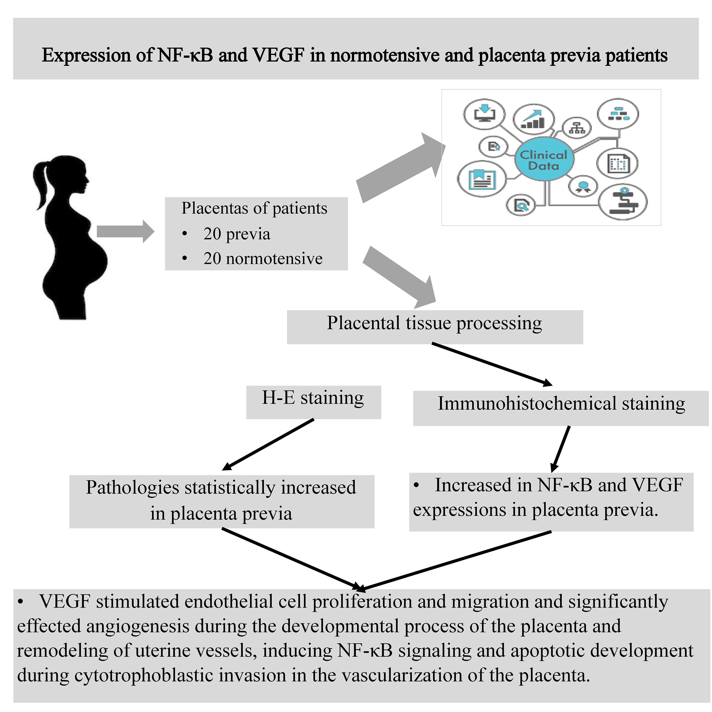
Objectives. The nuclear factor kappa-light-chain-enhancer of activated B cells (NF-κB) pathway and vascular endothelial growth factor (VEGF) are known to play crucial roles in inflammation and angiogenetic regulation. This study aimed to demonstrate the morphometric and immunohistochemical effects on inflammation and angiogenesis underlying placenta previa.
Materials and methods. Twenty pregnant patients with placenta previa and 20 healthy pregnant patients, all between 30 and 38 weeks gestational age, were included in the study. The gestational age of the pregnancies was determined according to the last date of menstruation and/or ultrasonographic measurements. Blood samples and clinical data were obtained from the prenatal patient groups. Samples were taken from the connecting stem region from both groups.
Results. The mean difference between the control and placenta previa patients was statistically significant for the parameters of blood vessels in villi, diameter of floating small villus, decidual cells, syncytial knots, congestion in blood vessels, fibrinoid accumulation, and inflammation. Significant degeneration and apoptotic changes in the syncytial cells of the root villi and an increase in syncytial nodes and bridges were observed in the placenta previa specimens. In the connecting stem region of the placenta previa samples, blood vessel dilatation, endothelial cell hyperplasia and a higher number of syncytial nodes were observed. In the immunohistochemical examination of the placenta previa samples, an increase in NF-κB and VEGF expression was observed in the endothelial cells, syncytial cells and Hofbauer cells.
Conclusions. Vascular endothelial growth factor was found to stimulate endothelial cell proliferation and migration, and to significantly affect angiogenesis during the developmental process of the placenta and remodeling of the uterine vessels, inducing NF-κB signaling and apoptotic development during cytotrophoblastic invasion in the vascularization of the placenta.
Key words: VEGF, NF-κB, placenta previa, chorionic villus
Background
Placenta previa is an obstetric condition in which there is atypical localization of the placenta around the internal cervical os. It is accompanied by painless vaginal bleeding in the 3rd trimester. It is a gynecological disease that can partially or completely obscure the cervical opening, with the placenta localized in the lower part of the uterus.1 Placenta previa is known to occur in 1 out of 200 pregnant women. It is important to detect this condition in the early stages of pregnancy because of the essential role of the follow-up. Placenta previa can complicate delivery and lead to life-threatening complications for the mother and baby, especially when it occurs close to the birth. There has been a remarkable increase in the incidence of placenta previa in recent years. It has been reported that cesarean section techniques, surgical interventions to the uterus, births at advanced age, high parity, tobacco and substance use, and in vitro techniques can trigger this disease.1, 2, 3
It is known that placenta previa can lead to cardiac problems due to pregnancy, prolapse of the placenta and accompanying mortality.4, 5 Dysfunction in arterial nutrition leads to the inability of the uterus and placenta to receive enough blood. These findings suggest that placental factors help in immune modulation, which is essentially required for successful pregnancy completion. There is a regulated balance between pro-inflammatory and anti-inflammatory factors. The emergence of regulatory T cells is induced with a suppressive effect against autoreactive T cell effectors. Indeed, a functioning neuropeptide system contributes to overall health, and changes in the expression of these neuropeptides and/or their receptors result in altered susceptibility to inflammatory and autoimmune diseases.6 Surgery may cause damage to the endometrium and myometrium of the uterus, increasing the risk of placenta previa.2
The signaling pathway leading to the eventual activation of nuclear factor kappa-light-chain-enhancer of activated B cells (NF-κB) plays an important role in immune and inflammatory responses.7 Regulation of maternal T cells during pregnancy is mediated by NF-κB p65; studies have reported that the degradation of p65 in maternal T cells is induced by the activation of fatty acid synthase (Fas). Placental exosomes expressing the Fas ligand (a type II membrane protein that belongs to the tumor necrosis factor superfamily) have been implicated as having an immunomodulatory function during pregnancy.8 In a study on congenital Zika syndrome in pregnant women, it was reported that Smaducin-6 (a membrane-bound Smad6-derived peptide) blocked the NF-κB activation mechanism via Pellino E3 ubiquitin protein ligase 1 but had no direct effect on congenital Zika syndrome.9 Olmos-Ortiz et al. observed that prolactin, toll-like receptor 4 (TLR4) expression, and mechanisms related to NF-κB phosphorylation provide and organize the upregulation of lipopolysaccharides. They reported that prolactin may play an important role in limiting inflammatory responses to lipopolysaccharides in the human placenta and inhibiting immune responses against infections that may threaten the outcomes of pregnancy. It is the first evidence of this mechanism for the anti-inflammatory activity of prolactin in the human placenta.10 They showed that infection of Listeria monocytogenes upregulates TLR2 and cytosolic DNA sensing pathways, as well as the downstream pro-inflammatory circuit in which NF-κB is involved.11 The effects of fatty acids on the placental inflammatory cytokines in relation to TLR4/NF-κB were investigated in a study in which trophoblast cells isolated from 14 normal-term human placentas were incubated with fatty acids. According to the study, the long-chain saturated fatty acids – stearic acid and palmitic acid – stimulated the secretion and synthesis of tumor necrosis factor-α (TNF-α) and interleukin (IL)-6 and 8 by trophoblast cells at a statistically significant level. The authors stated that unsaturated fatty acids do not change cytokine expression and that palmitate-induced inflammatory effects are accompanied by TLR4 activation, NF-κB phosphorylation and nuclear translocation.12 Pro-inflammatory stimulation of macrophage colony-stimulating factor (M-CSF) secreted by free 1st-trimester decidual cells in pregnant women and its relationship with macrophage development were investigated. It has been found that in free 1st-trimester decidual cells, IL-1β and TNF-α signal along the NF-κB pathway to induce M-CSF.13 It has been suggested that the interaction between Nestin and cyclin-dependent kinase 5 may lead to the progression of placenta previa by regulating NF-κB.14
In recent years, a great deal of research has focused on the relationship between abnormal angiogenesis and placenta previa.3 Vascular endothelial growth factor (VEGF) is known as a signaling protein that stimulates vasculogenesis and angiogenesis.15, 16 Chehroudi et al. stated that umbilical cord inflammatory cytokines and VEGF have not been extensively studied in terms of pregnancy disorders or maternal/neonatal causes. In that study, maternal and neonatal variables, as well as VEGF-A, VEGF-C and VEGF-D expression in umbilical cord lysates were examined. The authors determined that VEGF-A is strongly suppressed in adverse pregnancy cases.17 In another study, choroidal neovascularization was induced by laser and immune cells, and the neovascularization was analyzed using polymerase chain reaction (PCR). In the early inflammatory phase after the laser exposure, anti-placental growth factor, VEGF-A, was reported to be significantly upregulated. They observed that the increase in VEGF-A was limited to the scar, while anti-placental growth factor showed a wider spread.18 The expression of VEGF in hypertension, preeclampsia and preterm birth cases has been examined in various studies. It has been reported that the expression level of VEGF decreased due to hypertension,19 the level of VEGF receptors increased in preeclampsia,20 and the expression level of VEGF was very high in cases of premature delivery.21, 22
Vascular endothelial growth factor plays a role in angiogenic regulation and is secreted during pregnancy in sufficient amounts from cell groups outside the placental villi; it promotes proliferation, adhesion and/or invasion.23 Human deciduas from the 1st, 2nd and 3rd trimesters were examined via immunocytochemistry. The viability of total decidual cells and different cell isolates was not affected by triiodothyronine. In the 1st trimester, triiodothyronine was reported to reduce VEGF-A secretion by total decidual cells and to increase angiopoietin-2 secretion by stromal-depleted cells.24 They suggested that VEGF and soluble fms-like tyrosine kinase-1 levels are important for diagnosing placenta previa and can be considered additional parameters to differentiate between placenta accreta and increta.25 Moreover, the increase in the expression of high mobility group box protein 1 in the placenta may be related to the pathogenesis of placenta previa by regulating the expression of the proangiogenic factor VEGF.26 In another study, VEGF and leptin levels in 2nd-trimester amniotic fluid samples taken between 16 and 20 weeks of pregnancy were compared with a control group at 37 weeks. According to their results, amniotic fluid VEGF levels in the 2nd trimester predict preterm delivery better than leptin levels.27
The first blood vessel formation begins when angioblasts of mesenchymal origin gather in groups from the yolk sac of the embryo and develop the endothelial tube. The first developing veins reform an existing circulatory system, and both regional proliferation and regression, as well as branching and migration, are observed. With angiogenesis forming the new vascular system, mature endothelial cells divide and new capillaries are formed. It is known that VEGF signaling must be activated for vasculogenesis and angiogenesis to occur fully.28, 29
Objectives
This study aimed to determine the expression levels of NF-κB and VEGF in the placenta in order to understand the possible morphometric and immunohistochemical effects on inflammation and angiogenesis underlying placenta previa.
Materials and methods
Study population
This study was carried out at Van Özel Akdamar Hospital, Department of Obstetrics and Gynecology, between May 2021 and February 2022. It was approved by the ethics committee of Van Akdamar Hospital, Van, Turkey. The study included 20 pregnant patients with placenta previa and 20 healthy pregnant patients between 30 and 38 weeks gestational age.
The gestational ages of the pregnancies were determined by the last date of menstruation and/or ultrasonographic measurements. Blood samples and clinical data were obtained from the prenatal patient groups and compared. Placental samples from both the normal placenta and placenta previa groups were taken from under the connecting stem region and evaluated (Figure 1).
Histological analysis
For both groups, placental tissue samples (1 cm × 1 cm × 1 cm) were taken from around the umbilical cord under sterile conditions immediately after delivery and dissected. After soaking in 10% formol saline for at least 1 day, the samples were kept in 70% ethanol. The placental specimens were dehydrated in a routine ascending series of alcohol, cleared in terpineol, and then embedded in paraffin wax. For histological examination, 4–6-µm thick transverse sections were obtained and stained with hematoxylin and eosin (H&E).
Immunohistochemistry staining
The sections were left in distilled water and washed for 3–5 min in phosphate-buffered saline (PBS) (catalog No. 10010023; Thermo Fisher Scientific, Fremont, USA). Antigen uptake was performed in a microwave (700 W; Bosch®, Gerlingen, Germany) for 3 min at 90°C. For proteolysis, the sections were heated in a microwave oven at 700 W in a citrate buffer (pH 6) solution. The sections were washed for 3–5 min in PBS and incubated for 20 min in hydrogen peroxide (H2O2) (K-40677109, 64271; Merck, Darmstadt, Germany) (3 mL 30% H2O2 + 27 mL methanol). The sections were then washed for 3–5 min in PBS and blocked for 8 min with Ultra V Block (lot: PHL150128; Thermo Fisher Scientific). After drainage, primary antibodies (NF-κB antibodies, mouse monoclonal antibodies (1/100) and VEGF antibodies were applied. The sections were incubated and kept at 4°C overnight. Then, they were washed for 3–5 min in PBS and reincubated with applied secondary antibodies (Histostain-Plus Kit; Invitrogen, Carlsbad, USA) for 20 min. Next, the sections washed with PBS were exposed to streptavidin peroxidase (lot: PHL150128; Thermo Fisher Scientific) for 20 min. The sections were washed for 3–5 min in PBS and treated with 3,3’-diaminobenzidine (DAB) (lot: HD36221; Thermo Fisher Scientific) for 3–10 min (it was kept for minimal and maximal times between 3 and 10 min depending on the intensity of expression). The slides showing the reaction were stopped in PBS. After counterstaining with Harris Haematoxylin for 45 s, the slides were dried with residual alcohol and cleaned in xylene. The slides were mounted with Entellan (Sigma-Aldrich, St. Louis, USA), and histopathological examinations were performed using an Olympus BH-2 light microscope (Olympus Corp., Tokyo, Japan). Semiquantitative scaling of immunoreactivity was carried out. The intensity of staining was graded from 0 to 4 (0 – no staining, 1 – faint staining, 2 – moderate staining, 3 – intense staining, and 4 – highly intense staining). The values of 0–2 were considered one group, and the values of 3–4 were considered the other group.
Statistical analyses
The IBM SPSS Statistics for Windows v. 21.0 (IBM Corp., Armonk, USA) software was used for statistical analyses. Measured variables were presented as mean ± standard deviation (M ±SD) and median (interquartile range (IQR)). Categorical variables were presented as numbers and percentages (%). The conformity of the data to normal distribution was analyzed using the Shapiro–Wilk test and Q–Q plots. The Student’s t-test was used for normally distributed variables, the bootstrap test for means was used for not normally distributed variables, and the Welch’s t-test was used when equal variances were not assumed to compare the control and placenta previa groups. The Fisher’s exact test was used for the comparison of qualitative histologic parameter variables in the control and placenta previa groups. The hypotheses were taken in 2 directions, and p ≤ 0.05 was considered statistically significant.
Results
Histopathological examination
In the control group, the main root villi at cell junctions and syncytial cells in free villi were observed. Chromatin-rich nuclei were located in the cell center. Cytotrophoblast cells were oval in appearance, and the blood vessels in the villi were lined with squamous endothelial cells with regular lumens. There were a few syncytial nodes and bridges in the villi, and decidual cells with polygonal chromatin density were also observed polygonally (Figure 2A).
In the placenta previa group, a notable degeneration and apoptotic changes in the syncytial cells of the root villi and an increase in syncytial nodes and bridges were observed. Excessive expansion and occlusion in blood vessels, hyperplastic endothelial cells, and thinning and degenerative differences in the connective tissue fibers of the villi were observed (Figure 2B).
Immunohistochemical examination
of NF-κB expression
In the control group, the expression of NF-κB was mild in the syncytial cells of the root and free villi, and moderate in the vascular endothelial cells, macrophages and fibroblasts in the connective tissue cells (Figure 3A).
In the placenta previa group, an increase in the NF-κB expression was observed in the large root villi in the connecting stem region and in the syncytial nodes and bridges of mature chorionic villi. The NF-κB protein expression was significantly positive in Hofbauer cells, endothelial cells and connective tissue cells located in small-scale blood vessels (Figure 3B).
Immunohistochemical examination of VEGF expression
In the control group, the expression of VEGF was moderate in the root villi and blood vessel endothelial cells, syncytial cells, as well as some Hofbauer cells outside the decidual area region (Figure 3C).
In the placenta previa group, a significant increase in the VEGF expression was observed in the root villi, free villi, blood vessel endothelial cells, syncytial nodes and bridges, as well as Hofbauer cells (Figure 3D).
In the transverse section of the stem villus in the control group, cytotrophoblast cell nuclei in the heterochromatin structure, membrane thickness and organelle structures were normal, as well as mitochondria and endoplasmic reticulum structures in the decidual cell cytoplasm. The nuclei of the blood vessel endothelial cells protruded toward the heterochromatin and lumen, and Hofbauer cells were seen in a regular structure outside. Basement membrane thicknesses were normal (Figure 4A). In the placenta previa group, degenerative changes in cytotrophoblast cell nuclei were observed. The dilatation of endoplasmic reticulum structures in the cytoplasm, deterioration in the structure of mitochondrial cristae, and hyalinized areas in the villus connective tissue were observed (Figure 4B).
Statistical results
Statistical analysis of the parameters belonging to the control and placenta previa patients is shown in Table 1, Table 2. The mean differences between the control and placenta previa patients were statistically significant for the parameters of epithelial thickness in villi, diameter of floating small villi, diameter of blood vessels in villi, diameter of decidual cells, syncytial knots, congestion in blood vessels, fibrinoid accumulation, inflammation, degeneration in decidual cells, VEGF and NF-κB expression, maternal age, gravida, parity, and gestational age. Differences in significance levels between parameters are provided in detail in Table 1, Table 2.
The diameters of floating small villi, epithelial thickness in villi, diameter of blood vessels in villi, diameter of decidual cells, parity, gestational age, age, and gravida values of both groups are also given as a boxplot (Figure 5).
Discussion
It has been reported that inflammation is triggered by placental malformations, differentiation of the uterine endometrium and significant scarring in the uterus due to surgical approaches or other gynecological operations.15 It is known that the presence of scar tissue can lead to the development of inflammation in epithelial tissue, as well as vascular insufficiency in the cells of the placenta, such that the tissues cannot benefit from oxygen sufficiently.30 Inflammatory factors initiate the morphology of the uterine spiral vessels. In the lower segment of the uterus, the presence of pelvic adhesions causes atypical blastocyst implantation and dysperistalsis of uterine contraction to form placenta previa.31 Trophoblast cell infiltration and molecular regulation of placental angiogenesis play an important role in the molecular mechanisms underlying placenta previa.
The NF-κB has been shown to regulate many genes involved in important cellular responses, such as inflammation, migration, proliferation, and apoptosis. It has been noted that the early cytoplasmic events leading to the release and translocation of NF-κB are insufficient to explain the complex nature of NF-κB biology.32, 33 The NF-κB protein expression is also manifested in other diseases characterized by high oxidative stress and inflammatory factors, such as acquired immunodeficiency syndrome (AIDS), atherosclerosis, rheumatoid arthritis, osteoporosis, Alzheimer’s disease, and ischemia/reperfusion injury.34 In another clinical study, Atic and Deveci stated that endothelin-1 expression induced NF-κB levels in macrophage cells in the development of diabetic foot ulcers.35
It has been reported that NF-κB protein expression is lower in normal placentas compared to preeclamptic placentas. Also, the relationship between NF-κB expression and leukocytes and vascular tissue has been demonstrated, especially in preeclampsia.7, 36, 37 Another studies determined that an increase in the expression of NF-κB protein in the placenta can change the expression of different genes in preeclampsia and contribute to the pathogenesis of the disease. The activation of placental NF-κB in preeclampsia supports this observation, as it increases placental TNF-α, cyclooxygenase-2 and thromboxane levels.38, 39 Li et al. used quantitative PCR, western blot and immunofluorescence techniques to study the placental tissue of patients with diabetes mellitus and autophagy. They reported that women with gestational diabetes mellitus have an increased risk of neonatal infection through placental inflammation and autophagy. According to their quantitative PCR results, due to gestational diabetes mellitus, hypoxia-inducible factor-1α and the TLR4/myeloid differential protein-88/NF-κB pathway increase protein expression levels. In western blot results, they reported that NF-κB inhibitor alpha and NF-κB P65 expression in the placenta significantly increased due to gestational diabetes mellitus.40 Similarly, insulin resistance was investigated to understand morphological changes in gestational diabetes mellitus. The TLR4/ myeloid differential protein-88/NF-κB pathway can play an important role in the emergence of insulin resistance in the placentas of pregnant women with gestational diabetes mellitus.41 The TLR4/myeloid differential protein-88/NF-κB pathway has been examined in other experimental studies. It has been reported that these protein expressions are significantly increased in experimental animals due to diabetes mellitus.42, 43, 44 High-mobility group box 1, receptors for advanced glycation end products (AGEs), and the NF-κB p65 pathway, which are genes related to mRNA levels, were analyzed with PCR in 61 normotensive pregnant women and 64 women with severe preeclampsia. In the comparison between the 2 groups, the mRNA levels of high-mobility group box 1, receptors for AGEs, and NF-κB p65 were higher in the severe preeclamptic placentas.45
In this study, it was observed that NF-κB protein expression was prominent in the connective root region of placenta previa and in the large root villi in the syncytial nodes and bridges of mature chorionic villi. There was also a significant increase in the NF-κB protein expression in Hofbauer cells, endothelial cells and connective tissue cells located in small-scale blood vessels (Figure 3B). We determined that the increase in cytokine signaling that developed after the increased inflammatory reaction may be due to an increase in the inductive effect of macrophages.
Chantraine et al. compared the total area occupied by vessels in normal and placenta-increta placental beds. They reported that the vessels in placenta increta were significantly less frequent and larger. They stated that the vascular location is more heterogeneous, especially in placenta increta.46 Another study investigated the effects of maternal serum levels of VEGF, placental growth factor and soluble fms-like tyrosine kinase 1 on patients with and without complete placenta previa. According to the study findings, there was a decrease in the serum level of VEGF but an increase in the tissue level in the placenta. As a result of electrophoresis performed in placenta previa, VEGF level increased.25, 47 It has been reported that VEGF, which increases in response to placental hypoxia, is not functional during the pathogenesis of preeclampsia. Moreover, since the ligand-receptor binding is impaired, the amount of free VEGF in the preeclamptic placenta increases, impairing vascular function as a result of endothelial cell damage.48 A significant increase in VEGF expression was observed in the root villi, free villi, blood vessel endothelial cells, syncytial nodes and bridges, and Hofbauer cells due to the hypoxia effect after placenta previa.
It has been stated that cytotrophoblast cell degeneration can be explained by placental ischemia and that other etiological factors are not important. According to the histopathological findings of our study, there was a significant decrease in the transfer and synthetic activity of trophoblasts. Cellular respiration was also affected.49 Thickening of the basal lamina, increase in intervillus collagen and marked differentiation in mitochondrial structure were noted, with great damage to the multinucleated protoplasm masses, which lost their cell membranes under hypoxic conditions and became single cells. Significant degenerative changes in cytotrophoblast cell nuclei were observed in the placenta previa group in our study. Dilatation in endoplasmic reticulum structures in the cytoplasm, deterioration in the mitochondrial cristae structure and the appearance of hyalinized areas in the villus connective tissue were evaluated as negative images for cell and villus integrity disruption and trophoblast invasion, as well as for angiogenesis (Figure 4B). Faraji et al. evaluated maternal characteristics, VEGF and placental growth factor in the placenta accreta spectrum. The placental growth factor serum level was found to be statistically significantly higher in the placenta accreta spectrum and placenta accreta spectrum subgroups compared to the normal placenta group. They also stated that the previa status of the placenta accreta spectrum patients did not affect serum levels of VEGF and placental growth factor.50 Faraji et al. also stated that insufficient VEGF secretion and invasion of interstitial extravillus trophoblasts to a metastable cell phenotype and advanced myometrial invasion causes placenta previa. In a study of VEGF and phosphotyrosine immunostaining, it was reported that epithelial–mesenchymal invasion, especially the expression of vimentin and cytokeratin-7, was predominant in extravillus trophoblasts.47 Biberoglu et al. compared abnormal and normal placentation and data in terms of fms-like tyrosine kinase 1 and VEGF. They did not find a statistically significant result in maternal serum values of fms-like tyrosine kinase 1, placental growth factor, fms-like tyrosine kinase 1/placental growth factor ratio, and VEGF in the abnormal placenta groups.51 However, another study reported that amniotic fluid VEGF and leptin levels were the highest in women with placenta previa and the lowest in women with intrauterine growth retardation and pregnancy-induced hypertension.27
Limitations
The 2 main limitations are that the study focused on a relatively small group, and it was composed only of Turkish women. The NF-κB and VEGF signaling pathways were not detected at serum level. Examination at tissue levels was performed at immunohistochemistry and ultrastructural level. More extensive research is needed for a greater understanding of this phenomenon.
Conclusions
Vascular endothelial growth factor was found to stimulate endothelial cell proliferation and migration and significantly affect angiogenesis during the developmental process of the placenta and remodeling of the uterine vessels. Imbalances in VEGF can lead to aberrant placental vascular development. We determined that VEGF affects trophoblastic invasion by increasing NF-κB signaling as a result of hypoxia occurring after placenta previa and induces apoptotic development, which is critically important during cytotrophoblastic invasion in the vascularization of the placenta.


